Aim: This in vitro study investigates the limit of expansion forces and torque wrench forces developed by five skeletal bone expander designs (MICRO 2/4 expanders™) for clinical application. Material and Methods: A total of 30 skeletal expanders were placed in artificial bone blocks and mechanically tested, simulating maxillary expansion. Differences in jackscrew (Dentaurum™ [D], Superscrew™ [S] and Powerscrew™ [P]), number of orthodontic mini-implants (OMIs; two or four) and their placement inclinations (parallel 0° or 10° inclination) form five designs (D4/10°, S4/0°, S4/10°, P4/10° and P2/10°). Expansion forces and torque wrench values were registered, and radiographs were made initially and after 4 mm of expansion. Stress-strain curves were obtained after successive activations and the statistical analysis was performed as appropriate. Results: Plastic deformations in the OMIs and jackscrew occurred around the activation numbers 11–13, with torque wrench values in the range of 500–700 cN. The maximum expansion forces in expanders with four OMIs varied from 93.0 (D4/10°) to 166.6 N (P4/10°) whereas two OMI expanders (P2/10°) registered forces of 79.4 N. Radiographs revealed during loads bending forces (S4/00°, S4/10°) with jackscrew and OMIs deformation in a convex shape, and shear forces (P4/10°, P2/10°) demonstrated only OMIs deformation in a concave shape, providing 15% more expansive force. The jackscrew D4/10° did not have any deformation, but its wire key did not allow reliable activations from activation number 10 and compared to S4/10° and P4/10°, these expanders provided greater expansion forces ( P = 0.000 and P = 0.032, respectively). Conclusion: The different results obtained in stability and expansion forces indicate that if the activations are carried out under extreme conditions, they may have clinical importance with deformations and non-working expansion mechanics. Jackscrew designs play an important role in expansive forces and expander stability. Torque wrench values can be used clinically as a tool to asses the expansion forces and to avoid deformations.
Introduction Bone-borne miniscrew assisted palatal expansion (MAPE) is a common technique to improve maxillary transverse deficiency in young adolescents. Adult patients usually present a challenge, as they often require additional surgical assisted maxillary expansion (SARPE). There is still no clear statement about non-surgical expansion in adult patients using this technique. The aim of this study was to evaluate the success and complication rate of non-surgical palatal expansion in adults utilizing MAPE with a novel force-controlled polycyclic expansion protocol (FCPC). Methods This consecutive study consisted of 33 adult patients with an average age of 29.1 ± 10.2 years (min. 18 years, max. 58 years), including one dropout patient. First, four miniscrews were inserted and after 12-weeks latency, the expander was placed and the FCPC protocol was applied (MAPE group). In case of missing expansion, a SARPE was performed (SARPE group). After maximum expansion, a cone beam CT was made and widening of the midpalatal suture was measured. The outcome variables were successful non-surgical expansion and, with sample size power above 80%, the odds of failed non-surgical expansion and associated complications were evaluated. The primary predictor variable was age. Statistical analysis was performed using R (Version 3.1) to calculate power, to construct various models for measuring the odds of requiring surgical intervention/complications, and others. Results Successful non-surgical expansion was achieved in 27 patients (84.4%), ranging from 18 to 49 years. Mean age differed significantly between both groups (26.8 ± 8.2 years vs. 41.3 ± 9.9 years; p < 0.001). Mean expansion at the anterior and posterior palate for the MAPE group was 5.4 ± 1.5 mm and 2.5 ± 1.1 mm, respectively. Among these subjects’ complications were observed in 18.5%. Age significantly increased the odds of complications ( p = 0.019). Conclusions 1. The success rate of MAPE among individuals aged 18 to 49 years was 84.4%. 2. A V-shaped expansion pattern in the antero-posterior dimension was mostly observed. 3. Complications were significantly associated with age. 4. A careful expansion protocol seems to be beneficial to prevent unfavorable results in adult patients. Trial registration Consecutive cohort study, Review Board No. EK-2-2014/0016.
Purpose: The effects of orthognathic surgery (OGS) on the temporomandibular joint (TMJ) are still controversial. Based on a high-volume uniform collective, the function and clinical symptoms of the TMJ and the dysfunction index (Di) by Helkimo were evaluated prior and up to 2 years after OGS. Methods: A longitudinal cohort study was performed between 2006 and 2016. A comprehensive examination focusing on TMJ function, temporomandibular disorder (TMD) symptoms, and the Di had been performed preopertaively (T0) and postoperatively at 6 weeks (T1), 6 months (T2), 1 year (T3), and 2 years (T4). The predictor variables used were sex, age, skeletal class, type of surgery, and amount of dysfunction or number of TMD complaints before treatment. The outcome of this study was TMD symptoms and the Di. Descriptive and bivariate statistics were computed, and the significance level was set at P<.05. Results: The sample consisted of 375 patients (247 women, 128 men, mean age 28.1 +/- 9.4 years), 269 with skeletal Class II (71.7%) and 106 with skeletal Class III (28.3%) treated with sagittal split osteotomy bilateral sagittal split osteotomy (n = 173) or Le Fort I and bilateral sagittal split osteotomy (n = 202). Severe signs of dysfunction (Di 2/3) were seen in 5.9% of the patients before OGS and in 2.1% of the patients 2 years after OGS. The difference in Di (Di0/1 and Di2/3) between T0 and T1/T2 was statistically significant (P=.028; P=.011, respectively). TMD symptoms were significantly reduced 2 years after OGS (P=.028). Female gender (P=.013), skeletal Class II (P<.001), and counterclockwise rotation (P<.001) were the only risk factors significantly associated with the occurrence of TMD at T1. Conclusions: The results of this study suggest that in most cases TMD symptoms can be significantly reduced and only a few can be induced with OGS. No risk factors were found for long-term effects on the TMJ. (C) 2020 American Association of Oral and Maxillofacial Surgeons
Purpose: For surgically assisted rapid palatal expansion (SARPE), studies comparing different devices are missing. The aim was to evaluate expansion, relapse, and the complication rates for 3 different appliances. Patients and Methods: In this retrospective cohort study, tooth-borne (TB, n = 29), bone-borne (BB, n = 12), or orthodontic mini-implant (OMI)-borne (n = 13) appliances were used for SARPE in 2 clinical centers. Patients presenting with a maxillary width discrepancy greater than 5 mm were included. Measurements of cast models and radiographs were performed preoperatively, immediately after consolidation, and at 1 year postoperatively. All distances were measured between canines, premolars, and molars. Complications (tooth damage, infection, technical failure) were evaluated. The outcome variables were expansion, relapse, and complications. Type of appliance was selected as predictor variable. Withinand between-group differences regarding expansion, relapse, and complications for the 3 appliances were statistically analyzed. Results: In 54 patients (aged 28.8 +/- 8.6 years), mean expansion (+/- standard deviation) for the TB, BB, and OMI appliances immediately after consolidation was 5.29 +/- 1.39 mm, 5.22 +/- 1.72 mm, and 3.81 +/- 2.19 mm, respectively, at the dental level, and 5.51 +/- 1.40 mm, 4.66 +/- 2.03 mm, and 3.51 +/- 1.66 mm, respectively, at the bone level. Mean (+/- standard deviation) relapse for the TB, BB, and OMI appliances at 1 year postoperatively measured 1.44 +/- 1.01 mm, 0.76 +/- 1.37 mm, and 0.27 1.63 mm, respectively, at the tooth level and 1.19 +/- 0.93 mm, 0.71 +/- 0.96 mm, and 0.12 +/- 1.56 mm, respectively, at the bone level. Analysis of variance and the Tukey test showed significant differences between the TB and OMI appliances for expansion and relapse (P = .034 and P = .024, respectively). The overall complication rate differed significantly (P < .001), with rates of 17.2%, 66.7%, and 15.4% for the TB, BB, and OMI appliances, respectively. Conclusions: BB and OMI appliances resulted in less relapse. Owing to the high complication rate, the further use of BB appliances is questionable. (C) 2020 American Association of Oral and Maxillofacial Surgeons
Abstract Purpose The effects of orthognathic surgery (OGS) on the TMJ are still controversial. Based on a high-volume uniform collective, the function and clinical symptoms of the TMJ and the dysfunction index by Helkimo (Di) were evaluated prior and up to 2 years after OGS. Methods A longitudinal cohort study was performed between 2006 and 2016. A comprehensive examination focusing on TMJ function, TMD symptoms, and the Di had been performed pre- (T0) and postoperatively at 6 weeks (T1), 6 months (T2), 1 (T3), and 2 years (T4). The predictor variables used were sex, age, skeletal class, type of surgery, and amount of dysfunction or number of TMD complaints prior to treatment. The outcome of this study was TMD symptoms and the Di. Descriptive and bivariate statistics were computed, and the significance level was set at P Results The sample consisted of 375 patients (247 women, 128 men, mean 28.1 ± 9.4 years), 269 with skeletal class II (71.7%) and 106 with skeletal class III (28.3%) treated with sagittal split osteotomy (BSSO) (n = 173) or Le Fort I and BSSO (n = 202). Severe signs of dysfunction (Di2/3) were seen in 5.9% of the patients before and in 2.1% of the patients 2 years after OGS. The difference in Di (Di0/1 and Di2/3) between T0 and T1/T2 was statistically significant (P = .028; P = .011, respectively). TMD symptoms were significantly reduced 2 years after OGS (P = .028). Female gender (P = .013), skeletal class II (P Conclusion The results of this study suggest that in most cases TMD symptoms can be significantly reduced and only a few can be induced with OGS. No risk factors were found for long-term effects on the TMJ.
The aim of the present study was to evaluate the acceptance of a webinar series for continuing medical education hosted by the Austrian Society for Oral and Maxillofacial Surgery (ÖGMKG).A series of twelve webinars was streamed via the Internet and the participants’ satisfaction was evaluated by an online questionnaire.51 out of 140 participants (36.4%) completed the questionnaire completely and were included into the study for further analysis. The mean age of the participants was 37.9 ± 8.9 and did not vary significantly between gender (p = 0.53). The results of the questionnaire revealed a positive attitude of the participants towards this kind of webinar. The participants found that the webinars allowed for an adequate transfer of knowledge.Continuing medical education by webinars in oral and maxillofacial surgery is well accepted by the participants. Although both male and female participants had a positive attitude towards the webinars, females gave even better ratings than their male counterparts did.
Objectives: Dentofacial deformities can be analyzed by skeletal and soft tissue cephalometric analysis (CA). The aim was to evaluate the difference in reproducibility between both methods. Materials and methods: Lateral cephalograms of 112 patients (65 females and 47 males, 27.7 +/- 9.0 years) were oriented in natural head position (NHP) and digitized. The distances of skeletal (SNA, SNB, SnPog) and soft tissue (A', B' and Pog') landmarks relative to the respective norm values and the angles between the Nasion Sella line (NSL) and Frankfurt horizontal (FH) to NHP were measured for statistical evaluation and compared with respective data of an adult control group (CG) with class I occlusion and harmonic facial balance. Results: The mean differences (mm +/- SD) of skeletal and soft tissue landmarks were -2.4 +/- 4.4 (A), -7.0 +/- 9.3 (B), -6.3 +/- 11.2 (Pog),-0.9 +/- 1.8 (A'),-4.7 +/- 6.2 (B'), and-6.1 +/- 7.8 (Pog'), respectively. Pearsons's correlation (r) between the measurements of SNA/A', SNB/B' and SNPog/Pog' were r =.158 (p =.092), r =.662 (p<.001) and r =.655 (p<.001), respectively. The mean (+/- SD) angles between NSL and FH to NHP were -9.8 degrees +/- 5 and 0.0 degrees +/- 3.9, respectively. Conclusion: Variability of cranial-based measurements could give a possible explanation for the high variation and the low reproducibility of skeletal cephalometric analysis with soft tissue measurements. Soft-tissue cephalometric analysis would probably improve facial analysis and treatment planning.
Purpose: To evaluate piezosurgery for bilateral sagittal split osteotomy (BSSO) for its duration and inferior alveolar nerve (IAN) perturbation.Patients and Methods: In this prospective randomized study, the authors evaluated 100 BSSO procedures in 50 patients. Piezoelectric (group I) and conventional (group II) osteotomies were carried out on each side of the mandible of a patient by 2 specialists. The surgeons had at least 1 year of experience using piezosurgery. The period from incision to complete splitting of the mandibular bone was recorded (ie, procedure duration). The intraoperative status (visibility and relocation) of the IAN also was recorded. The neurosensory function of the IAN was measured by the 2-point discrimination threshold and static light touch methods before surgery and postoperatively (1, 3, and 6 weeks and 6 and 12 months). Parameters were compared between the test groups by the paired t, nonparametric Wilcoxon, or chi(2) test.Results: Intergroup comparison showed the mean duration of osteotomy was significantly shorter for group I (17 +/- 6 vs 25 +/- 9 minutes; P <.001). The rate of intraoperative exposures of the IAN was slightly lower for group I (68%) compared with group II (81%). However, the difference was not relevant. Neurosensory disturbance and recovery of the IAN did not differ between groups.Conclusion: Piezoelectric osteotomy requires considerably less time than conventional mechanical approaches, but shows no advantage in preventing neurosensory perturbation. (C) 2017 American Association of Oral and Maxillofacial Surgeons
In a case report in the May 2016 issue, Carlson et al reported the use of a microimplant-assisted rapid palatal expansion (MARPE) appliance to correct transverse maxillary deficiency in an adult (Carlson C, Sung J, McComb RW, Machado AW, Moon W. Microimplant-assisted rapid palatal expansion appliance to orthopedically correct transverse maxillary deficiency in an adult. Am J Orthod Dentofacial Orthop 2016;149:716-28). The report was interesting and very systematic, but it was missing some important details. As the authors stated in their introduction, true skeletal expansion is free of dental and alveolar side effects, and only pure skeletally anchored expanders can achieve this. However, the appliance used in this case report, a MARPE appliance, is a dentally and skeletally anchored expander with evident dental (molar) side effects. MARPE means mini-implant "assisted" rapid maxillary expansion, which expresses skeletal anchorage in additional to dental anchorage. These appliances are subsumed under the "hybrid" type of expanders as reported by Wilmes and Drescher1Wilmes B. Drescher D. A miniscrew system with interchangeable abutments.J Clin Orthod. 2008; 42: 574-580PubMed Google Scholar and Ludwig et al.2Ludwig B.G. B Zorkun B. Wilmes B. Kinzinger G. Lisson J. Forcierte Gaumennahterweiterung mit skelettalem Kraftansatz: die Hybrid-GNE.Kieferorthop. 2009; 23: 267-274Google Scholar Hybrid expanders use mini-implants in the lateral anterior palate and the maxillary first molars as anchorage. The MARPE is a hybrid expander because it is anchored on 4 mini-implants in the lateral posterior palatal region and the maxillary first molars. To clarify anchorage definitions, a term such as BAME should be used, for pure "bone-anchored maxillary expanders." The letter R, for "rapid," should be omitted, because thanks to solid anchorage, these expansions can also be performed in a slow way. This technique was presented with the Micro6-expander in 2013 (Fig).3Winsauer H. Vlachojannis J. Winsauer C. Ludwig B. Walter A. A bone-borne appliance for rapid maxillary expansion.J Clin Orthod. 2013; 47: 375-381PubMed Google Scholar Explaining the outcomes of their rapid palatal expansion, the authors described "uprighting" of the maxillary first molars. The change in degrees indicates buccal tipping of those molars, a highly undesirable side effect with a high risk for periodontal harm or root damage. Pure bone-borne expanders3Winsauer H. Vlachojannis J. Winsauer C. Ludwig B. Walter A. A bone-borne appliance for rapid maxillary expansion.J Clin Orthod. 2013; 47: 375-381PubMed Google Scholar, 4Walter A. Wendl B. Ploder O. Mojal S. Puigdollers A. Stability determinants of bone-borne force-transmitting components in three RME hybrid expanders—an in vitro study.Eur J Orthod. 2016 Apr 1; ([Epub ahead of print])Google Scholar do not show any translational dental movements or buccal tipping5Winsauer H, Ploder O, Katsaros C, Puigdollers A, Walter A. Skeletal changes after polycyclic maxillary expansion with a pure bone-borne device without SARPE in adult patients. Poster presentation at the 91st Congress of The European Orthodontic Society in Stockholm, Sweden; June 11, 2016.Google Scholar because no teeth are involved. After the expansion, they even show minor spontaneous palatal tipping5Winsauer H, Ploder O, Katsaros C, Puigdollers A, Walter A. Skeletal changes after polycyclic maxillary expansion with a pure bone-borne device without SARPE in adult patients. Poster presentation at the 91st Congress of The European Orthodontic Society in Stockholm, Sweden; June 11, 2016.Google Scholar of the buccal teeth, as Carlson et al described for the maxillary canines only. In this case report, the mini-implant- and molar-anchored expander was opened 10 mm, achieving an overall palatal widening of 6 mm between the first molars. Now the question arises, what has happened to the missing 4 mm? Four millimeters of anchorage loss (2 mm per side) in this young adult would mean either that the implants have moved through the bone, or the implants have tipped or deformed, or even both. A cone-beam computed tomography cross-section image of this region could have answered this question and given visual information about the bone height and the situation around the mini-implants, although as stated in the Treatment progress section, an image with the appliance in place had been taken. Why was it not presented in this case report? Why is there is not a single photo of the appliance before or after expansion in this presentation? The estimated average bone height in this region (16 mm behind the incisive foramen and 7 mm lateral of the midline) is around 3 mm.6Winsauer H. Vlachojannis C. Bumann A. Vlachojannis J. Chrubasik S. Paramedian vertical palatal bone height for mini-implant insertion: a systematic review.Eur J Orthod. 2014; 36: 541-549Crossref PubMed Scopus (35) Google Scholar How much higher is the anchorage loss in older adults (>25 y) with more resistance against expansion due to more mature bone?7Knaup B. Yildizhan F. Wehrbein H. Age-related changes in the midpalatal suture. A histomorphometric study.J Orofacial Orthop. 2004; 65: 467-474Crossref PubMed Scopus (94) Google Scholar According to the examinations by Walter et al,4Walter A. Wendl B. Ploder O. Mojal S. Puigdollers A. Stability determinants of bone-borne force-transmitting components in three RME hybrid expanders—an in vitro study.Eur J Orthod. 2016 Apr 1; ([Epub ahead of print])Google Scholar directly loaded titanium mini-implants with a diameter of 1.5 mm under these circumstances will undergo major deformations with the risk of breakage. According to this study, hybrid expanders with dental anchorage and the assistance of 2 to 4 mini-implants should be used only in adolescent patients, not in adults.4Walter A. Wendl B. Ploder O. Mojal S. Puigdollers A. Stability determinants of bone-borne force-transmitting components in three RME hybrid expanders—an in vitro study.Eur J Orthod. 2016 Apr 1; ([Epub ahead of print])Google Scholar In a study of 33 adults with pure BAME, 90% of the patients between 23 and 33 years (n = 11) had successful palatal widening without SARPE and no dental side effects.8Winsauer H, Walter A, Muchitsch AP, Winsauer C, Jaeschke D, Katsaros C, et al. Pure bone borne maxillary expansion with 4 mini-implants in adults with and without SARPE: a consecutive study of 35 patients. Oral presentation at the 91st Congress of The European Orthodontic Society in Venice, Italy; June 13, 2015.Google Scholar In this letter, we have focused on different types of anchorage for palatal expanders. Thorough treatment decisions should be made according to the patients age in regard to appliance design and opening protocol (rapid vs slow vs polycyclic). The challenges, however, are to prevent dental side effects and to enable palatal widening in even mature adult patients without SARPE. Authors' responseAmerican Journal of Orthodontics and Dentofacial OrthopedicsVol. 151Issue 1PreviewWe agree that the term "hybrid" is appropriate if the dentition is used with the implants for anchorage for the expansion force. However, this does not mean that incorporating the dentition in an appliance design always makes it a hybrid or dental expander. If the purpose of the dentition in the appliance is not anchorage for the expansion force but something else, then the term "true skeletal anchorage" can be applied to the appliance. The expander we used, which we now officially call the "maxillary skeletal expander" (MSE), was originally designed to deliver the expansion force to 4 implants inserted deeply, engaging both layers of the cortical bone (palatal and nasal layers); the first molars were used to stabilize the position of the jackscrew during the expansion rather than for anchorage. Full-Text PDF
Background: The aim was to test which component [wire arm, connecting abutment attachment, and orthodontic mini-implant (OMI)] of the force-transmitting system (FTS) in the anterior palate of three commonly used hybrid expanders (HEs; WILMES-HE, LUDWIG-HE, and WINSAUER-HE) deforms under increasing load.Materials and methods: Crude single and double wire arms were tested individually. Non-opening of the maxillae halves was simulated in artificial bone blocks with single wire and double wire FTS specimens. OMIs were inserted 8 mm and underwent 6 mm of continuous static lateral loading. Deformation angles were measured (X-ray, n = 6) at 0, 3 and 6 mm feed. OMIs and abutments were scan electron microscope (SEM) evaluated.Results: After 1.0 mm of loading, the single wire arm of all FTS deformed between 63.4 (16.5) N and 76.2 (18.4) N, and the double wire arm of reinforced FTS (wires positioned 'side by side') deformed after 1.0 mm between 110.0 (18.4) N and 134.8 (22.3) N. The crude single wire resisted 89 (5.1) N until plastic deformation, whereas the crude double wire positioned 'on top of each other' resisted 438 (21.3) N. At 6 mm loading, the reinforced WINSAUER-HE FTS withstood a maximum load of 320.9 (31.1) N and the reinforced LUDWIG-HE FTS 19% less, both under great deformation of double wires and OMIs. The screw-fixated WILMES-HE FTS abutment attachment (overlapping OMI head 34%) detached around 250 N. The bonded WINSAUER-HE and LUDWIG-HE abutment attachments did not detach. Nor did the modified bonded plus the modified screw-fixated WILMESHE abutment attachment when overlapping 100%.Conclusion: Early OMI and single wire arm deformation in HEs are crucial for unsuccessful RME in more mature maxillae. Double wire arms should be obligatory. OMIs with inner diameter greater 1.36 mm are recommended. One hundred per cent overlapping abutment attachments do not detach.
PURPOSE:The purpose of this study was to measure and compare the working time for virtual surgical planning (VSP) in orthognathic surgery in a largely office-based workflow in comparison with conventional surgical planning (CSP) regarding the type of surgery, staff involved, and working location. MATERIALS AND METHODS:This prospective cohort study included patients treated with orthognathic surgery from May to December 2016. For each patient, both CSP with manual splint fabrication and VSP with fabrication of computer-aided design-computer-aided manufacturing splints were performed. The predictor variables were planning method (CSP or VSP) and type of surgery (single or double jaw), and the outcome was time. Descriptive and analytic statistics, including analysis of variance for repeated measures, were computed. RESULTS:The sample was composed of 40 patients (25 female and 15 male patients; mean age, 24.6 years) treated with single-jaw surgery (n = 18) or double-jaw surgery (n = 22). The mean times for planning single-jaw surgery were 145.5 ± 11.5 minutes for CSP and 109.3 ± 10.8 minutes for VSP, and those for planning double-jaw surgery were 224.1 ± 11.2 minutes and 149.6 ± 15.3 minutes, respectively. Besides the expected result that the working time was shorter for single-versus double-jaw surgery (P < .001), it was shown that VSP shortened the working time significantly versus CSP (P < .001). The reduction of time through VSP was relatively stronger for double-jaw surgery (P < .001 for interaction). All differences between CSP and VSP regarding profession (except for the surgeon's time investment) and location were statistically significant (P < .01). The surgeon's time to plan single-jaw surgery was 37.0 minutes for CSP and 41.2 minutes for VSP; for double-jaw surgery, it was 53.8 minutes and 53.6 minutes, respectively. CONCLUSIONS:Office-based VSP for orthognathic surgery was significantly faster for single- and double-jaw surgery. The time investment of the surgeon was equal for both methods, and all other steps of the workflow differed significantly compared with CSP.
Two techniques to separate the lower incisors prior to mandibular symphyseal distraction osteogenesis (MSDO) were evaluated with respect to avoiding tooth damage.METHODS:Fifty patients (20.2 ± 7.0 years) requiring MSDO were treated with a tooth-borne appliance by utilizing two preoperative protocols to separate the central incisors: i) brackets and a V-bent wire with an open coil spring (two-step; TS; n = 24) and ii) a wire attached from the appliance to the central incisors with subsequent dento-alveolar expansion prior to surgery (one-step; OS; n = 26). The distance between the lower incisors was measured preoperatively on radiographs and measurements at the cast models were performed. Complications and radiographs were analyzed.RESULTS:The mean distance (±SD) between the lower central incisors for OS and TS prior to surgery was 3.44 ± 1.05 and 3.18 ± 1.13 mm, respectively. The mean expansion for OS and TS was 4.3 ± 2.9 and 4.3 ± 2.7 mm at the dental level and 3.8 ± 3.2 and 4.0 ± 2.1 mm at the bone level, respectively. Four patients undergoing the TS and one patient undergoing the OS showed transient dental complications.CONCLUSION:Pre-surgical dento-alveolar expansion by utilizing a one-step technique to separate the lower central incisors reduces the risk of permanent tooth damage and weakens the mandibular bone in the midline.
Background: The long-term stability of the segments is the goal of transverse maxillary expansion.
The aim was to evaluate three different biodegradable polylactic acid- (PLA-) based osteosynthesis materials (OM). These OM (BioSorb, LactoSorb, and Delta) were used in 64 patients of whom 55 (85.9%) had fractures of the zygoma, five (7.8%) in the LeFort II level, two of the frontal bone (3.1%), and two of the maxillary sinus wall (3.1%). In addition to routine follow-up (FU) at 3, 6, and 12 months (m) (T1, T2, and T3) all patients were finally evaluated at a mean FU after 14.1 m for minor (e.g., nerve disturbances, swelling, and pain) and major (e.g., infections and occlusal disturbances) complications. Out of all 64 patients 38 presented with complications; of these 28 were minor (43.8%) and 10 major (15.6%) resulting in an overall rate of 59.4%. Differences in minor complications regarding sensibility disturbance at T1 and T3 were statistically significant (P=0.04). Differences between the OM were not statistically significant. Apart from sufficient mechanical stability for clinical use of all tested OM complications mostly involved pain and swelling probably mainly related to the initial bulk reaction attributable to the drop of pH value during the degradation process. This paper includes a review of the current aspects of biodegradable OM.
Objectives: The aim was to investigate the image quality of dual-energy computed-tomography (DECT) compared to single-energy images at 80 kV and 140 kV in oral tumors.Materials and methods: Forty patients underwent a contrast-enhanced DECT scan on a definition flash-CT. Four reconstructions (80 kV, 140 kV, mixed (M), and optimum-contrast (OC)) were assessed by four blinded readers for subjective image quality (10-point scale/10 = best). For objective quality assessment, linear attenuation measurements (line density profiles (LDP)) were positioned at the tumor margin, and the difference between minimum and maximum was calculated. Signal-to-noise ratios (SNR) were measured in the tongue.Results: The mean image quality for all readers was 5.1 +/- 0.3, 8.4 +/- 0.3, 8.1 +/- 0.2, and 8.3 +/- 0.2 for the 140 kV, 80 kV, M, and OC, respectively (P < 001 between 140 kV and all others). The mean difference between the minimum and maximum within the LDP was 139.4 +/- 59.0, 65.7 +/- 29.5, 105.1 +/- 46.5, and 118.7 +/- 59.4 for the 80 kV, 140 kV, M, and OC, respectively (P < 001). The SNR for the tongue was 3.8 +/- 2.1, 3.8 +/- 2.1, 4.2 +/- 2.4, and 4.1 +/- 2.3 for the 80 kV, 140 kV, M, and OC, respectively.Discussion: DECT of oral tumors offers high image quality, with subjectively rated image quality and attenuation contrast at the tumor margin similar to that of 80 kV; DECT, however, provides a significantly higher SNR compared to 80 kV. (C) 2013 Elsevier Ltd. All rights reserved.
In orthognathic treatment planning for the correction of dentofacial deformities imaging and planning are important steps prior to surgery. Based on the natural head orientation, the anterio-posterior and vertical position of the upper incisor tip is one of the key elements in aesthetic planning. Gum exposure and facial and smile harmony are factors that influence this position. After definition of the upper incisor tip position, the maxillomandibular complex (MMC) can be altered according to the treatment needs (severe class II deficiency, reduced airway space etc.). Depending on the pivot point and the amount of rotation of the MMC, the hard and soft tissue changes can vary. Placement of the pivot point anteriorly (anterior nasal spine or upper incisor tip) results in an increase of the posterior face height and in extensive advancement of the lower face (up to 25 mm). In contrast, placement of the pivot point posteriorly reduces the anterior face height and has limited effect to the lower face profile. The advancement of the lower face is influenced by the location of the pivot point, the amount of rotation of the MMC (up to 5 degree), the amount of overjet corrected with sagittal split osteotomy and the amount of advancement of genioplasty. Careful analysis of the cephalograms and the face and implementation of these data into the planning are important to optimize occlusal function and facial aesthetics after orthognathic surgery. The concept of rotation of the MMC in either way widens the spectrum in treatment planning.
Der MMDO-Hinge-Expander wurde entwickelt, um die mediane mandibuläre Distraktionsosteogenese des Unterkiefers zu optimieren. Die Apparatur besteht aus einer lingual platzierten Expansionsschraube und 2 Kappenschienen, in denen bis zu 5 Zähne gefasst sind. Innovativ ist die scharniergelenkartige, kippstabile Verbindung zwischen Schraube und Haltearmen, die eine für das Kiefergelenk schonende Expansion im anterioren Teil der Mandibula ermöglicht. Zusätzlich zur Zementierung werden die Kappenschienen mittels Federbolzenschrauben gesichert. Mithilfe von Brackets und einem elastischen Teilbogen (evtl. mit Druckfeder) wird präoperativ ein etwa 3 mm breites Diastema geschaffen und der Hinge-Expander eingesetzt. In Narkose wird nach einem vertikalen Schleimhautschnitt von 10 mm Länge eine mediane bikortikale Osteotomie durchgeführt. Die vollständige Trennung der Unterkieferhälften wird intraoperativ durch Aufdrehen der Distraktionsschraube verifiziert. 5 Tage nach dem minimal invasiven Eingriff wird der Hinge-Expander mit 0,6 mm/Tag aktiviert. Infolge der Rigidität der Apparatur und ihrer sicheren Befestigung an den Seitenzähnen erfolgt eine parallele Öffnung des Distraktionsspaltes. Nach Erreichen der gewünschten Expansion (bis 10 mm) bleibt er für weitere 8–10 Wochen zur Retention im Mund. Mit einer abnehmbaren Halteplatte wird anschließend für mindestens 8 Wochen weiter retiniert. Die Vorteile für den Patienten umfassen: verbesserte Gesichtsästhetik durch Reduktion der bukkalen Korridore, kurze Gesamtbehandlungszeit, geringste Morbidität durch minimal invasiven Zugang, Vermeiden von Narben und die spontane Auflösung des Frontengstandes ohne Behandlungsgerät im Anschluss an die Distraktion.
The MMDO hinge expander was developed to optimize median mandibular distraction osteogenesis.The appliance consists of a lingually placed expansion screw and two splints, each covering up to 5 teeth. The innovation is a connection between screw and retention arms, which act like a hinge axis with sufficient stability against tipping of the buccal teeth. This enables mandibular expansion in the anterior region without altering the intercondylar distance. For safety reasons, in addition to cementation, the slints are also fixated with 2 springlocked connecting pins on each side.Prior to osteotomy and the placement of the hinge expander, a diastema of 3 mm between the 2 lower central incisors is created with 2 brackets, a short segmented wire and eventually an open coil spring. After creation of a vertical 10 mm incision a mandibular midline osteotomy is performed in general anesthesia. The complete separation of the mandibular halves is intraoperatively verified by partially opening the expansion screw. 5 days later the hinge expander is activated 0.6 mm per day. Due to its rigidity and its reliable connection with the buccal teeth the opening of the distraction gap is parallel. Once the desired expansion (up to 10 mm) is reached, the appliance remains in the mouth for retention for another 8-10 weeks. Further retention is carried out with a removable plate for at least 8 weeks. The advantages for the patients include: improved facial esthetics by reducing buccal corridors, short total treatment time, very low morbidity because of the minimal invasive procedure, lack of scars and spontaneous leveling of the anterior teeth without the need of further treatment.
 Rolf Ewers合作论文数CMF Institut Wien23
 Clemens Klug合作论文数16A Wagner合作论文数14
 Heinz Winsauer合作论文数14
 Andreas Kolk合作论文数12
 Voracek Martin合作论文数Fakultät für Psychologie, Universitat Wien11
 Georg Enislidis合作论文数10
 Andre Walter合作论文数7
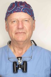 Michael Truppe合作论文数Karl Landsteiner Institute for Biotelematics, Danube University Krems7
 Winfried Mayr合作论文数Center for Biomedical Engineering and Physics Austria, Medical University Vienna6
 Robert Kohnke合作论文数5Arnulf Baumann合作论文数5
 Ewald Unger合作论文数5R. Koehnke合作论文数5M Oeckher合作论文数4G. Koller合作论文数4C. Lechner合作论文数4A. De Vries合作论文数4
 Werner Millesi合作论文数4Georg Burggasser合作论文数4Gerhard Undt合作论文数4
 Kurt Schicho合作论文数4U Losert合作论文数3
 Bernhard Haller合作论文数Institute of AI and Informatics in Medicine, Technical University of Munich3
 Hanns Plenk合作论文数3
 Rudolf Seemann合作论文数University Hospital for Craniomaxillofacial and Oral Surgery3Arne Wagner合作论文数3c winsauer合作论文数2W Backfrieder合作论文数2C Czerny合作论文数2Brigitte Wendl合作论文数2Katharina Juengling合作论文数2Laura Burger-Krebes合作论文数2Edzard Wiener合作论文数2
 Adegbayi Adeola Adekunle合作论文数Department of Oral and Maxillofacial Surgery, 291389Lagos University Teaching Hospital2Fabian Kanz合作论文数Center for Forensic Medicine, Medical University of Vienna2J. E. Vlachojannis合作论文数2Muchitsch Alfred Peter合作论文数2
 Oliver Bissinger合作论文数2Puigdollers Andreu合作论文数2
 Guido R Sigron合作论文数2Joszef Piffko合作论文数2F. Boehler合作论文数2Kaan C. Yerit合作论文数2H. Plenk Jr.合作论文数2Manfred Tschabitscher合作论文数2
 Klaus-Dietrich Wolff合作论文数2Ch. Kermer合作论文数2




