Blood-borne factors are essential to maintain neuronal synaptic plasticity and cognitive resilience throughout life. One such factor is osteocalcin (OCN), a hormone produced by osteoblasts that influences multiple physiological processes, including hippocampal neuronal homeostasis. However, the mechanism through which this blood-borne factor communicates with neurons remains unclear. Here we show the importance of a core primary cilium (PC) protein-autophagy axis in mediating the effects of OCN. We found that the OCN receptor GPR158 is present at the PC of hippocampal neurons and mediates the regulation of autophagy machinery by OCN. During aging, autophagy and PC core proteins are reduced in neurons, and restoring their levels is sufficient to improve cognitive impairments in aged mice. Mechanistically, the induction of this axis by OCN is dependent on the PC-dependent cAMP response element-binding protein signaling pathway. Altogether, this study demonstrates that the PC-autophagy axis is a gateway to mediate communication between blood-borne factors and neurons, and it advances understanding of the mechanisms involved in age-related cognitive decline.
BACKGROUND: The ability to respond to mechanical forces is a basic requirement for maintaining endothelial cell (ECs) homeostasis, which is continuously subjected to low shear stress (LSS) and high shear stress (HSS). In arteries, LSS and HSS have a differential impact on EC autophagy processes. However, it is still unclear whether LSS and HSS differently tune unique autophagic machinery or trigger specific autophagic responses in ECs. METHODS: Using fluid flow system to generate forces on EC and multiscale imaging analyses on ApoE −/− mice whole arteries, we studied the cellular and molecular mechanism involved in autophagic response to LSS or HSS on the endothelium. RESULTS: We found that LSS and HSS trigger autophagy activation by mobilizing specific autophagic signaling modules. Indeed, LSS-induced autophagy in endothelium was independent of the class III PI3K (phosphoinositide 3-kinase) VPS34 (vacuolar sorting protein 34) but controlled by the α isoform of class II PI3K (phosphoinositide 3-kinase class II α [PI3KCIIα]). Accordingly, reduced PI3KCIIα expression in ApoE −/− mice (ApoE −/− PI3KCIIα +/− ) led to EC dysfunctions associated with increased plaque deposition in the LSS regions. Mechanistically, we revealed that PI3KCIIα inhibits mTORC1 (mammalian target of rapamycin complex 1) activation and that rapamycin treatment in ApoE −/− PI3KCIIα +/− mice specifically rescue autophagy in arterial LSS regions. Finally, we demonstrated that absence of PI3KCIIα led to decreased endothelial primary cilium biogenesis in response to LSS and that ablation of primary cilium mimics PI3KCIIα-decreased expression in EC dysfunction, suggesting that this organelle could be the mechanosensor linking PI3KCIIα and EC homeostasis. CONCLUSIONS: Our data reveal that mechanical forces variability within the arterial system determines EC autophagic response and supports a central role of PI3KCIIα/mTORC1 axis to prevent EC dysfunction in LSS regions.
Insulin-producing pancreatic β cells play a crucial role in the regulation of glucose homeostasis, and their failure is a key event for diabetes development. Prolonged exposure to palmitate in the presence of elevated glucose levels, termed gluco-lipotoxicity, is known to induce β cell apoptosis. Autophagy has been proposed to be regulated by gluco-lipotoxicity in order to favor β cell survival. However, the role of palmitate metabolism in gluco-lipotoxcity-induced autophagy is presently unknown. We therefore treated INS-1 cells for 6 and 24 h with palmitate in the presence of low and high glucose concentrations and then monitored autophagy. Gluco-lipotoxicity induces accumulation of LC3-II levels in INS-1 at 6 h which returns to basal levels at 24 h. Using the RFP-GFP-LC3 probe, gluco-lipotoxicity increased both autophagosomes and autolysosmes structures, reflecting early stimulation of an autophagy flux. Triacsin C, a potent inhibitor of the long fatty acid acetyl-coA synthase, completely prevents LC3-II formation and recruitment to autophagosomes, suggesting that autophagic response requires palmitate metabolism. In contrast, etomoxir and bromo-palmitate, inhibitors of fatty acid mitochondrial β-oxidation, are unable to prevent gluco-lipotoxicity-induced LC3-II accumulation and recruitment to autophagosomes. Moreover, bromo-palmitate and etomoxir potentiate palmitate autophagic response. Even if gluco-lipotoxicity raised ceramide levels in INS-1 cells, ceramide synthase 4 overexpression does not potentiate LC3-II accumulation. Gluco-lipotoxicity also still stimulates an autophagic flux in the presence of an ER stress repressor. Finally, selective inhibition of sphingosine kinase 1 (SphK1) activity precludes gluco-lipotoxicity to induce LC3-II accumulation. Moreover, SphK1 overexpression potentiates autophagic flux induced by gluco-lipotxicity. Altogether, our results indicate that early activation of autophagy by gluco-lipotoxicity is mediated by SphK1, which plays a protective role in β cells.
Macroautophagy is a lysosomal degradative pathway for intracellular macromolecules, protein aggregates, and organelles. The formation of the autophagosome, a double membrane‐bound structure that sequesters cargoes before their delivery to the lysosome, is regulated by several stimuli in multicellular organisms. Pioneering studies in rat liver showed the importance of amino acids, insulin, and glucagon in controlling macroautophagy. Thereafter, many studies have deciphered the signaling pathways downstream of these biochemical stimuli to control autophagosome formation. Two signaling hubs have emerged: the kinase mTOR, in a complex at the surface of lysosomes which is sensitive to nutrients and hormones; and AMPK, which is sensitive to the cellular energetic status. Besides nutritional, hormonal, and energetic fluctuations, many organs have to respond to mechanical forces (compression, stretching, and shear stress). Recent studies have shown the importance of mechanotransduction in controlling macroautophagy. This regulation engages cell surface sensors, such as the primary cilium, in order to translate mechanical stimuli into biological responses.
Shear stress induced by urinary flow stimulates macroautophagy (hereafter referred to as autophagy) in kidney proximal tubule epithelial cells. Autophagy and selective degradation of lipid droplets by lipophagy contribute to tubule homeostasis by the production of ATP and control of epithelial cell size. Autophagy/lipophagy is controlled by a signaling cascade emanating from the primary cilium, localized at the apical side of epithelial cells. Downstream of the primary cilium, AMPK controls mitochondrial biogenesis on the one hand and autophagy/lipophagy on the other hand, which together increase fatty acid production that fuels oxidative phosphorylation to increase energy production. Recently, we reported that the co-transcriptional factors YAP1 and WWTR1/TAZ act downstream of AMPK to control autophagy. In fact, YAP1 and the transcription factor TEAD control the expression of RUBCN/rubicon. Under shear stress, YAP1 is excluded from the nucleus in a SIRT1-dependent manner to favor autophagic flux by downregulating the expression of RUBCN. When simulating in vitro a pathological urinary flow in murine proximal tubule kidney epithelial cells, we observe the nuclear retention of YAP1 and, consequently, high expression of RUBCN and inhibition of autophagic flux. Importantly, these findings were confirmed in biopsies of patients suffering from diabetic nephropathy, a major cause of chronic kidney disease.
In recent years, primary familial brain calcification (PFBC), a rare neurological disease characterized by a wide spectrum of cognitive disorders, has been associated to mutations in the sodium (Na)-Phosphate (Pi) co-transporter SLC20A2. However, the functional roles of the Na-Pi co-transporters in the brain remain still largely elusive. Here we show that Slc20a1/(PiT-1) and Slc20a2/(PiT-2) are the most abundant Na-Pi co-transporters expressed in the brain and are involved in the control of hippocampal-dependent learning and memory. We reveal that Slc20a1 and Slc20a2 are differentially distributed in the hippocampus and associated with independent gene clusters, suggesting that they influence cognition by different mechanisms. Accordingly, using a combination of molecular, electrophysiological and behavioral analyses, we show that while PiT-2 favors hippocampal neuronal branching and survival, PiT-1 promotes synaptic plasticity. The latter relies on a likely Otoferlin-dependent regulation of synaptic vesicle trafficking, which impacts the GABAergic system. These results provide the first demonstration that Na-Pi co-transporters play key albeit distinct roles in the hippocampus pertaining to the control of neuronal plasticity and cognition. These findings could provide the foundation for the development of novel effective therapies for PFBC and cognitive disorders.
Supplementary Materials, Figures 1-5 from Disruption of Sphingosine 1-Phosphate Lyase Confers Resistance to Chemotherapy and Promotes Oncogenesis through Bcl-2/Bcl-xL Upregulation
Supplementary Figure Legends 1-2 from c-Jun NH2-Terminal Kinase Activation Is Essential for DRAM-Dependent Induction of Autophagy and Apoptosis in 2-Methoxyestradiol–Treated Ewing Sarcoma Cells
Supplementary Figure 2 from c-Jun NH2-Terminal Kinase Activation Is Essential for DRAM-Dependent Induction of Autophagy and Apoptosis in 2-Methoxyestradiol–Treated Ewing Sarcoma Cells
Shear stress generated by urinary fluid flow is an important regulator of renal function. Its dysregulation is observed in various chronic and acute kidney diseases. Previously, we demonstrated that primary cilium-dependent autophagy allows kidney epithelial cells to adapt their metabolism in response to fluid flow. Here, we show that nuclear YAP/TAZ negatively regulates autophagy flux in kidney epithelial cells subjected to fluid flow. This crosstalk is supported by a primary cilium-dependent activation of AMPK and SIRT1, independently of the Hippo pathway. We confirm the relevance of the YAP/TAZ-autophagy molecular dialog in vivo using a zebrafish model of kidney development and a unilateral ureteral obstruction mouse model. In addition, an in vitro assay simulating pathological accelerated flow observed at early stages of chronic kidney disease (CKD) activates YAP, leading to a primary cilium-dependent inhibition of autophagic flux. We confirm this YAP/autophagy relationship in renal biopsies from patients suffering from diabetic kidney disease (DKD), the leading cause of CKD. Our findings demonstrate the importance of YAP/TAZ and autophagy in the translation of fluid flow into cellular and physiological responses. Dysregulation of this pathway is associated with the early onset of CKD.
Background and Aims : The ability to generate and respond to mechanical forces is a basic requirement for maintaining cell homeostasis. This is of particular importance for the cardiovascular system which is subjected to different mechanical forces such as Low and High shear stresses (LSS and HSS). Autophagy is involved in endothelial cells (ECs) response to HSS generated by blood flow. However, it is still unclear whether the variability of shear stress triggers specific autophagic response in ECs. Methods: We used fluid flow chamber system to generate different mechanical forces on ECs in vitro and we specifically studied LSS and HSS artery territories, referred as aortic arch and descending aorta respectively, in mice susceptible to atherosclerosis. Results: We found that LSS and HSS mobilize two different signalling modules relying on class II PI3K (PI3KCIIα) and III PI3K/Vps34 respectively to induce autophagy. We demonstrated that LSS-induced autophagy required the primary cilium and is critical to prevent ECs permeability and integrity. Accordingly, atherogenic stress or reduced PI3KCIIα expression led to a decrease in the ciliated ECs associated with decreased autophagic flux and accelerated plaque deposition in the LSS regions. Furthermore, we revealed that PI3KCIIα controls mTORC1 activation. Notably, mTORC1 inhibition impaired atherosclerosis development in ApoE-/- PI3KCIIα+/- mice and led to primary cilium restoration suggesting a crosstalk between primary cilium biogenesis and autophagy in LSS context. Conclusions: Our data reveal that mechanical forces variability within arterial system determine ECs autophagic response and support a central role of PI3KCIIα/mTOR axis in EC mechanosensing in atheroprone regions.
Kidney mass and function are sexually determined, but the cellular events and the molecular mechanisms involved in this dimorphism are poorly characterized. By combining female and male mice with castration/replacement experiments, we showed that male mice exhibited kidney overgrowth from five weeks of age. This effect was organ specific, since liver and heart weight were comparable between males and females, regardless of age. Consistently, the androgen receptor was found to be expressed in the kidneys of males, but not in the liver. In growing mice, androgens led to kidney overgrowth by first inducing a burst of cell proliferation and then an increase of cell size. Remarkably, androgens were also required to maintain cell size in adults. In fact, orchiectomy resulted in smaller kidneys in a matter of few weeks. These changes paralleled the changes of the expression of ornithine decarboxylase and cyclin D1, two known mediators of kidney growth, whereas, unexpectedly, mTORC1 and Hippo pathways did not seem to be involved. Androgens also enhanced kidney autophagy, very likely by increasing transcription factor EB nuclear translocation. Functionally, the increase of tubular mass resulted in increased sodium/phosphate transport. These findings were relevant to humans. Remarkably, by studying living gender-paired kidney donors-recipients, we showed that tubular cell size increased three months after transplantation in men as compared to women, regardless of the donor gender. Thus, our results identify novel signaling pathways that may be involved in androgen-induced kidney growth and homeostasis and suggest that androgens determine kidney size after transplantation.
Naked mole-rats (NMR) are subterranean rodents characterized by an unusual longevity coupled with an unexplained resistance to aging. In the present study, we performed extensive in situ analysis and single-cell RNA-sequencing comparing young and older animals. At variance with other species, NMR exhibited a striking stability of skin compartments and cell types, which remained stable over time without aging-associated changes. Remarkably, the number of stem cells was constant throughout aging. We found three classical cellular states defining a unique keratinocyte differentiation trajectory that were not altered after pseudo-temporal reconstruction. Epidermal gene expression did not change with aging either. Langerhans cell clusters were conserved, and only a higher basal stem cell expression of Igfbp3 was found in aged animals. In accordance, NMR skin healing closure was similar in young and older animals. Altogether, these results indicate that NMR skin is characterized by peculiar genetic and cellular features, different from those previously demonstrated for mice and humans. The remarkable stability of the aging NMR skin transcriptome likely reflects unaltered homeostasis and resilience.
Organs and cells must adapt to shear stress induced by biological fluids, but how fluid flow contributes to the execution of specific cell programs is poorly understood. Here we show that shear stress favours mitochondrial biogenesis and metabolic reprogramming to ensure energy production and cellular adaptation in kidney epithelial cells. Shear stress stimulates lipophagy, contributing to the production of fatty acids that provide mitochondrial substrates to generate ATP through β-oxidation. This flow-induced process is dependent on the primary cilia located on the apical side of epithelial cells. The interplay between fluid flow and lipid metabolism was confirmed in vivo using a unilateral ureteral obstruction mouse model. Finally, primary cilium-dependent lipophagy and mitochondrial biogenesis are required to support energy-consuming cellular processes such as glucose reabsorption, gluconeogenesis and cytoskeletal remodelling. Our findings demonstrate how primary cilia and autophagy are involved in the translation of mechanical forces into metabolic adaptation.
The ability to adapt the proteolysis rates based on fluctuations in essential amino acid (EAA) availability is essential for life. The GCN2-eIF2α-ATF4 signaling pathway is involved in the adaptive response to EAA deprivations. Our previous results demonstrated that activation of this pathway is involved in upregulating the expression of many autophagy genes at the transcriptional level thanks to the transcription factor ATF4. In the present study, we investigated whether the kinase GCN2 is also involved in early upregulation of the autophagic process in response to EAA deficiencies, thereby contributing to a rapid increase in the proteolysis rate. We observed that a one-hour leucine deprivation upregulated autophagy in both cultured cells and in vivo in mouse liver, as reflected by an increase in both [S278]-ATG16L1 phosphorylation and LC3B conversion, and decreased p62 protein level. Using cells and mice with genetic ablation of Gcn2 as well as genetic reconstitution experiments in vitro, data showed that GCN2 was required for this upregulation of autophagy. Downstream GCN2, the phosphorylation of eIF2α was necessary, while ATF4 was not. Overall, these findings revealed a major role of GCN2-eIF2α signaling in the regulation of autophagy in response to short-term EAA deprivation.
The maintenance of cellular homeostasis in response to extracellular stimuli, i.e., nutrient and hormone signaling, hypoxia, or mechanical forces by autophagy, is vital for the health of various tissues. The primary cilium (PC) is a microtubule-based sensory organelle that regulates the integration of several extracellular stimuli. Over the past decade, an interconnection between autophagy and PC has begun to be revealed. Indeed, the PC regulates autophagy and in turn, a selective form of autophagy called ciliophagy contributes to the regulation of ciliogenesis. Moreover, the PC regulates both mitochondrial biogenesis and lipophagy to produce free fatty acids. These two pathways converge to activate oxidative phosphorylation and produce ATP, which is mandatory for cell metabolism and membrane transport. The autophagy-dependent production of energy is fully efficient when the PC senses shear stress induced by fluid flow. In this review, we discuss the cross-talk between autophagy, the PC and physical forces in the regulation of cell biology and physiology.
Influenza virus infections are major public health threats due to their high rates of morbidity and mortality. Upon influenza virus entry, host cells experience modifications of endomembranes, including those used for virus trafficking and replication. Here we report that influenza virus infection modifies mitochondrial morphodynamics by promoting mitochondria elongation and altering endoplasmic reticulum-mitochondria tethering in host cells. Expression of the viral RNA recapitulates these modifications inside cells. Virus induced mitochondria hyper-elongation was promoted by fission associated protein DRP1 relocalization to the cytosol, enhancing a pro-fusion status. We show that altering mitochondrial hyper-fusion with Mito-C, a novel pro-fission compound, not only restores mitochondrial morphodynamics and endoplasmic reticulum-mitochondria contact sites but also dramatically reduces influenza replication. Finally, we demonstrate that the observed Mito-C antiviral property is directly connected with the innate immunity signaling RIG-I complex at mitochondria. Our data highlight the importance of a functional interchange between mitochondrial morphodynamics and innate immunity machineries in the context of influenza viral infection.
Despite the great advances in autophagy research in the last years, the specific functions of the four mammalian Atg4 proteases (ATG4A-D) remain unclear. In yeast, Atg4 mediates both Atg8 proteolytic activation, and its delipidation. However, it is not clear how these two roles are distributed along the members of the ATG4 family of proteases. We show that these two functions are preferentially carried out by distinct ATG4 proteases, being ATG4D the main delipidating enzyme. In mammalian cells, ATG4D loss results in accumulation of membrane-bound forms of mATG8s, increased cellular autophagosome number and reduced autophagosome average size. In mice, ATG4D loss leads to cerebellar neurodegeneration and impaired motor coordination caused by alterations in trafficking/clustering of GABA A receptors. We also show that human gene variants of ATG4D associated with neurodegeneration are not able to fully restore ATG4D deficiency, highlighting the neuroprotective role of ATG4D in mammals.
The cyclin-dependent kinase inhibitor p27Kip1 (p27) has been involved in promoting autophagy and survival in conditions of metabolic stress. While the signaling cascade upstream of p27 leading to its cytoplasmic localization and autophagy induction has been extensively studied, how p27 stimulates the autophagic process remains unclear. Here, we investigated the mechanism by which p27 promotes autophagy upon glucose deprivation. Mouse embryo fibroblasts (MEFs) lacking p27 exhibit a decreased autophagy flux compared to wild-type cells and this is correlated with an abnormal distribution of autophagosomes. Indeed, while autophagosomes are mainly located in the perinuclear area in wild-type cells, they are distributed throughout the cytoplasm in p27-null MEFs. Autophagosome trafficking towards the perinuclear area, where most lysosomes reside, is critical for autophagosome–lysosome fusion and cargo degradation. Vesicle trafficking is mediated by motor proteins, themselves recruited preferentially to acetylated microtubules, and autophagy flux is directly correlated to microtubule acetylation levels. p27−/− MEFs exhibit a marked reduction in microtubule acetylation levels and restoring microtubule acetylation in these cells, either by re-expressing p27 or with deacetylase inhibitors, restores perinuclear positioning of autophagosomes and autophagy flux. Finally, we find that p27 promotes microtubule acetylation by binding to and stabilizing α-tubulin acetyltransferase (ATAT1) in glucose-deprived cells. ATAT1 knockdown results in random distribution of autophagosomes in p27+/+ MEFs and impaired autophagy flux, similar to that observed in p27−/− cells. Overall, in response to glucose starvation, p27 promotes autophagy by facilitating autophagosome trafficking along microtubule tracks by maintaining elevated microtubule acetylation via an ATAT1-dependent mechanism.
 M Aubery合作论文数49
 Eric Ogier-Denis合作论文数INSERM U410, Institut de la National de la Santé et de la Recherche Médicale42C Bauvy合作论文数36Dupont Nicolas合作论文数36
 Etienne Morel合作论文数31Bauvy Chantal合作论文数30
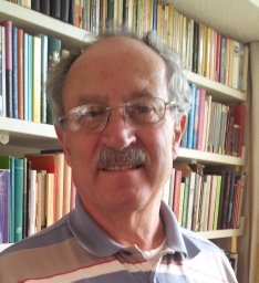 Alfred J. Meijer合作论文数B.C.P. Jansen Institute, University of Amsterdam24
 Maryam Mehrpour合作论文数INSERM U756, Faculté de pharmacie;Université;Faculté de pharmacie, Université Paris-Sud F-92296 Chatenay Malabry24
 Sophie Pattingre合作论文数22Mojgan Djavaheri-Mergny合作论文数Institut Bergonié, Université de Bordeaux, INSERM22J Botti合作论文数17Joëlle Botti合作论文数17
 Germain Trugnan合作论文数17Hamaï Ahmed合作论文数16
 Guido Kroemer (吉多·科罗默)合作论文数Faculty of Medicine, University of Paris Cité16C Sapin合作论文数14Lorin Séverine合作论文数14
 Isabelle Beau合作论文数14Anna Chiara Nascimbeni合作论文数12Gérard Pierron合作论文数12Francesco Cecconi合作论文数12R Ghidoni合作论文数11J FONT合作论文数11Proikas-Cezanne Tassula合作论文数11
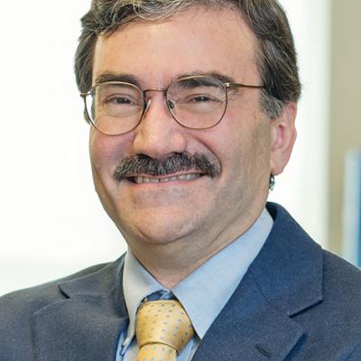 Daniel Klionsky合作论文数Life Sciences Institute, University of Michigan;Department of Molecular, Cell & Developmental Biology, College of Literature, Science, and the Arts, University of Michigan11
 Patricia Boya合作论文数University of Fribourg11Ma Doyennettemoyne合作论文数10
 Fulvio Reggiori合作论文数Department of Biomedicine, Aarhus University10
 Kevin Ryan合作论文数Cancer Research UK Scotland Institute;School of Cancer Sciences, University of Glasgow;Beatson Institute, University of Glasgow10Audrey Esclatine合作论文数10J J Houri合作论文数9Thierry Levade合作论文数9
 Frank Madeo合作论文数Institut für Molekulare Biowissenschaften, Universität Graz9
 Ciro Isidoro合作论文数Laboratorio di Patologia Molecolare;Dipartimento di Scienze Mediche;Laboratorio di Patologia Molecolare|Dipartimento di Scienze Mediche9
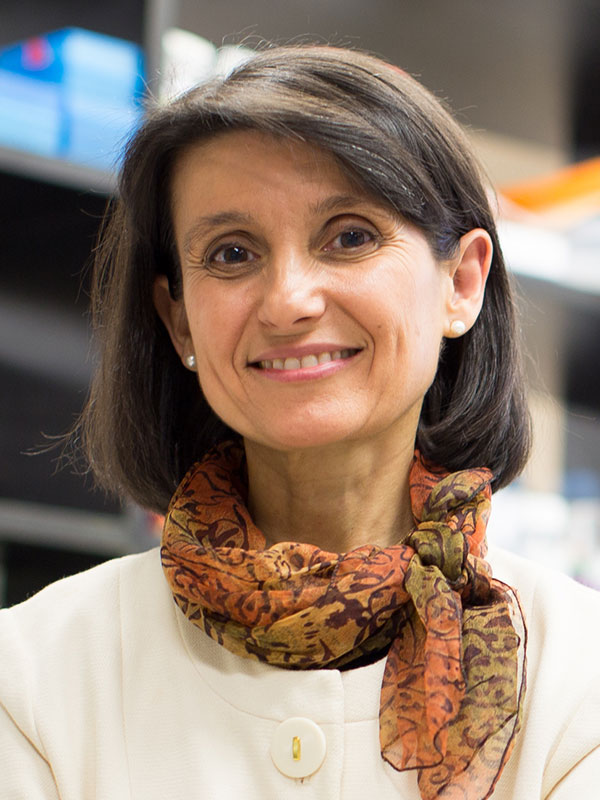 Ana Maria Cuervo合作论文数Department of Developmental & Molecular Biology, Albert Einstein College of Medicine;Department of Medicine, Albert Einstein College of Medicine;Institute for Aging Research, Albert Einstein College of Medicine9R Moutsita合作论文数8Martine Biard-Piechaczyk合作论文数8Francesca Scarlatti合作论文数8
 G Friedlander合作论文数Institut National de la Santé et de la Recherche Médicale U1151, Université de Paris8
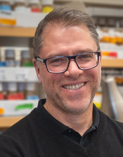 Eric H. Baehrecke合作论文数Department of Molecular, Cell and Cancer Biology, University of Massachusetts Medical School8Souquere Sylvie合作论文数8
 Rautou Pierre-Emmanuel合作论文数Département Hépato‐gastroentérologie, Centre De Recherche Sur L’inflammation, UMR 1149 Inserm – Université Paris Cité;Hepatology Department, Beaujon Hospital;University Paris Diderot8
 Federica Roccio合作论文数7Christine Leroy合作论文数7
 Sharon A. Tooze合作论文数London Research Institute;The Francis Crick Institute7
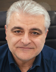 Nektarios Tavernarakis合作论文数School of Medicine, University of Crete;Institute of Molecular Biology and Biotechnology, Foundation for Research and Technology-Hellas7Maiuri Maria Chiara合作论文数7
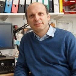 David Rubinsztein合作论文数Department of Medical Genetics, Cambridge Institute for Medical Research, University of Cambridge;UK Dementia Research Institute, University of Cambridge7
 Elazar Zvulun合作论文数Dept Biol Chem, Weizmann Inst Sci7




