A genetic predisposition to PML is now substantially supported by case reports of patients molecularly diagnosed with an inborn error of immunity (IEI) and progressive multifocal leukoencephalopathy (PML). Over the past 10 years, 4 IEI genes linked to PML has now grown to 26 as of 2025. Of these 26 genes believed to be causal of an IEI and PML, 24 (92%) are also linked with hemophagocytic lymphohistiocytosis/macrophage activation syndrome (HLH/MAS)—a severe hyper-inflammation syndrome associated with several IEI genes, most notably in 4 genes (PRF1, STX11, STXBP2, UNC13D) causing familial forms of the syndrome. Many HLH-linked genes are associated with life-threatening Epstein–Barr virus infections, which analogously suggests JC virus infection plus presence of a pathogenic variant in an HLH-linked IEI gene also increases risk of PML. PML also occurs as a serious adverse event for a subset of immunosuppressive therapies (e.g., natalizumab and rituximab) used to treat patients with immune disorders (e.g., multiple sclerosis and hematological malignancies). Recently, 4 PML risk variants were reported for use in a PML risk test to screen patients who are considering treatment with PML-linked therapies. Interestingly, of the 4 genes with a PML risk variant, 2 (LY9 and STXBP2) cause or are linked to HLH. The aim of our review is two-fold: (1) raise awareness among researchers and clinicians (e.g., neurologists, oncologists, and rheumatologists) that patient genetics are a key risk factor for PML, and (2) further reinforce the rationale for screening at-risk patients for PML risk variants before prescribing a PML-linked drug.
Progressive multifocal leukoencephalopathy (PML) is a rare demyelinating disorder of the brain caused by reactivation of the JC virus (JCV), a polyomavirus that infects at least 60% of the population but is asymptomatic or results in benign symptoms in most people. PML occurs as a secondary disease in a variety of disorders or as a serious adverse event from immunosuppressant agents, but is mainly found in three groups: HIV-infected patients, patients with hematological malignancies, or multiple sclerosis (MS) patients on the immunosuppressant therapy natalizumab. It is severely debilitating and is deadly in ~50% HIV cases, ~90% of hematological malignancy cases, and ~24% of MS-natalizumab cases. A PML risk prediction test would have clinical utility in all at risk patient groups but would be particularly beneficial in patients considering therapy with immunosuppressant agents known to cause PML, such as natalizumab, rituximab, and others. While a JC antibody test is currently used in the clinical decision process for natalizumab, it is suboptimal because of its low specificity and requirement to periodically retest patients for seroconversion or to assess if a patient's JCV index has increased. Whereas a high specificity genetic risk prediction test comprising host genetic risk variants (i.e., germline variants occurring at higher frequency in PML patients compared to the general population) could be administered one time to provide clinicians with additional risk prediction information that is independent of JCV serostatus. Prior PML case reports support the hypothesis that PML risk is greater in patients with a genetically caused immunodeficiency disorder. To identify germline PML risk variants, we performed exome sequencing on 185 PML cases (70 in a discovery cohort and 115 in a replication cohort) and used the gnomAD variant database for interpretation. Our study yielded 19 rare variants (maximum allele frequency of 0.02 in gnomAD ethnically matched populations) that impact 17 immune function genes (10 are known to cause inborn errors of immunity). Modeling of these variants in a PML genetic risk test for MS patients considering natalizumab treatment indicates that at least a quarter of PML cases may be preventable.
BackgroundThe nucleotide binding protein-like (NUBPL) gene was first reported as a cause of mitochondrial complex I deficiency (MIM 613621, 618242) in 2010. To date, only eight patients have been reported with this mitochondrial disorder. Five other patients were recently reported to have NUBPL disease but their clinical picture was different from the first eight patients. Here, we report clinical and genetic findings in five additional patients (four families).MethodsWhole exome sequencing was used to identify patients with compound heterozygous NUBPL variants. Functional studies included RNA-Seq transcript analyses, missense variant biochemical analyses in a yeast model (Yarrowia lipolytica) and mitochondrial respiration experiments on patient fibroblasts.ResultsThe previously reported c.815-27T>C branch-site mutation was found in all four families. In prior patients, c.166G>A [p.G56R] was always found in cis with c.815-27T>C, but only two of four families had both variants. The second variant found in trans with c.815-27T>C in each family was: c.311T>C [p.L104P] in three patients, c.693+1G>A in one patient and c.545T>C [p.V182A] in one patient. Complex I function in the yeast model was impacted by p.L104P but not p.V182A. Clinical features include onset of neurological symptoms at 3–18 months, global developmental delay, cerebellar dysfunction (including ataxia, dysarthria, nystagmus and tremor) and spasticity. Brain MRI showed cerebellar atrophy. Mitochondrial function studies on patient fibroblasts showed significantly reduced spare respiratory capacity.ConclusionWe report on five new patients with NUBPL disease, adding to the number and phenotypic variability of patients diagnosed worldwide, and review prior reported patients with pathogenic NUBPL variants.
In an unbiased genome-wide screen for copy number variants (CNVs) on a cohort of Parkinson's disease (PD) patients, we identified in one patient a complex chromosomal rearrangement involving the nucleotide binding protein-like (NUBPL) gene on chromosome 14q12. We noted that mutations in the NUBPL gene had been reported as causing autosomal recessive (AR) mitochondrial Complex I (CI) deficiency in children. The precise breakpoints of the rearrangement in our PD case were found to be identical to those described in a patient with AR CI deficiency who also harbored a second pathogenic mutation in NUBPL. Mitochondrial dysfunction has long been considered a strong contributor to PD, and there is substantial evidence that decreased CI activity plays a central role in PD pathogenesis. We hypothesize that pathogenic NUBPL variants may increase the risk for PD analogous to variants in the glucosylceramidase beta (GBA) gene that increase the risk of developing PD in heterozygous carriers.
The "Iowa kindred," a large Iowan family with autosomal-dominant Parkinson's disease, has been followed clinically since the 1920s at the Mayo Clinic. In 2003, the genetic cause was determined to be a 1.7 Mb triplication of the alpha-synuclein genomic locus. Affected individuals present with an early-onset, severe parkinsonism-dementia syndrome. Here, we present a descendant of the Iowa kindred with novel, disease-associated non-motor findings of reduced heart rate variability, complete anosmia, and a rare skin condition called colloid milium. At autopsy, key neuropathological findings were compatible with diffuse Lewy body disease. Using high-resolution comparative genomic hybridization (CGH) array analysis to fine-map the genomic breakpoints, we observed two independent recombination events of the SNCA locus that resulted in a genomic triplication of twelve genes, including SNCA, and the disruption of two genes, HERC6 and CCSER1, at the genomic breakpoints. In conclusion, we provide further evidence that the mere two-fold overexpression of alpha-synuclein leads to a fulminant alpha-synucleinopathy with rapid progression and severe clinical and neuropathological features.
Background The pseudoautosomal short stature homeobox-containing (SHOX) gene encodes a homeodomain transcription factor involved in cell-cycle and growth regulation. SHOX/SHOX enhancers deletions cause short stature and skeletal abnormalities in a female-dominant fashion; duplications appear to be rare. Neurodevelopmental disorders (NDDs), such as autism spectrum disorders (ASDs), are complex disorders with high heritability and skewed sex ratio; several rare (< 1% frequency) CNVs have been implicated in risk.Methods We analysed data from a discovery series of 90 adult ASD cases, who underwent clinical genetic testing by array-comparative genomic hybridisation (CGH). Twenty-seven individuals harboured CNV abnormalities, including two unrelated females with microduplications affecting SHOX. To determine the prevalence of SHOX duplications and delineate their associated phenotypic spectrum, we subsequently examined array-CGH data from a follow-up sample of 26 574 patients, including 18 857 with NDD (3541 with ASD).Results We found a significant enrichment of SHOX microduplications in the NDD cases (p=0.00036; OR 2.21) and, particularly, in those with ASD (p=9.18x10(-7); OR 3.63) compared with 12 594 population-based controls. SHOX duplications affecting the upstream or downstream enhancers were enriched only in females with NDD (p=0.0043; OR 2.69/p=0.00020; OR 7.20), but not in males (p=0.404; OR 1.38/p=0.096; OR 2.21).Conclusions Microduplications at the SHOX locus are a low penetrance risk factor for ASD/NDD, with increased risk in both sexes. However, a concomitant duplication of SHOX enhancers may be required to trigger a NDD in females. Since specific SHOX isoforms are exclusively expressed in the developing foetal brain, this may reflect the pathogenic effect of altered SHOX protein dosage on neurodevelopment.
The proline dehydrogenase (PRODH) gene maps to 22q11.2 in the region deleted in the velo‐cardio‐facial syndrome (VCFS). A moderate to severe reduction (>50%) in PRODH activity resulting from recessive deletions and/or missense mutations has been shown to cause type 1 hyperprolinemia (HPI). Autistic features have been reported as a common clinical manifestation of HPI. Here we studied the frequency of a recurrent small 22q11.2 deletion encompassing PRODH and the neighboring DGCR6 gene in three case‐control studies, one comprising HPI patients (n = 83), and the other two comprising autism spectrum disorder (ASD) patients (total of n = 2800), analyzed with high‐resolution microarrays. We found that the PRODH deletion is a strong risk factor for HPI (OR = 50.7; 95%CI = 7.5–2147) but not for ASD (P = 0.4, OR = 0.6–3.3). This result indicates either that the suggested association between ASD and HPI is spurious and results from a bias leading to the preferential inclusion of patients with autistic features in HPI series, or that HPI is present in only a very small subset of ASD patients. In this latter case, a very large sample size would be required to detect an association between the PRODH deletion and ASD in a case‐control study. © 2016 Wiley Periodicals, Inc.
Annals of NeurologyVolume 78, Issue 4 p. 663-664 Letters/Replies Comparative genomic hybridization solves a 14-year-old PARKIN mystery Birgitt Schüle MD, Birgitt Schüle MD Parkinson's Institute and Clinical Center, Sunnyvale, CASearch for more papers by this authorEli Hatchwell MD, PhD, Eli Hatchwell MD, PhD Population Diagnostics, Inc., Melville, NYSearch for more papers by this authorPeggy S. Eis PhD, Peggy S. Eis PhD Population Diagnostics, Inc., Melville, NYSearch for more papers by this authorJ. William Langston MD, J. William Langston MD Parkinson's Institute and Clinical Center, Sunnyvale, CASearch for more papers by this author Birgitt Schüle MD, Birgitt Schüle MD Parkinson's Institute and Clinical Center, Sunnyvale, CASearch for more papers by this authorEli Hatchwell MD, PhD, Eli Hatchwell MD, PhD Population Diagnostics, Inc., Melville, NYSearch for more papers by this authorPeggy S. Eis PhD, Peggy S. Eis PhD Population Diagnostics, Inc., Melville, NYSearch for more papers by this authorJ. William Langston MD, J. William Langston MD Parkinson's Institute and Clinical Center, Sunnyvale, CASearch for more papers by this author First published: 14 July 2015 https://doi.org/10.1002/ana.24478Citations: 2Read the full textAboutPDF ToolsRequest permissionExport citationAdd to favoritesTrack citation ShareShare Give accessShare full text accessShare full-text accessPlease review our Terms and Conditions of Use and check box below to share full-text version of article.I have read and accept the Wiley Online Library Terms and Conditions of UseShareable LinkUse the link below to share a full-text version of this article with your friends and colleagues. Learn more.Copy URL Share a linkShare onEmailFacebookTwitterLinkedInRedditWechat No abstract is available for this article. References 1 Farrer M, Chan P, Chen R, et al. Lewy bodies and parkinsonism in families with parkin mutations. Ann Neurol 2001; 50: 293–300. 2 Lincoln S, Wiley J, Lynch T, et al. Parkin-proven disease: common founders but divergent phenotypes. Neurology 2003; 60: 1605–1610. 3 Tan LC, Tanner CM, Chen R, et al. Marked variation in clinical presentation and age of onset in a family with a heterozygous parkin mutation. Mov Disord 2003; 18: 758–763. 4 Lücking CB, Dürr A, Bonifati V, et al. Association between early-onset Parkinson's disease and mutations in the parkin gene. N Engl J Med 2000; 342: 1560–1567. 5 Vasson A, Leroux C, Orhant L, et al. Custom oligonucleotide array-based CGH: a reliable diagnostic tool for detection of exonic copy-number changes in multiple targeted genes. Eur J Hum Genet 2013; 21: 977–987. Citing Literature Volume78, Issue4October 2015Pages 663-664 ReferencesRelatedInformation
Rare copy number variants (CNVs) disrupting ASTN2 or both ASTN2 and TRIM32 have been reported at 9q33.1 by genome-wide studies in a few individuals with neurodevelopmental disorders (NDDs). The vertebrate-specific astrotactins, ASTN2 and its paralog ASTN1, have key roles in glial-guided neuronalmigration during brain development. To determine the prevalence of astrotactin mutations and delineate their associated phenotypic spectrum, we screened ASTN2/TRIM32 and ASTN1 (1q25.2) for exonic CNVs in clinical microarray data from 89 985 individuals across 10 sites, including 64 114 NDD subjects. In this clinical dataset, we identified 46 deletions and 12 duplications affecting ASTN2. Deletions of ASTN1 were much rarer. Deletions near the 3' terminus of ASTN2, which would disrupt all transcript isoforms (a subset of these deletions also included TRIM32), were significantly enriched in the NDD subjects (P = 0.002) compared with 44 085 population-based controls. Frequent phenotypes observed in individuals with such deletions include autism spectrum disorder (ASD), attention deficit hyperactivity disorder (ADHD), speech delay, anxiety and obsessive compulsive disorder (OCD). The 3'-terminal ASTN2 deletions were significantly enriched compared with controls in males with NDDs, but not in females. Upon quantifying ASTN2 human brain RNA, we observed shorter isoforms expressed from an alternative transcription start site of recent evolutionary origin near the 3' end. Spatiotemporal expression profiling in the human brain revealed consistently high ASTN1 expression while ASTN2 expression peaked in the early embryonic neocortex and postnatal cerebellar cortex. Our findings shed new light on the role of the astrotactins in psychopathology and their interplay in human neurodevelopment.
The identification of rare inherited and de novo copy number variations (CNVs) in human subjects has proven a productive approach to highlight risk genes for autism spectrum disorder (ASD). A variety of microarrays are available to detect CNVs, including single-nucleotide polymorphism (SNP) arrays and comparative genomic hybridization (CGH) arrays. Here, we examine a cohort of 696 unrelated ASD cases using a high-resolution one-million feature CGH microarray, the majority of which were previously genotyped with SNP arrays. Our objective was to discover new CNVs in ASD cases that were not detected by SNP microarray analysis and to delineate novel ASD risk loci via combined analysis of CGH and SNP array data sets on the ASD cohort and CGH data on an additional 1000 control samples. Of the 615 ASD cases analyzed on both SNP and CGH arrays, we found that 13,572 of 21,346 (64%) of the CNVs were exclusively detected by the CGH array. Several of the CGH-specific CNVs are rare in population frequency and impact previously reported ASD genes (e.g., NRXN1, GRM8, DPYD), as well as novel ASD candidate genes (e.g., CIB2, DAPP1, SAE1), and all were inherited except for a de novo CNV in the GPHN gene. A functional enrichment test of gene-sets in ASD cases over controls revealed nucleotide metabolism as a potential novel pathway involved in ASD, which includes several candidate genes for follow-up (e.g., DPYD, UPB1, UPP1, TYMP). Finally, this extensively phenotyped and genotyped ASD clinical cohort serves as an invaluable resource for the next step of genome sequencing for complete genetic variation detection.
Cytogenetic analysis of acute myeloid leukemia (AML) cells has accelerated the identification of genes important for AML pathogenesis. To complement cytogenetic studies and to identify genes altered in AML genomes, we performed genome-wide copy number analysis with paired normal and tumor DNA obtained from 86 adult patients with de novo AML using 1.85 million feature SNP arrays. Acquired copy number alterations (CNAs) were confirmed using an ultra-dense array comparative genomic hybridization platform. A total of 201 somatic CNAs were found in the 86 AML genomes (mean, 2.34 CNAs per genome), with French-American-British system M6 and M7 genomes containing the most changes (10-29 CNAs per genome). Twenty-four percent of AML patients with normal cytogenetics had CNA, whereas 40% of patients with an abnormal karyotype had additional CNA detected by SNP array, and several CNA regions were recurrent. The mRNA expression levels of 57 genes were significantly altered in 27 of 50 recurrent CNA regions <5 megabases in size. A total of 8 uniparental disomy (UPD) segments were identified in the 86 genomes; 6 of 8 UPD calls occurred in samples with a normal karyotype. Collectively, 34 of 86 AML genomes (40%) contained alterations not found with cytogenetics, and 98% of these regions contained genes. Of 86 genomes, 43 (50%) had no CNA or UPD at this level of resolution. In this study of 86 adult AML genomes, the use of an unbiased high-resolution genomic screen identified many genes not previously implicated in AML that may be relevant for pathogenesis, along with many known oncogenes and tumor suppressor genes.
Genetic contributions to human cognition and behavior are clear but difficult to define. Williams syndrome (WS) provides a unique model for relating single genes to visual-spatial cognition and social behavior. We defined a approximately 1.5 Mb region of approximately 25 genes deleted in >98% of typical WS and then rare small deletions, showing that visual-spatial construction (VSC) in WS was associated with the genes GTF2IRD1 and GTF2I. To distinguish the roles of GTF2IRD1 and GTF2I in VSC and social behavior, we utilized multiple genomic methods (custom high resolution oligonucleotide microarray, multicolor FISH and somatic cell hybrids analyzed by PCR) to identify individuals deleted for either gene but not both. We analyzed genetic, cognitive and social behavior in a unique individual with WS features (heart defects, small size, facies), but with an atypical deletion of a set of genes that includes GTF2IRD1, but not GTF2I. The centromeric breakpoint localized to the region 72.32-72.38 Mb and the telomeric breakpoint to 72.66 Mb, 10 kb downstream of GTF2IRD1. Cognitive testing (WPPSI-R, K-BIT, and PLS-3) demonstrated striking deficits in VSC (Block Design, Object Assembly) but overall performance 1.5-3 SD above WS means. We have now integrated the genetic, clinical and cognitive data with previous reports of social behavior in this subject. These results combine with previous data from small deletions to suggest the gene GTF2IRD1 is associated with WS facies and VSC, and that GTF2I may contribute to WS social behaviors including increased gaze and attention to strangers.
 Eli Hatchwell合作论文数16
 Rebecca R. Selzer合作论文数10SM Olson合作论文数7
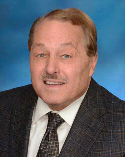 Joseph R. Lakowicz合作论文数School of Medicine, University of Maryland7
 Todd A Richmond合作论文数7Victor I. Lyamichev合作论文数7Michelle Curtis合作论文数6Robert W. Kwiatkowski合作论文数6Hatim T. Allawi合作论文数6Jeff G. Hall合作论文数6Ip Hon S合作论文数5
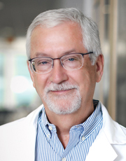 Timothy J. Ley合作论文数Division of Oncology, John T. Milliken Department of Medicine, School of Medicine, Washington University in St. Louis5
 Michael L. Johnson合作论文数Pharmacology,University of Virginia Health System5Tsetska Takova合作论文数5Kevin L. Vedvik合作论文数5marilyn c olsonmunoz合作论文数4christian tor bartholomay合作论文数4james j schaefer合作论文数4Bruce Neri合作论文数4Graubert Timothy A合作论文数4Ma W合作论文数4
 David P Millar合作论文数4Luanne Chehak合作论文数4
 Wiesław Wiczk合作论文数4
 Howard L. McLeod合作论文数Center for Precision Medicine & Functional Genomics, Utah Tech University;Clarified Precision Medicine4Andrew A. Lukowiak合作论文数4Michael W. Kaiser合作论文数4
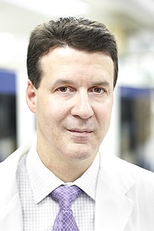 Stephen Scherer合作论文数Department of Molecular Genetics, Temerty Faculty of Medicine, University of Toronto;The Centre for Applied Genomics, The Hospital for Sick Children;Institute of Medical Science, University of Toronto;The McLaughlin Centre, Faculty of Medicine, University of Toronto4Rw Kwiatkowski合作论文数3William D. Shannon合作论文数3
 Christina R. Chow合作论文数3
 Christopher D Bruno合作论文数3Bhooma Thiruvahindrapuram合作论文数3
 Patrick Cahan合作论文数Department of Biomedical Engineering, Whiting School of Engineering, Johns Hopkins University3
 Birgitt Schuele合作论文数Department of Pathology, School of Medicine, Stanford University;Wu Tsai Neurosciences Institute, Stanford University;Center for Definitive and Curative Medicine, School of Medicine, Stanford University3
 Krzysztof Mrózek合作论文数Comprehensive Cancer Center, The Ohio State University3
 Mark A. Watson合作论文数Department of Pathology, Washington University School of Medicine3
 Elaine R. Mardis合作论文数Departments of Pediatrics, College of Medicine, The Ohio State University;Institute for Genomic Medicine, Nationwide Children's Hospital3Daniel C. Link合作论文数3
 Clara D. Bloomfield合作论文数Comprehensive Cancer Center, The Ohio State University3
 Richard K. Wilson合作论文数Institute for Genomic Medicine, Nationwide Children's Hospital;Ohio State University College of Medicine3Jack D. Baty合作论文数3Sharon Heath合作论文数3
 James Langston合作论文数Parkinson’s Institute and Clinical Center;Stanford Neuroscience Health Center;Department of Pathology, School of Medicine, Stanford University3
 Joel M Geoghegan合作论文数Center for Cancer Research, National Cancer Institute3
 James E. Dahlberg合作论文数Department of Physiological Chemistry,University of Wisconsin Madison3Matthew J. Walter合作论文数NINDS, NIH3Marilyn C. Olson合作论文数3T Richmond合作论文数2




