We previously reported that pre-ischemic vagus nerve stimulation (VNS) protects against myocardial ischemia, resembling classical ischemic preconditioning (cPC). This study investigates the time course and mechanisms underlying VNS-induced cardioprotection. Male FVB/N mice (3–5 months) underwent 30 min regional myocardial ischemia followed by 120 min reperfusion (IR). Ten-minute right-sided cervical VNS was delivered at defined intervals before ischemia to assess early and delayed protective windows. Risk area (RA) and infarct size (IS) were quantified using Evans Blue/tetrazolium staining. Myocardial phosphorylation of protein kinase B (Akt), glycogen synthase kinase-3β (GSK-3β), inducible nitric oxide synthase (iNOS), and mitochondrial respiration were evaluated. IR controls exhibited an IS of 57 ± 7
Backgrounds: Breast cancer metastasis remains the leading cause of mortality and frequently targets the bone. Breast cancer cells release soluble factors and extracellular vesicles that disrupt bone marrow (BM)/bone homeostasis, promoting osteoclastogenesis and the accumulation of senescent cells. In line with updated cancer hallmarks, senescent mesenchymal stem/ stromal cells (MSCs), osteoblasts, and osteocytes contribute to remodeling of the BM microenvironment, thereby favoring pre-metastatic niche (PMN) formation and subsequent bone metastasis. We previously demonstrated that untreated stage III-B breast cancer patients (BCPs) exhibit increased oxidative stress and elevated reactive oxygen species (ROS) levels, accompanied by senescent and functionally impaired BM-MSCs- key regulators of BM/bone homeostasis. In the present study, we sought to identify the molecular targets affected by oxidative stress that drive MSC senescence in these patients. Methods: BM-MSCs were isolated from untreated stage III-B BCPs and healthy volunteers (HVs). Oxidative stress responses were evaluated by quantitative real-time PCR (qRT-PCR) analysis of stress-and antioxidant-related genes. Oxidative damage to DNA, proteins, and lipids was assessed using alkaline comet assay, chromosomal aberration (CAs) analysis, micronuclei (MN) and nuclear blebs (NBs) quantification, protein carbonyl content, and detection of 4-hydroxynonenal (4-HNE) adducts. The MSC secretome was analyzed by label-free quantitative proteomics followed by Gene Ontology enrichment analysis. Results: Our results show that elevated oxidative stress in BCPs induces the overexpression of oxidative stress-related and antioxidant response genes in BM-MSCs; however, this response is insufficient to prevent extensive ROS-induced damage to deoxyribonucleic acid (DNA), proteins, and lipids. In addition, proteomic analysis of the BM-MSC secretome revealed a distinct protein expression profile in BCPs compared with HVs. Conclusions: Together, these findings highlight oxidative stress-induced MSC damage as a key mechanism contributing to PMN formation and suggest potential therapeutic targets to mitigate bone metastasis in advanced breast cancer.
It has been reported that exposure to indoor PM impairs redox metabolism and promotes inflammation, which might aggravate respiratory diseases. Lung epithelial cells are suggested to play a central role, since they produce inflammatory and oxidative stress mediators following PM uptake. We aimed to study the pathways leading to redox metabolism alterations and NLRP3 inflammasome activation in A549 cells and EpiAlveolar 3D tissue model exposed to Indoor Dust (ID) at the concentration of 25 and 100 μg/mL for up to 24 h. TEM images showed deposits of ID particles inside the cells at 3 and 24 h. In parallel, cells were exposed to ID, showed an increase in intracellular oxidative stress. Moreover, oxidative damage to lipids measured as 4-HNE protein adducts was observed after exposure. Additionally, dose-and time-dependent NFkB nuclear translocation and NLRP3-inflammasome activation was evidenced by the increased IF signal of ASC and NLRP3 after ID exposure. Moreover, an increase in the expression of ASC and NLRP3 was also observed, consistently with an increase in IL-1β levels. Finally, on A549 cells, alteration in wound closure process was observed compared to control. When evaluating EpiAlveolar 3D tissue model exposed to ID an impairment in barrier integrity was found, as indicated by TEER measurement together with an increase in HO-1 and 4-HNE signal. In addition, colocalization of both inflammasome components NLRP3 and ASC, were also found as well as the increased IL-1β levels. Our findings contribute to the understanding of the mechanisms by which ID promotes inflammation and oxidative stress in lung tissues.
Background/Objectives: Glaucoma is the leading cause of irreversible blindness worldwide and oxidative stress is considered to play a key role in its development. While antioxidants offer a promising approach to mitigating oxidative stress, their clinical application is often hindered by bioavailability and absorption challenges. Entrapment antioxidants within liposomes may overcome these issues, enhancing their stability and delivery. The aim of this study was to develop a novel composite liposomal formulation for glaucoma treatment, designed to enhance lipoic acid bioavailability and administration through its incorporation into the lipid bilayer. Methods: Liposomes were prepared via lipid film hydration and extrusion. To characterize them, the following analyses were performed: FTIR spectroscopy, liposomal bilayer melting temperature (Tm), TEM, DLS, Z-potential, antioxidant activity, and cytotoxicity assays. Results: The efficient incorporation of lipoic acid into the liposomes’ lipid bilayer was confirmed by FTIR. This incorporation resulted in an increase in the Tm, from 37.0 °C for liposomes to 40.0 °C for liposomes with lipoic acid (L-LA). TEM images confirmed that the spherical morphology of the lipid vesicles remained unchanged following LA incorporation. Dynamic Light Scattering analysis revealed effective diameters of 423 ± 36 nm for L liposomes and 404 ± 62 nm for L-LA liposomes. Notably, the Z-potential shifted from +4.7 ± 0.4 mV (L) to −0.4 ± 0.3 mV (L-LA). Furthermore, L-LA exhibited significant antioxidant activity (31.6 ± 0.4%) compared with L (5.3 ± 0.3%) and biocompatibility, suggesting its potential for therapeutic applications. Conclusions: In summary, biocompatible composite liposomes with antioxidant capacity were successfully developed, resulting in promising candidates for neuroprotective glaucoma therapy.
espanolIntroduccion: Se ha propuesto que el precondicionamiento isquemico remoto (PCr) reduce el tamano del infarto a traves de la activacionde una via neuronal parasimpatica. Sin embargo, los mecanismos intracelulares responsables de esta proteccion no se conocencompletamente.Objetivo: Describir algunas de las senales intracelulares activadas a nivel cardiaco por el PCr antes de la isquemia miocardica y queparticipan en la proteccion.Material y metodos: Corazones aislados de ratas fueron sometidos a 30 minutos de isquemia global, seguidos de 120 minutos dereperfusion (I/R). En un segundo grupo, antes del aislamiento del corazon se realizo un protocolo de PCr (tres ciclos de isquemia/reperfusion en la arteria femoral izquierda); una vez finalizado, se repitio el protocolo del grupo I/R. Adicionalmente, se estudiaroncuatro grupos experimentales, en los que antes del PCr se realizo una vagotomia cervical bilateral [SV (seccion vagal)] o se administroatropina (bloqueante de los receptores muscarinicos), L-NAME (inhibidor de la sintesis de NO) y 5-HD (bloqueante de los canalesmK+ATP), respectivamente. Se midieron el tamano del infarto y la fosforilacion de la eNOS en los grupos I/R, PCr y SV. Por otro lado,se midio la produccion mitocondrial de H2O2.Resultados: El PCr redujo significativamente el tamano del infarto y este efecto fue abolido por la SV y con los tratamientos conatropina, L-NAME y 5-HD. Ademas, el PCr incremento la fosforilacion de la eNOS y este efecto fue abolido por la SV. Finalmente, elPCr produjo un aumento de la produccion de H2O2 mitocondrial, hecho que tambien fue abolido con la SV.Conclusiones: El PCr activa una via muscarinica vagal, que involucra la fosforilacion de la eNOS, la apertura de los canales mK+ATPde la mitocondria y un aumento de la produccion de H2O2 mitocondrial. EnglishBackground: Remote ischemic preconditioning (rIPC) has been suggested to reduce infarct size through the activation of a parasympatheticneural pathway. However, the intracellular mechanisms responsible for this protection remain unclear.Objective: The aim of this study was to describe some of the intracellular protective signals activated at the cardiac level by rIPCprior to myocardial ischemia.Methods: Isolated rat hearts were subjected to 30 minutes of global ischemia and 120 minutes of reperfusion (I/R). In a secondgroup, before the isolation of the heart, a rIPC protocol (three cycles of left femoral artery ischemia/reperfusion) was performed,followed by the I/R protocol. Additionally, four experimental groups were studied, in which prior to the rIPC protocol a bilateralcervical vagotomy [VS (vagal section)] was performed or atropine (muscarinic receptor blocker), L-NAME (NO synthesis inhibitor),and 5-HD (mK+ATP channel blocker) was administered, respectively. Infarct size and eNOS phosphorylation were measured in I/R,rIPC, and VS groups. Finally, mitochondrial H2O2 production was assessed.Results: Remote ischemic preconditioning significantly decreased infarct size and this effect was abolished by VS and atropine,L-NAME, and 5-HD treatments. Furthermore, rIPC increased eNOS phosphorylation and this effect was abolished by VS. Finally,rIPC increased the mitochondrial H2O2 production, and this effect was also abolished by VS.Conclusions: Remote ischemic preconditioning activates a muscarinic vagal pathway involving eNOS phosphorylation, opening ofmitochondrial mK+ATP channels, and the production of mitochondrial H2O2.
Objective: Previous studies have implicated air pollution fine particulate matter (PM2.5) in various cardiovascular cardiometabolic and renal disease states. However, the molecular mechanisms by which these pollutants mediate these comorbidities have not been fully elucidated. Dysregulation of the renin-angiotensin system (RAS) may be one potential mechanism. This study aimed to investigate the participation of the RAS in air pollution-associated pathologies. Design and method: To study the impact of PM2.5 on systemic and tissue components of the RAS, male 8-week-old Balb/C mice were exposed to filtered air (FA) or urban air (UA) from Buenos Aires City, in whole-body exposure chambers for 14 weeks. Levels of main RAS components including angiotensin-converting enzyme (ACE) and ACE2, as well as AT1, AT2, and Mas receptors (R) abundance, were determined in kidney, heart, and lung tissue by Western Blotting (WB) and their corresponding mRNA expression was detected by RT-qPCR. Circulating angiotensin (Ang) II levels were determined by radioimmunoassay. Results: Exposure to air pollution resulted in increased mRNA levels of ACE and MasR, and increased protein levels of ACE in the kidney; upregulated mRNA and protein abundance of cardiac and pulmonary AT2R and MasR, together with increased levels of proteins nitrated at Tyr residues in both kidney and lung homogenates, indicative of nitrosative stress in these tissues. In addition, exposure to PM2.5 was associated with increased levels of circulating Ang II, indicative of an exacerbation of the RAS. Conclusions: Our findings indicate that chronic exposure to air pollution induces an altered expression of both tissue and systemic components of the RAS. Given that both the AT2R and the MasR have been ascribed to participate in tissue-repair mechanisms, the upregulation of these receptors detected in mouse heart and lung after chronic exposure to polluted air could represent a mechanism of tissue protection against damage induced by PM2.5.
The present investigation was focused on the development of Soluplus (R)-based nanomicelles (NMs) (10 % w/v) loaded with Efavirenz (EFV) (5 mg/mL) and Curcumin (natural bio-enhancer) (CUR) (5, 10 and 15 mg/mL) to improve the oral bioavalability of EFV. Micellar formulations were obtained employing an acetone-diffusion technique. Apparent aqueous solubility was increased up to similar to 1250-fold and 25,000-fold for EFV and CUR, respectively. Drug-loaded nanoformulations showed an excellent colloidal stability with unimodal size distribution and PDI values < 0.30. In vitro drug release was 41.5 % (EFV) and 2.6 % (CUR) from EFV-CUR-NMs over 6 h in simulated gastrointestinal fluids. EFV-CUR-loaded NMs resulted as safe nanoformulations according to the in vitro cytocompatibility assays in Caco-2 cells. Furthermore, CUR bio-enhancer activity was demonstrated for those nanoformulations. A CUR concentration of 15 mg/mL produced a significant (p < 0.05) increment (2.64-fold) of relative EFV oral bioavailability. Finally, the active role of the lymphatic system in the absorption process of EFV, after its oral administration was assessed in a comparative pharmacokinetic study in presence and absence of cycloheximide, a lymphatic transport inhibitor. Overall our EFV-CUR-NMs denoted their potential as a novel nanotechnological platform, representing a step towards an optimized "nano-sized" therapy for AIDS patients.
The skin is the largest organ of the body, and it acts as a protective barrier against external factors. Chronic wounds affect millions of people worldwide and are associated with significant morbidity and reduced quality of life. One of the main factors involved in delayed wound healing is oxidative injury, which is triggered by the overproduction of reactive oxygen species. Oxidative stress has been implicated in the pathogenesis of chronic wounds, where it is known to impair wound healing by causing damage to cellular components, delaying the inflammatory phase of healing, and inhibiting the formation of new blood vessels. Thereby, the treatment of chronic wounds requires a multidisciplinary approach that addresses the underlying causes of the wound, provides optimal wound care, and promotes wound healing. Among the promising approaches to taking care of chronic wounds, antioxidants are gaining interest since they offer multiple benefits related to skin health. Therefore, in this review, we will highlight the latest advances in the use of natural polymers with antioxidants to generate tissue regeneration microenvironments for skin wound healing.
Significance: The growing importance of mitochondria in the immune response and inflammation is multifaceted. Unraveling the different mechanisms by which mitochondria have a relevant role in the inflammatory response beyond the energy management of the process is necessary for improving our understanding of the host immune defense and the pathogenesis of various inflammatory diseases and syndromes. Critical Issues: Mitochondria are relevant in the immune response at different levels, including releasing activation molecules, changing its structure and function to accompany the immune response, and serving as a structural base for activating intermediates as NLRP3 inflammasome. In this scientific journey of dissecting mitochondrial mechanisms, new questions and interesting aspects arise, such as the involvement of mitochondrial-derived vesicles in the immune response with the putative role of preventing uncontrolled situations. Recent Advances: Researchers are continuously rethinking the role of mitochondria in acute and chronic inflammation and related disorders. As such, mitochondria have important roles as centrally positioned signaling hubs in regulating inflammatory and immune responses. In this review, we present the current understanding of mitochondrial mechanisms involved, beyond the largely known mitochondrial dysfunction, in the onset and development of inflammatory situations. Future Directions: Mitochondria emerge as an interesting and multifaceted platform for studying and developing pharmaceutical and therapeutic approaches. There are many ongoing studies aimed to describe the effects of specific mitochondrial targeted molecules and treatments to ameliorate the consequences of exacerbated inflammatory components of pathologies and syndromes, resulting in an open area of increasing research interest.
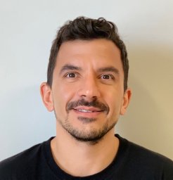 Timoteo Marchini合作论文数University Heart Center, Cardiology and Angiology I, University of Freiburg;and Department of Pneumology, University of Freiburg;University of Freiburg, University of Freiburg62
 Magnani Natalia D合作论文数School of Pharmacy and Biochemistry, University of Buenos Aires-CONICET61
 Silvia Alvarez合作论文数School of Pharmacy and Biochemistry, University of Buenos Aires48
 Mariana Garcés合作论文数37
 Virginia Vanasco合作论文数School of Pharmacy and Biochemistry, University of Buenos Aires31
 Lourdes Caceres合作论文数Inst Bioquim & Med Mol IBIMOL, Univ Buenos Aires27Calabró Valeria合作论文数22Susana Llesuy合作论文数21
 Tamara Vico合作论文数Dept Ciencias Quim, Univ Buenos Aires20
 Deborah Tasat合作论文数20
 Ricardo J. Gelpi合作论文数Department of Pathology, Faculty of Medicine, Univesity of Buenos Aires;Cardiovascular Physiopathology Institute, Univesity of Buenos Aires19Giuseppe Valacchi合作论文数Department of Physiology, University of Siena15Valeria Tripodi合作论文数Faculty of Pharmacy and Biochemistry, University of Buenos Aires15Agustina Freire合作论文数14
 Martin Donato合作论文数12
 Sandra M. Ferreira合作论文数11
 Manuela Martinefski合作论文数11
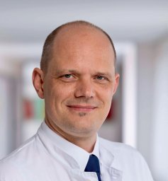 Dennis Wolf合作论文数Atherogenesis Research Group, University of Freiburg10Sofía Reynoso合作论文数9
 Virginia Perez合作论文数9
 Buchholz Bruno合作论文数9
 Alessandra Pecorelli合作论文数Plants for Human Health Institute, N.C. State University9Laura Caltana合作论文数9
 Juan Santiago Adan Arean合作论文数8Verónica D'Annunzio合作论文数7
 Ailen Gala Hvozda Arana合作论文数7
 Lasagni Vitar Romina M合作论文数School of Pharmacy and Biochemistry, University of Buenos Aires7Alberto Boveris合作论文数7
 Martin Desimone合作论文数Facultad de Farmacia y Bioquimica, Universidad de Buenos Aires;Conicet7
 Alejandro Berra合作论文数7Claudia Reides合作论文数7
 Marcelo L. Wagner合作论文数Cátedra de Botánica y Museo Botánico Juan A. Dominguez, Universidad de Buenos Aires7Beatriz González-Flecha合作论文数Harvard School of Public Health Physiology Program, Department of Environmental Health Boston Massachusetts 021157Christian Höcht合作论文数School of Pharmacy and Biochemistry, University of Buenos Aires6
 Diego A Chiappetta合作论文数National Science Research Council (CONICET),6
 S. Fabian Lerner合作论文数5Sara Malchow合作论文数5
 Franco Cervellati合作论文数5
 Marcela A. Moretton合作论文数5
 Daniel H Gonzalez Maglio合作论文数Pharmacy and Biochemistry School, University of Buenos Aires5Diamela T. Paez合作论文数4María B Guglielmotti合作论文数4
 Tamara Mazo合作论文数4
 Daniel Gustavo Olmedo合作论文数4
 Olaf Groß合作论文数Univ Lausanne, Dept Biochem, CH-1066 Epalinges, Switzerland4
 Ezequiel Bernabeu合作论文数4
 Julieta del Mauro合作论文数4
 Cecilia Dobrecky合作论文数4




