Abstract Background Our aim was to train and test a deep learning-based algorithm for automatically segmenting kidneys and renal cysts in patients with autosomal dominant polycystic kidney disease (ADPKD). Methods We retrospectively selected all ADPKD patients who underwent renal MRI with coronal T2-weighted imaging at our institution from 2008 to 2022. The 20 most recent examinations constituted the test dataset, to mimic pseudoprospective enrolment. The remaining ones constituted the training dataset to which eight normal renal MRIs were added. Kidneys and cysts ground truth segmentations were performed on coronal T2-weighted images by a junior radiologist supervised by an experienced radiologist. Kidneys and cysts of the 20 test MRIs were segmented by the algorithm and three independent human raters. Segmentations were compared using overlap metrics. The total kidney volume (TKV), total cystic volume (TCV), and cystic index (TCV divided by TKV) were compared using Bland–Altman analysis. Results We included 164 ADPKD patients. Dice similarity coefficients ranged from 85.9% to 87.4% between the algorithms and the raters’ segmentations and from 84.2% to 86.2% across raters’ segmentations. For TCV assessment, the biases ± standard deviations (SD) were 3–19 ± 137–151 mL between the algorithm and the raters, and 22–45 ± 49–57 mL across raters. The algorithm underestimated TKV and TCV in two outliers with TCV > 2800 mL. For cystic index assessment, the biases ± SD were 2.5–6.9% ± 6.7–8.3% between the algorithm and the raters, and 2.1–9.4 ± 7.4–11.6% across raters. Conclusion The algorithm’s performance fell within the range of inter-rater variability, but large TKV and TCV were underestimated. Relevance statement Accurate automated segmentation of the renal cysts will enable the large-scale evaluation of the prognostic value of TCV and cystic index in ADPKD patients. If these biomarkers are prognostic, then automated segmentation will facilitate their use in daily routine. Key Points Cystic volume is an emerging biomarker in ADPKD. The algorithm’s performance in segmenting kidneys and cysts fell within interrater variability. The segmentation of very large cysts, under-represented in the training dataset, needs improvement. Graphical Abstract
Real-time imaging is required to guide minimally invasive catheter-based cardiac interventions. While transesophageal echocardiography allows for high-quality visualization of cardiac anatomy, X-ray fluoroscopy provides excellent visualization of devices. We have developed a novel image fusion system that allows real-time integration of 3-D echocardiography and the X-ray fluoroscopy. The system was validated in the following two stages: 1) preclinical to determine function and validate accuracy; and 2) in the clinical setting to assess clinical workflow feasibility and determine overall system accuracy. In the preclinical phase, the system was assessed using both phantom and porcine experimental studies. Median 2-D projection errors of 4.5 and 3.3 mm were found for the phantom and porcine studies, respectively. The clinical phase focused on extending the use of the system to interventions in patients undergoing either atrial fibrillation catheter ablation (CA) or transcatheter aortic valve implantation (TAVI). Eleven patients were studied with nine in the CA group and two in the TAVI group. Successful real-time view synchronization was achieved in all cases with a calculated median distance error of 2.2 mm in the CA group and 3.4 mm in the TAVI group. A standard clinical workflow was established using the image fusion system. These pilot data confirm the technical feasibility of accurate real-time echo-fluoroscopic image overlay in clinical practice, which may be a useful adjunct for real-time guidance during interventional cardiac procedures.
Colorectal cancer is the third most common cancer in both men and women. It is estimated that in 2004, nearly 147 000 cases of colon and rectal cancer will be diagnosed in the USA, and approximately 57 000 people would die from the disease; however, only 44% of the eligible population undergoes any type of colorectal cancer screening. Many reasons have been identified for non-compliance, with key ones being patient comfort, bowel preparation and cost. Virtual colonoscopy derived from computed tomography (CT) images is gaining broader acceptance as a screening method for colorectal neoplasia. Our research suggests that computer-aided detection (CAD) as a second reader has great potential in improving polyp detection. The ColonCAD prototype presented in this paper was developed and tested on cases representative of the variability and quality in true clinical practice. Results of this study with 150 patients demonstrate that: the developed algorithm generalises well: the sensitivity for polyps >= 6 mm is on average 90%; and the median false positive rate is a manageable 3 per volume.
The use of ultrasound imaging for guidance of cardiac interventional procedures is limited by the small field of view of the ultrasound volume. A larger view can be created by image-based registration of several partially overlapping volumes, but automatic registration is likely to fail unless the registration is initialized close to the volumes' correct alignment. In this article, we use X-ray images to track a transesophageal ultrasound probe and thereby provide initial position information for the registration of the ultrasound volumes. The tracking is possible using multiple X-rays or just a single X-ray for each probe position. We test the method in a phantom experiment and find that with at least 50% overlap, 88% of volume pairs are correctly registered when tracked using three X-rays and 86% when using single X-rays. Excluding failed registrations with errors greater than 10 mm, the average registration accuracy is 2.92 mm between ultrasound volumes and 4.75 mm for locating an ultrasound volume in X-ray space. We conclude that the accuracy and robustness of the registrations are sufficient to provide useful images for interventional guidance.
PURPOSE X-ray fluoroscopically guided cardiac electrophysiology (EP) procedures are commonly carried out to treat patients with arrhythmias. X-ray images have poor soft tissue contrast and, for this reason, overlay of a three-dimensional (3D) roadmap derived from preprocedural volumetric images can be used to add anatomical information. It is useful to know the position of the catheter electrodes relative to the cardiac anatomy, for example, to record ablation therapy locations during atrial fibrillation therapy. Also, the electrode positions of the coronary sinus (CS) catheter or lasso catheter can be used for road map motion correction. METHODS In this paper, the authors present a novel unified computational framework for image-based catheter detection and tracking without any user interaction. The proposed framework includes fast blob detection, shape-constrained searching and model-based detection. In addition, catheter tracking methods were designed based on the customized catheter models input from the detection method. Three real-time detection and tracking methods are derived from the computational framework to detect or track the three most common types of catheters in EP procedures: the ablation catheter, the CS catheter, and the lasso catheter. Since the proposed methods use the same blob detection method to extract key information from x-ray images, the ablation, CS, and lasso catheters can be detected and tracked simultaneously in real-time. RESULTS The catheter detection methods were tested on 105 different clinical fluoroscopy sequences taken from 31 clinical procedures. Two-dimensional (2D) detection errors of 0.50 ± 0.29, 0.92 ± 0.61, and 0.63 ± 0.45 mm as well as success rates of 99.4%, 97.2%, and 88.9% were achieved for the CS catheter, ablation catheter, and lasso catheter, respectively. With the tracking method, accuracies were increased to 0.45 ± 0.28, 0.64 ± 0.37, and 0.53 ± 0.38 mm and success rates increased to 100%, 99.2%, and 96.5% for the CS, ablation, and lasso catheters, respectively. Subjective clinical evaluation by three experienced electrophysiologists showed that the detection and tracking results were clinically acceptable. CONCLUSIONS The proposed detection and tracking methods are automatic and can detect and track CS, ablation, and lasso catheters simultaneously and in real-time. The accuracy of the proposed methods is sub-mm and the methods are robust toward low-dose x-ray fluoroscopic images, which are mainly used during EP procedures to maintain low radiation dose.
Two-dimensional (2D) X-ray imaging is the dominant imaging modality for cardiac interventions. However, the use of X-ray fluoroscopy alone is inadequate for the guidance of procedures that require soft-tissue information, for example, the treatment of structural heart disease. The recent availability of three-dimensional (3D) trans-esophageal echocardiography (TEE) provides cardiologists with real-time 3D imaging of cardiac anatomy. Increasingly X-ray imaging is now supported by using intra-procedure 3D TEE imaging. We hypothesize that the real-time co-registration and visualization of 3D TEE and X-ray fluoroscopy data will provide a powerful guidance tool for cardiologists. In this paper, we propose a novel, robust and efficient method for performing this registration. The major advantage of our method is that it does not rely on any additional tracking hardware and therefore can be deployed straightforwardly into any interventional laboratory. Our method consists of an image-based TEE probe localization algorithm and a calibration procedure. While the calibration needs to be done only once, the GPU-accelerated registration takes approximately from 2 to 15s to complete depending on the number of X-ray images used in the registration and the image resolution. The accuracy of our method was assessed using a realistic heart phantom. The target registration error (TRE) for the heart phantom was less than 2mm. In addition, we assess the accuracy and the clinical feasibility of our method using five patient datasets, two of which were acquired from cardiac electrophysiology procedures and three from trans-catheter aortic valve implantation procedures. The registration results showed our technique had mean registration errors of 1.5-4.2mm and 95% capture range of 8.7-11.4mm in terms of TRE.
In this paper, we propose to create a rich database of synthetic time series of 3D echocardiography (US) images using simulations of a cardiac electromechanical model, in order to study the relationship between electrical disorders and kinematic patterns visible in medical images. From a real 4D sequence, a software pipeline is applied to create several synthetic sequences by combining various steps including motion tracking and segmentation. We use here this synthetic database to train a machine learning algorithm which estimates the depolarization times of each cardiac segment from invariant kinematic descriptors such as local displacements or strains. First experiments on the inverse electrokinematic learning are demonstrated on the synthetic 3D US database and are evaluated on clinical 3D US sequences from two patients with Left Bundle Branch Block.
 Raoul Florent合作论文数10
 Luca Bogoni合作论文数Carestream9Olivier Nempont合作论文数7
 Nicholas Ayache合作论文数Inria;AI Institute “3IA Côte d’Azur”6K.S. Rhode合作论文数Division of Imaging Sciences
School of Medicine, King's College London 6Geert Gijsbers合作论文数5gogin nicolas p b合作论文数5
Nicholas Ayache合作论文数Inria;AI Institute “3IA Côte d’Azur”6K.S. Rhode合作论文数Division of Imaging Sciences
School of Medicine, King's College London 6Geert Gijsbers合作论文数5gogin nicolas p b合作论文数5 Nicolas Villain合作论文数5
 Aldo Rinaldi合作论文数Cardiovascular Centre, Guy's and St Thomas' NHS Foundation Trust;Faculty of Life Sciences & Medicine, King’s College London5Senthil Periaswamy合作论文数AstraZeneca5Jonathan Stoeckel合作论文数5
 Aruna Arujuna合作论文数King's College London5Patrick Etyngier合作论文数5
 Anna Jerebko合作论文数5
 Sarang Lakare合作论文数InTouchApp5Matthias Wolf合作论文数4
 R. James Housden合作论文数4
 Reza Razavi合作论文数Faculty of Life Sciences & Medicine, King's College London;School of Biomedical Engineering & Imaging Sciences, King's College London;Department of Cardiovascular Imaging, King's College London;British Heart Foundation Centre, King's College London4
 Adityo Prakosa合作论文数Asclepios Research Project, INRIA Sophia-Antipolis4Jaswinder S. Gill合作论文数4
 Murat Dundar合作论文数Department of Computer & Information Science, Indiana University - Purdue University4
 Yingliang Ma合作论文数School of Computing Sciences, University of East Anglia4Saloux Eric合作论文数4M Macari合作论文数3
 Jianming Liang合作论文数3
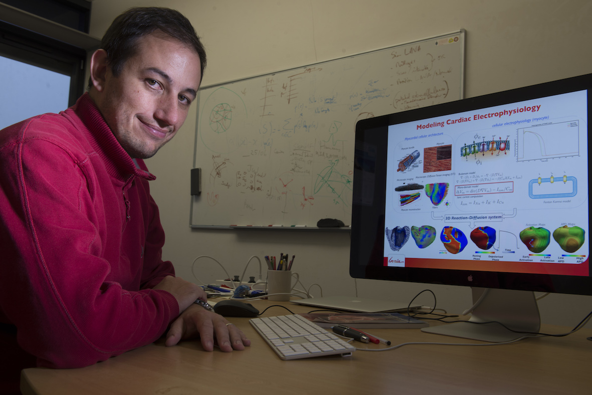 Maxime Sermesant合作论文数INRIA Sophia Antipolis,France3Gang Gao合作论文数3Roland Bullens合作论文数3Xiangwei Zhang合作论文数3
 Jean-François Mangin合作论文数CEA;CATI3Niels Nijhof合作论文数3Martyn Thomas合作论文数2
 McLennan Geoffrey合作论文数2Graeme P. Penney合作论文数2
 Stefan Wörz合作论文数2
 Eric A. Hoffman合作论文数Department of Radiology, Carver College of Medicine, University of Iowa2Karl Rohr合作论文数2
 Hervé Delingette合作论文数Inria;Université Côte d’Azur2
 Pascal ALLAIN合作论文数2
 Arnaud Cachia合作论文数CEA, Service Hospitalier Frédéric Joliot2Jane Hancock合作论文数2
 Milan Sonka合作论文数Department of Ophthalmology and Visual Sciences, Carver College of Medicine, The University of Iowa;Department of Electrical and Computer Engineering, College of Engineering, The University of Iowa;Iowa Institute for Biomedical Imaging, The University of Iowa2Ezequiel Geremia合作论文数2Dennis Caulfield合作论文数2Victoria Parish合作论文数2
 Mark O Neill合作论文数School of Biomedical Engineering & Imaging Sciences, Faculty of Life Sciences & Medicine, King’s College London;St. Thomas' Hospital;Guy's and St Thomas' NHS Foundation Trust2
 Olivier Clatz合作论文数Ministère des Solidarités et de La Santé2
 Herve Delingette合作论文数ASCLEPIOS;CardioSense3D Large Initiative Action2Michael Cooklin合作论文数2




