The objective of this study was to evaluate the effect of seletracetam (SEL), a potent modulator of synaptic vesicle glycoprotein 2A (SV2A), in patients with photoparoxysmal EEG response (PPR) to intermittent photic stimulation (IPS) as proof-of-principle of efficacy in patients with epilepsy. In this multicenter, single-blind Phase II study, adults with photosensitive epilepsy, with/without concomitant antiseizure medication therapy, underwent IPS under 3 eye conditions (at eye closure, eyes closed and eyes open) after a single oral dose of placebo (day - 1) or SEL (day 1; 0.5, 1, 2, 4, 10, or 20 mg). Complete suppression was a standardized photosensitivity range reduction to 0 over >= 1 time points for all eye conditions. Partial suppression was a >= 3-point reduction over >= 3 testing times vs the same time points on day - 1 in >= 1 eye condition. In addition, pharmacokinetics and safety were assessed. Of 27 evaluable patients, 9 reentered to receive a 2nd dosing 1-6 months later, providing a total of 36 individual exposures. At all doses administered - even the lowest -, several subjects reached a complete abolishment of PPR, with a rapid onset of effect. Overall, complete abolishment of PPR was obtained in 40-71 % of the patients; the effect increasing with the dose. In terms of effective doses to suppress PPR, SEL was at least 1,500 times more potent than levetiracetam and 10-20 times more potent than brivaracetam. Adverse events of SEL, including dizziness and somnolence, were mild to moderate. Pharmacokinetics of SEL demonstrated rapid absorption and a linear dose:plasma level relationship. This proof-of-principle study demonstrates that - based on our own experience - SEL is the most potent compound ever tested in the photosensitivity model.
In stroke units, diagnosing seizures may be difficult, especially in aphasic patients. We discuss herein our systematic 4-hour video EEG monitoring of 61 patients with aphasia within the first 72hours after the onset of ischaemic stroke. Five electrographic seizures were identified in 3 patients, with no clinical signs apparent on the video and no symptoms reported by patients. We did not record status epilepticus nor generalized seizure. Comparative analyses disclosed a higher risk of early seizures in patients with haemorrhagic transformation. Video EEG monitoring detected electrographic seizures in 5% of stroke patients with aphasia. This monitoring could be useful for selected patients, especially those with haemorrhagic transformation.
Stroke, whether ischemic or hemorrhagic, is the main etiology of epilepsy in the elderly. However, incidences and outcomes differ according to stroke subtype and delay of onset following the stroke. While the medical literature is extensive, it is not always consistent, and many questions still remain regarding risk factors and management of vascular epilepsy. Thus, the present report here is an overview of the clinical aspects of vascular epilepsy using a practical approach that integrates data from meta-analyses and the more recently published expert recommendations.
EEG recordings are necessary beyond epileptological situations. They are also useful for ICU doctors for adults and children (coma, confusion, anoxia, hypothermia, etc.), emergency doctors and anesthetists (delayed awakening from anesthesia). The question of the conditions of care of such clinical status, especially after working hours in hospitals as well as ICU and emergency services, arises. What are the solutions? – Inter-hospitals collaboration Advantages: performing EEGs will be cheaper for hospitals. Better confidence among professionals, establishment of a regional network in France and hope for medical job creation or financial means. Drawbacks: transferring an EEG is not always secured. Lack of medical technicians, long-medical procedure. Electro-physiological laboratories will also be overloaded with work. – Calling on private companies Advantages: immediate use of an operating service. EEG interpretation performed by a network of health professionals, who comply with interpretation specifications. Data security imposed and regulated by public institutions (ARS, CNIL). Patient monitoring, access to recordings by hospitals who requested EEGs. Drawbacks: what about medical data? Confidence problem easily solved by a use agreement. Neurophy, which is led by Pr. Hervé Vespignani and which counts a group of French interpreters, who comply with an EEG interpretation charter, was created in accordance with the SNPC recommendations on EEG. Neurophy shares its experience based on the 10, 000 EEGs collected from more than 60 clients partners and around 1300 doctors using this method. Thanks to the 10,000 EEGs, we are able to design a profile of different patients using this service and to show different emergency indications on EEGs. We are able to provide a service to hospitals in a continuum of care. The further development of telemedicine applied to EEG is to be encouraged as well as its rigorous evaluation for improving the patient care.
Purpose. - To analyze the semiological characteristics of the language and speech disorders arising during epileptic seizures, and to describe the patterns of language and speech disorders that can predict laterality of the epileptic focus.Method. - This study retrospectively analyzed 95 consecutive videos of seizures with language and/or speech disorders in 44 patients admitted for diagnostic video-EEG monitoring. Laterality of the epileptic focus was defined according to electro-clinical correlation studies and structural and functional neuroimaging findings. Language and speech disorders were analyzed by a neurologist and a speech therapist blinded to these data.Results. - Language and/or speech disorders were subdivided into eight dynamic patterns: pure anterior aphasia; anterior aphasia and vocal; anterior aphasia and "arthria''; pure posterior aphasia; posterior aphasia and vocal; pure vocal; vocal and arthria; and pure arthria. The epileptic focus was in the left hemisphere in more than 4/5 of seizures presenting with pure anterior aphasia or pure posterior aphasia patterns, while discharges originated in the right hemisphere in almost 2/3 of seizures presenting with a pure vocal pattern. No laterality value was found for the other patterns.Conclusion. - Classification of the language and speech disorders arising during epileptic seizures into dynamic patterns may be useful for the optimal analysis of anatomo-electro-clinical correlations. In addition, our research has led to the development of standardized tests for analyses of language and speech disorders arising during seizures that can be conducted during video-EEG sessions. (C) 2017 Elsevier Masson SAS. All rights reserved.
Nous rapportons le cas d'un patient accueilli en alerte thrombolyse pour épisodes régressifs à répétition d'aphasie et paralysie faciale droite au décours d'une injection intra-thécale d'Iohexol. Un homme de 86 ans, droitier, présentant comme principaux antécédents une cardiopathie ischémique, une hypertension artérielle, et porteur d'un pacemaker, a été adressé en alerte thrombolyse au CHU de Bicêtre suite à trois épisodes stéréotypés d'aphasie associée à une paralysie faciale centrale droite, d'une durée de 5 minutes, avec perte de contact. À l'admission, le patient était asymptomatique (score NIHSS 0). La TDM cérébrale retrouvait une hyper densité de la quasi-totalité des espaces sous arachnoïdiens, infra et sus-tentoriels. Le doppler en urgence ne montrait pas de signe de sténose carotidienne ou sylvienne gauche. L'anamnèse révélera la réalisation la veille d'une saccoradiculographie à l'Iohexol, dans un bilan de probable canal lombaire étroit. Le patient n'a pas présenté de nouveau symptôme durant son hospitalisation, et l'imagerie de contrôle effectuée à 48 heures des symptômes a pu montrer la régression des hyper densités et l'absence d'anomalie parenchymateuse. L'EEG était normal. Aucun traitement de fond n'a été introduit. L'injection intrathécale de produit de contraste est une cause rapportée, mais rare d'épilepsie, responsable également de déficits neurologiques focaux ou de méningo-encéphalite. Les symptômes présentés par le patient semblent correspondre à des crises d'épilepsie partielles en lien avec la neurotoxicité du produite de contraste. L'injection intrathécale de produit de contraste dans le cadre d'une myélographie est une cause rare mais possible de crise d'épilepsie, pouvant simuler des AIT, à ne pas méconnaître.
Developmental venous anomalies (DVAs) are congenital anatomical variants of normal venous drainage of normal brain. Although DVAs are often discovered on the occasion of a seizure, their involvement in epilepsy is poorly studied. Our objective was to determine whether DVA can cause seizures, in the cases where there is no associated lesion, including no cavernoma or dysplasia. Based on clinical history, cerebral MRI, EEG recording, and 18F-FDG PET, we report 4 patients with DVA revealed by seizures. The first patient had a convulsive seizure caused by a hemorrhagic infarction due to thrombosis of her DVA. The second patient had a left temporo-parietal DVA next to a nonspecific lesion, possibly a sequelae of a venous infarction. The last two patients disclosed an isolated and uncomplicated DVA with a concordant epileptic focus confirmed on ictal video EEG recording. We reviewed literature and identified 21 other published cases of seizures caused by complications of a DVA and 9 patients that may have a direct link between epilepsy and an isolated and uncomplicated DVA. Seizures are linked to a DVA in two main situations: presence of an associated epileptogenic lesion, such as cavernoma or dysplasia, and occurrence of a complication of the DVA. Before concluding that a seizure is caused by a DVA, it is essential to perform full MRI protocols to search them. It remains rare and uncertain that isolated and uncomplicated DVA can cause seizures. In this last situation, physiopathological processes are probably different in each patient.
ABSTRACTIntroductionMany patients treated with intravenous immunoglobulin (IVIg) are >60 years of age. Tolerability has yet to be demonstrated in this age group.MethodsThis is a retrospective study of adverse reactions among consecutive patients treated with IVIg for neurological disorders. Risk factors were recorded. Correlation and relative risks were calculated for age, risk factors, IVIg course, daily dose, concentration, preparation, and duration of treatment. An infusion and monitoring protocol was applied.ResultsTwo hundred forty‐four patients were reviewed, including 62% who were ≥60 years of age (total dose 1.8 ± 0.4 g/kg body weight, daily dose 30.3 ± 2.0 g). Sixty‐nine percent received sugar‐stabilized IVIg. Forty‐nine percent presented with >1 risk factor. Adverse reactions occurred in 35% and led to treatment discontinuation in 5%, with a similar incidence among age groups. In patients ≥60 years old, sucrose‐free IVIg administration was an independent predictor of adverse reactions, including renal failure.ConclusionIn the elderly, IVIg infusions are safe. Adverse reactions mainly depend on IVIg preparation and administration. Renal failure is not uncommon with sugar‐free IVIg. Muscle Nerve 53: 683–689, 2016
Le crisi epilettiche possono essere provocate da stimoli visivi. La fotosensibilità è il fattore patologico principale di queste crisi o epilessie riflesse. Essa è determinata geneticamente e si osserva preferenzialmente intorno alla pubertà e nel 5% degli epilettici. La sua evidenziazione richiede una tecnica rigorosa di stimolazione luminosa intermittente realizzata durante la registrazione elettroencefalografica. La fotosensibilità può essere isolata o associata a un'epilessia, il più delle volte di tipo idiopatico. Prima dell'avvento degli schermi piatti, la televisione era il principale fattore causale delle crisi. La fisiopatologia è poco nota. Il trattamento dipende da ogni caso; si ha la scelta tra delle regole di igiene di vita e dei farmaci antiepilettici.
La remédiation cognitive (CRT) est une technique apparaissant de plus en plus intéressante pour le traitement de l'anorexie mentale chez l'adulte. Nous proposons d'adapter cette technique à un groupe d'enfants et d'adolescents âgés de 12 à 17 ans hospitalisés dans notre unité pour anorexie mentale.Nous avons proposé à 10 patients hospitalisés pour anorexie mentale, des séances hebdomadaires de CRT d'une heure chacune pendant 10 semaines afin d'en évaluer d'abord la faisabilité et l'acceptabilité. Une évaluation neuropsychologique a été effectuée avant et, pour ceux ayant suivi la totalité des séances, après traitement. Des lettres de feedback ont été recueillies après chaque séance afin d'améliorer le déroulement et le contenu du programme en vue de son application thérapeutique et de recherches futures.Les résultats préliminaires suggèrent que cette technique est acceptable pour ce type de population. Les résultats aux tests neuropsychologiques sont encourageants, et le traitement des lettres de feedback nous a permis de modifier certains points du programme.Dans le futur, les études nécessiteront un échantillon plus large, des méthodes d'évaluation plus sensibles, ainsi qu'une nouvelle évaluation du mode de déroulement et du contenu du programme.Cognitive remediation therapy (CRT) seems to be increasingly interesting in the treatment of anorexia nervosa for adult patients. We attempted to apply this support to a group of young inpatients, initially to assess its feasibility and acceptability, and then to improve its content for therapeutic application and future research.Ten 12- to 17-year-old inpatients with primary DSM-IV diagnosis of anorexia nervosa participated in a 10-week intervention program with a one-hour group session of CRT per week. All 10 patients were assessed before the intervention and those who completed the 10 sessions were assessed after. Assessment included a clinical examination by a psychiatrist, a battery of clinical inventories, and set-shifting tests. Moreover, each patient wrote a letter providing feedback on the intervention for subsequent analysis.Only two patients completed all 10 sessions, the other eight who were discharged from the hospital in the meantime could not attend the sessions for practical reasons. After the 10 sessions, an improvement in BMI and in measured levels of some psychopathological symptoms was observed in our two patients. Most neuropsychological task performances were improved after cognitive remediation. Feedback from the 10 patients was generally positive.This preliminary investigation suggests that cognitive remediation therapy is acceptable and feasible in this population. Replication of these findings requires a larger sample, improvement of the trial design, more sensitive measures, and another training format to avoid loss of so many participants.
Background Homocystinuria caused by cystathionine beta synthase (CBS) deficiency is most often diagnosed in childhood and has a variable expressivity. The most frequent abnormalities include intellectual disability, ectopia lentis, myopia, skeletal abnormalities or thromboembolism. Objective To report a case of homocystinuria unraveled by cerebral venous thrombosis (CVT). Observation A 17 year old female was admitted in our department of neurology for subacute headache and presented seizures in the emergency room. Cerebral imaging revealed CVT. Severe hyperhomocysteinemia was found and led to the diagnosis of homocystinuria due to composite heterozygous mutations in the CBS gene. Further investigations disclosed lens subluxation in association with myopia, mild scoliosis and osteopenia. The patient was treated by heparin followed by warfarin, vitamin therapy and dietary methionine restriction. Total homocysteine and methionine levels became normal in a few weeks and the patient had a complete recovery. Conclusion In patients with CVT, plasma total homocysteine measurement as part of the etiologic work up may reveal severe hyperhomocysteinemia due to CBS or remethylation defects that require specific treatment and management including perhaps protein-restricted diet and/or vitamin therapy for life.
A new class of drugs, the nonimidazole histamine 3 receptor (H3R) antagonists, has been developed in the past decade for treatment of various brain diseases. Pitolisant is such a drug. We studied the pharmacodynamic effect of pitolisant in patients with epilepsy in early Phase II, using the photosensitivity proof of concept model. A total of 14 adult patients (11 females and 3 males; 5 drug naïve) were studied for three days to evaluate the effect of a single oral dose of pitolisant on EEG photosensitivity ranges. All patients showed repeatedly a generalized photoparoxysmal response (PPR) prior to drug administration on placebo Day 1. A statistically significant suppressive effect (standardized photosensitive response [SPR] reduction as measured with paired t-tests) for 20-, 40-, or 60-mg doses of pitolisant was seen in 9/14 (64%) patients of whom 6/14 (43%) showed abolition of the response to intermittent photic stimulation (IPS). Patients on the highest dosage (60 mg) showed the strongest effect with an effect lasting up to 28 h. Thus, full-scale Phase II studies with this novel H3R antagonist, pitolisant, in patients with epilepsy are warranted.
Objective To report the clinical features, causes and outcome of cerebral cortical border-zone infarcts BZI (C-BZI). Methods The authors prospectively included patients with MRI-confirmed C-BZI among individuals consecutively admitted in Stroke Unit. Results Forty-five patients presented C-BZI out of 589 with MRI-confirmed cerebral infarcts (7.6%). Particular clinical characteristics existed in C-BZI in comparison with other cerebral infarctions as a whole, including: (1) frequent transient symptoms at onset (27% vs 9%; p<0.001) and low severity score (NIHSS=3.1±3.0 vs 5.2±6.1; p=0.02); (2) early seizures in first 2 weeks (7/45 (15.6%) vs 12/544 (2.2%); p<0.001), even when focusing only on other infarctions involving the cerebral cortex (15.6% vs 4.3%; p<0.01); (3) heterogeneous clinical presentation but specific transcortical aphasia allowing a clinical suspicion of BZI before MRI; and (4) frequently associated internal carotid disease (69%), with subsequent early surgery in 75% of the cases. Following adapted care in stroke unit, C-BZIs' prognosis appeared good (Rankin score ≤2 at D90) for 82% of the patients. Conclusion Some clinical features are overrepresented in such infarctions, including initial transient symptoms preceding the onset of a completed deficit, transcortical aphasia and early seizures. Despite lower initial severity, C-BZIs justify early management in stroke unit, often followed by carotid surgery, leading to an overall good prognosis.
Paroxysmal dysarthria-ataxia syndrome (PDA) is a rare neurological disorder that can be either primary or symptomatic of acute neurological dysfunction. Episodes of symptomatic PDA are poorly documented and there are no video reports. We describe the cases of two patients with symptomatic PDA related to demyelinating diseases. Detailed studies of the patients' speech disorders showed that the dysarthria and gait disorders were of the ataxic type in both cases. Both patients had midbrain lesions at or below the level of the red nucleus, confirming that this area is critically involved in PDA. The best clinical signs for distinguishing between symptomatic and primary PDA are adult onset and short (<1 min) episodes in the former. If these signs are present, brain MRI should be used to identify a cause of symptomatic PDA.
PURPOSE:Diffusion tensor imaging (DTI) can provide quantitative information of brain abnormalities in patients with temporal lobe epilepsy (TLE) that are not detectable with conventional magnetic resonance imaging (MRI). METHODS:Seventeen patients with medically TLE were selected for the study. The patients and ten healthy subjects underwent 25 directions DTI acquisition. The patients were separated into two groups based on the MRI findings: eight TLE MRI-negative patients with no signal abnormalities on conventional MRI and nine TLE patients with hippocampal sclerosis (HS). Fractional anisotropy (FA), mean diffusivity (MD), and the three diffusivities (lambda(1), lambda(2) and lambda(3)) were measured in bilateral hippocampi of controls, MRI-negative, and HS patients. Comparisons between the three groups were performed for hippocampi ipsi- and contralateral to epileptogenic zone. RESULTS:The ipsilateral hippocampus of MRI-negative patients presented statistical increased anisotropy and no significant difference in diffusivities versus controls. Significant differences in anisotropy and diffusivities were detected between the ipsilateral hippocampus of HS when compared with controls. CONCLUSION:DTI depicted hippocampal abnormalities in TLE patients with a normal conventional MRI different from those found in patients with HS. Diffusivity and anisotropy indices provide significant differences inside hippocampus and should be jointly considered to improve the DTI measurements specificity in TLE patients.
 Bouilleret Viviane合作论文数8D Ducreux合作论文数7Pierre Genton合作论文数7
 Edouard Hirsch合作论文数Université de Strasbourg;University Hospital in Strasbourg7Christian Denier合作论文数7Dominique Parain合作论文数6C Denier合作论文数5Gagnepain J-P合作论文数4David Adams合作论文数3M Dreyfus合作论文数3C Kaplan合作论文数3Mariana Sarov合作论文数3Felten D合作论文数3
 Pierre Lasjaunias合作论文数Departement de Neuroradiologie, Hopital de Bicetre3Armel Stockis合作论文数SGS Biopharma S.A.3Francine Chassoux合作论文数3D. Liacu合作论文数3P. Ducreux合作论文数3Bernhard J. Steinhoff合作论文数3M. Quirins合作论文数2Dgakn Trenite合作论文数2Jean-Paul Gagnepain合作论文数2P Jami-Ceccomori合作论文数2I. An合作论文数2
 Marie Theaudin合作论文数2
 Giovanni De Marco合作论文数2Amal Fouad合作论文数2G Said合作论文数2Valerie Proulle合作论文数2Nadine Ajzenberg合作论文数2
 Jacqueline A. French合作论文数Department of Neurology, Grossman School of Medicine, New York University2
 Constance Flamand-Roze合作论文数ST, Centre Hospitalo-Universitaire de Bicêtre2
 Dorothee G.a. Kasteleijn-Nolst Trenite合作论文数Faculty of Medicine and Psychology, Sapienza University2Archambaud Frédérique合作论文数2Cauquil Cecile合作论文数2Cauquil-Michon C合作论文数2Join-Lambert Claire合作论文数2Guedj Thierry合作论文数2Not Adeline合作论文数2Pierre Lozeron合作论文数2
 Jean-Pierre Cartron合作论文数2I. Idy-Peretti合作论文数2
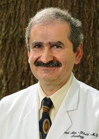 Bassel W. Abou Khalil合作论文数Department of Neurology, School of Medicine, Vanderbilt University2J. Renoux合作论文数2N Badinand-Hubert合作论文数2
 Fabrice Bartolomei合作论文数Aix-Marseille University2JF Savet合作论文数1J Bouillat合作论文数1H Guinot合作论文数1




