Contemporary implant dentistry achieves remarkable survival rates, yet the rising prevalence of peri-implantitis and increasing patient complexity reveal the limits of a technically centered model. This narrative review synthesizes current evidence supporting the four pillars of the 4P framework — predictive, preventive, personalized, and participatory — and proposes an integrated patient-centered clinical algorithm for implant dentistry. A narrative literature search was conducted in PubMed, Scopus, and the Cochrane Library (January 2000 to June 2026), prioritizing systematic reviews, meta-analyses, randomized controlled trials, and international consensus statements. Reporting followed the Scale for the Assessment of Narrative Review Articles (SANRA). Predictive tools — resonance frequency analysis, genomic biomarkers, artificial intelligence algorithms, and radiomic analysis — improve individual risk stratification. Preventive strategies anchored in a four-level continuum reduce peri-implantitis frequency from 43.9% to 18% in maintenance-compliant patients. Personalized approaches optimize osseointegration according to each patient's biological profile. Participatory implantology, through shared decision-making and patient-reported outcome measures, improves the therapeutic alliance and long-term adherence. The 4P model constitutes a coherent and clinically applicable framework. Its novel contribution is a sequential decision-making logic distinguishing pre-placement gate-keeping from post-placement dynamic pillar reorientation, providing clinicians with a structured algorithm applicable at each phase of implant treatment.
This study aims to compare the incidence of sinus membrane perforation between osseodensification and osteotome crestal sinus lift techniques when 5 mm of residual bone is present beneath the sinus floor. Additionally, it seeks to identify when the perforation most commonly occurs-during instrumentation, bone grafting, or implant insertion. A split-mouth design using 20 sinuses from 10 fresh human cadaver heads were employed. One side underwent a bone-added osteotome crestal sinus lift, while the other side received an osseodensification lift. Membrane perforation was recorded through video analysis. Statistical analysis was performed. Sinus membrane perforation occurred in 40% of the osteotome group and 50% of the osseodensification group, with no significant difference between the two (P = .564). Most perforations occurred during implant placement (20% in the control group and 40% in the test group), but this difference was not statistically significant (P = .480). Perforations during drilling were observed in 10% of test sinuses and 20% of the control group, but this difference was not statistically significant (P = .317). No perforations occurred during bone graft placement. There was no significant difference in the incidence of membrane perforation between the osseodensification and osteotome groups. However, a higher incidence during implant placement suggests potential limitations in the predictability of crestal sinus lift procedures with 5 mm of residual bone. Further research is needed to optimize outcomes and predictability in crestal sinus lift procedures.
Maxillary sinus floor augmentation (MSFA) is one of the most predictable hard tissue augmentation procedures performed to support long-term dental implant survival and success. However, risks and complications still exist with this procedure including pain, bleeding, infection, oroantral communication, and inadequate bone regeneration for implant placement. To decrease some of these potential complications and improve outcomes, biologics are becoming more widely used in maxillary sinus augmentation. Autologous blood concentrates (ABCs) can be utilized as a membrane to seal a sinus membrane perforation or as "sticky bone" to mix with particulate material to make a congealed bone graft that will stay in place and be less likely to migrate from the site of placement or into the sinus cavity. Although the data is heterogeneous and somewhat conflicting, there is some evidence to support the use of ABCs for bone graft consolidation and overall wound healing due to the growth factors contained in the ABC. In addition, clotting factors in the plasma aid in hemostasis, which is an essential first step in the wound healing cascade. Individual growth factors such as rhBMP-2 and rhPDGF-BB are also clinically available for use in bone grafting procedures such as MSFA. rhBMP-2 is FDA approved for maxillary sinus augmentation, and clinical trials demonstrate de novo bone formation to facilitate dental implant placement. However, studies do not show a significant improvement over autogenous bone or bone substitutes such as xenograft or allografts. rhPDGF-BB has limited studies in this area, but may decrease residual particulate graft material and aid in increasing vital bone when bone substitutes are used. However, the use for MSFA is "off label" and few studies are available. Finally, enamel matrix derivative (EMD) has no data, and therefore, limited use for MFSA.
Purpose: To achieve a consensus among international experts regarding the management of postoperative complications after maxillary sinus floor elevation. Materials and methods: A total of 32 experts were enrolled and divided into dental implant providers (21), experts with a well-established reputation as sinus specialists (8), ear, nose and throat specialists (2), and experts with a well-established reputation as ear, nose and throat specialists (1). Before starting, a systematic literature search was conducted on the topic, and a list of articles was sent to the panel. The development group formulated 20 statements, which were sent out in the form of a survey. After each round, the statements upon which a consensus was not reached were reformulated based on anonymous comments from participants. A total of three rounds were planned. Results: After the third round, a consensus was reached on 15 key statements regarding the management of postoperative complications following sinus floor elevation. Agreement was established on issues including common postoperative symptoms, use of radiographic assessments, the necessity of surgical interventions such as partial or total graft removal, and the potential need for functional endoscopic sinus surgery. Near-consensus was achieved on additional points concerning normal postoperative symptoms, timing of total graft removal and approaches to late graft infections. Conclusions: The present Delphi consensus suggests that postoperative symptoms such as pain and swelling are generally manageable with appropriate pharmacological treatment. It also outlines conditions where radiographic evaluation is recommended for further assessment. Surgical options, including partial or total graft removal and functional endoscopic sinus surgery, are recommended based on the clinical scenario and response to initial treatments. Variability in practices, particularly regarding antibiotic use and specific intervention timing, suggests a need for further research to be conducted in order to standardise treatment protocols and address gaps in evidence.
Maxillary sinus floor elevation is usually performed in two different ways: the lateral approach involves the creation of a bony window on the maxillary sinus lateral wall, providing direct access to the sinus cavity for membrane elevation and subsequent graft placement, and the transcrestal approach is considered less invasive. The aim of this article is to describe, based on the literature, how to anticipate, avoid, and manage the intraoperative complications that can occur with both approaches. For both approaches, the most common complication is the sinus membrane perforation. For the lateral approach, an average frequency ranging from 15.7% to 23.1% is reported, but because of the better visibility, their management will be easier compared to the transcrestal approach. Mean perforation rate reported for the transcrestal approach is lower (3.1%-6.4%), but it should be noted that a significant number of perforations cannot be detected and managed given the blind nature of this technique. Anatomical parameters such as sinus width and buccal wall thickness may be a risk factor for one approach and not the other. As it is impossible to assess the resistance of the Schneiderian membrane, the transcrestal approach is more likely to lead to infectious complications in the event of perforation. Others, such as the risk of vascular damage, are encountered only with the lateral approach, which can be prevented easily by dissecting the alveolo-antral artery. For both approaches, prevention is essential and consists in analyzing the anatomy, mastering the surgical technique, and collaborating with the ENT to manage the essentially infectious consequences of intraoperative complications.
PURPOSE:To establish consensus-driven guidelines that could support the clinical decision-making process for implant-supported rehabilitation of the posterior atrophic maxilla and ultimately improve long-term treatment outcomes and patient satisfaction.MATERIALS AND METHODS:A total of 33 participants were enrolled (18 active members of the Italian Academy of Osseointegration and 15 international experts). Based on the available evidence, the development group discussed and proposed an initial list of 20 statements, which were later evalu-ated by all participants. After the forms were completed, the responses were sent for blinded ana-lysis. In most cases, when a consensus was not reached, the statements were rephrased and sent to the participants for another round of evaluation. Three rounds were planned.RESULTS:After the first round of voting, participants came close to reaching a consensus on six statements, but no consensus was achieved for the other fourteen. Following this, nineteen statements were rephrased and sent to participants again for the second round of voting, after which a consensus was reached for six statements and almost reached for three statements, but no consensus was achieved for the other ten. All 13 statements upon which no consensus was reached were rephrased and included in the third round. After this round, a consensus was achieved for an additional nine statements and almost achieved for three statements, but no consensus was reached for the remaining statement.CONCLUSION:This Delphi consensus highlights the importance of accurate preoperative planning, taking into consideration the maxillomandibular relationship to meet the functional and aesthetic requirements of the final restoration. Emphasis is placed on the role played by the sinus bony walls and floor in providing essential elements for bone formation, and on evaluation of bucco-palatal sinus width for choosing between lateral and transcrestal sinus floor elevation. Tilted and trans-sinus implants are considered viable options, whereas caution is advised when placing pterygoid implants. Zygomatic implants are seen as a potential option in specific cases, such as for completely edentulous elderly or oncological patients, for whom conventional alternatives are unsuitable.
After tooth loss in the posterior area of the maxilla, sinus floor elevation is often required to compensate the vertical bone loss due to sinus pneumatization. This narrative review reports on the potential benefits of autologous platelet concentrates (APCs) during this procedure. As for transcrestal approach, APCs have been used as "sole" substitute/graft. However, because of the low number of clinical trials available with PRGF, and even none for PRP, no definitive conclusions can be made regarding their efficacy. The number of studies on the use of L-PRF were outnumbered indicating good feasibility for vertical bone gain, with a high implant survival rate and a low degree of complications. PRP and PRGF have not been studied as a "single/sole" substitute for a one-stage lateral window approach, probably because of the weak physical characteristics of the membranes. L-PRF alone appears to be a predictable grafting material for lateral maxillary sinus grafting and a reduced RBH should not be considered as a risk factor. Compared to a "standard" bone substitute L-PRF shows slightly less vertical bone gain (consider enough membrane application and use of bony window as new sinus floor roof over the implant apices), enhanced early resorption (first 6 months after application), but a similar stable bone gain afterward. For a two-stage lateral window approach, APCs "alone" cannot be recommended, due to their weak withstand to the sinus pneumatization forces. APCs combined with bone substitutes seem to accelerate bone formation, without any additional benefits on the long-term new bone gain. The use of L-PRF membranes for the treatment of perforations appears to be an effective treatment option, but further clinical studies are needed to confirm this. Even though the abovementioned statements are based on large numbers of studies, additional RCTs comparing APCs with different types of grafting procedures for sinus elevation are needed.
Management of peri-implantitis is becoming an increasing issue for implantologists and periodontists. The need for bone augmentation is more and more frequent, especially in the posterior maxilla requiring sinus augmentation. Peri-implantitis represents a real danger for implants, but to this day, the available literature concerning the impact of this disease on regenerated bone and on maxillary sinus pathology is very limited. This report presents two cases showing bone alterations due to peri-implantitis and its possible impact on maxillary sinus health. In both cases, the causal implant was removed regardless of prior functional endoscopic surgery to restore sinus health, and it was not necessary to implement any reconstruction procedure because bone regeneration occurred naturally. Further research will be necessary to confirm these initial findings.
Maxillary sinus augmentation with a lateral approach is known to present more postoperative complications than other atrophic posterior maxilla treatment modalities because it is more invasive. These complications include infections that occur in the form of chronic or acute sinusitis. According to the literature, the frequency of these complications ranges from 3% to 5%. They can result from an inadequate management of intraoperative complications or from a poor evaluation of maxillary sinus particularities and pathology before the surgery. Therefore, the prevention of postoperative complications lies in the selection of cases that will allow for the identification and evaluation of infectious risk. Only a multidisciplinary approach that includes an implantologist, a rhinologist, and the treating physician will allow this. On the other hand, in infectious complication cases, the intervention of the otorhinolaryngologist (ENT) specialist is necessary. Based on the available literature and the author's experience, the methodology described in this article will allow for the prevention and management of postoperative complications related to this surgical technique.
The lateral approac for maxillary sinus grafting has become a routine technique with an implant survival rate over 96% in the maxillary posterior region. However, this technique may be associated with some complications that may occur at different time points and influence short and long-term implant survival rate. This article is dedicated to intra operative complications which are mainly due to anatomical variations such as the shape of the sinus, the presence of septa and other particularities such as the intraosseous passage of the antral alveolar artery which may interfere with the position of the vestibular window. In order to identify the risk and prevent the complications, a perfect knowledge of the anatomy is therefore essential. If a complication does occur, it must be treated in an efficient manner to prevent the postoperative complications that may follow, in the form of chronic or acute sinusitis. The prevalence of intra operative complications is conversely proportional to the surgeon’s skill and experience without an influence on implant survival if they are properly managed.
Sinus augmentation has become an integrated surgical phase in posterior maxillary implant prosthesis reconstruction. Since the residual alveolar bony height usually requires additional volume particularly at this anatomical region, sinus floor augmentation is advocated routinely. Over the years, Implant success rate is proved to be comparable to the one in the pristine bone, which is well documented in the literature. Anatomical aspects as well as surgeon skills are at most importance to achieve predictable outcome. In this narrative review, the different osteotomy techniques, the indications toward 1 or 2-stage approaches, the control of the Schneiderian membrane integrity as well as the management of intra- and post-operative complications are thoroughly discussed according the current data. In light of the excellent long-term implant success rate concurrent with the application of contemporary advanced techniques of the sinus augmentation via the lateral wall osteotomy approach, reduce invasiveness and less complication occurrences are well documented. A well-codified patient selection involving the rhinologist as an integral medical team would be significantly beneficial toward early diagnosis. In-depth knowledge of the anatomy, execution of a well standardized surgical technique, and understanding the complication etiology and their management are prerequisites for reducing patient morbidity to minimal discomfort and predictable successful outcome.
PURPOSE The aim of the present retrospective clinical study was to evaluate the outcome of a maxillary sinus lateral window augmentation protocol, which sought to shorten the treatment time. MATERIALS AND METHODS This protocol entailed sinus augmentation with deproteinized bovine bone minerals (DBBM) and simultaneous implant placement in patients with minimal residual bone height. A total of 89 sinus augmentation procedures were performed in 74 patients, in whom 160 implants were placed between 2005 and 2013. The mean residual bone height was 2.6 ± 0.6 mm. The healing time before loading was 4.18 ± 0.63 months. RESULTS In a first evaluation in 2014 the early implant survival rate (EIsR) was 96.8% after a mean period of 5.4 ± 2.2 years. A second evaluation in 2019 after a mean period of 10.4 ± 2.2 years showed a late implant survival rate (LIsR) of 83.1%. The failures after 2014 were all caused by peri-implantitis, which affected 14.6% and 16.8% of patients and implants, respectively. This prevalence of peri-implantitis does not appear to be higher than that usually observed in nonaugmented sites. CONCLUSION This reduction in the duration of treatment compared to the usual duration of 9 to 12 months does not seem to affect the predictability of the technique.
The aim of the present study was to generate an international and multidisciplinary consensus on the clinical management of implant protrusion into the maxillary sinuses and nasal fossae. A total of 31 experts participated, 23 of whom were experts in implantology (periodontologists, maxillofacial surgeons and implantologists), 6 were otolaryngologists and 2 were radiologists. All the participants were informed of the current scientific knowledge on the topic based on a systematic search of the literature. A list of statements was created and divided into three surveys: one for all participants, one for implant providers and radiologists and one for otolaryngologists and radiologists. A consensus was reached on 15 out of 17 statements. According to the participants, osseointegrated implants protruding radiographically into the maxillary sinus or nasal fossae require as much monitoring and maintenance as implants fully covered by bone. In the event of symptoms of sinusitis, collaboration between implant providers and otolaryngologists is required. Implant removal should be considered only after pharmacological and surgical management of sinusitis have failed.
AIMS:The objective of the present case series is to report on the rationale, surgical technique and outcome of a protocol for peri-implant mucosal phenotype modification therapy, referred to as "fibrin immobilization vestibular extension (FIVE)".MATERIAL AND METHODS:The protocol utilized entailed apical positioning and stabilization of peri-implant flap with modular screws. The screws were also used for the immobilization of solid matrix platelet-rich fibrin to fill the gap created between apically positioned flap and the crestal margin of the flap.RESULTS:A total of 30 patients (12 male, 18 females) with 93 implants were treated with FIVE protocol for various indications, including for vestibular extension following alveolar ridge augmentation (N = 6), preprosthetic (N = 9), postprosthetic (N = 2), and peri-implantitis (N = 13). The keratinized mucosal width preoperatively was 1.67 mm with 95% confidence interval [CI] (1.46, 1.88). Immediately following FIVE surgery, the vestibule was extended to 9.10 with 95% CI (8.44, 9.76). At 3 months, 4.9 mm (95% CI: 4.5-5.2 mm) of peri-implant keratinized mucosal width was present. The keratinized mucosal width remained relatively stable thereafter and was 4.0 mm (95% CI: 3.5-4.5 mm) at 3 years post-FIVE surgery. When overall group means across all time points were analyzed, maxilla had mean of 6.1 mm (95% CI: 5.8-6.5) versus mandible exhibited mean of 5.1 mm (95% CI: 4.6-5.6 mm). The mean of maxilla was significantly higher than that of the mandible (p < 0.0001) across all time points. Treatment of peri-implantitis with FIVE lead to significant pocket reduction and wide band of keratinized mucosa. Seven of 38 implants in 3 of 13 peri-implantitis patients were removed due to advanced peri-implantitis.DISCUSSION:The present case series provides proof-of-principle data for efficacy of FIVE for peri-implant phenotype modification therapy that generated attached keratinized mucosa in a variety of applications. This protocol provides an alternative to procedures involving harvesting of autogenous mucosal graft.
AIM:The aim of this study is to determine the incidence of obliterated osteomeatal complex (OMC) due to the presence of anatomic variants.SETTINGS AND DESIGN:Retrospective Study.MATERIALS AND METHODS:In this retrospective study, a total of 71 patients, 34 males and 37 females, aged 35-65 years were included in the study. Cone beam computed tomography (CBCT) scans of patients were assessed to identify the status of the OMC in the presence of anatomic variants and their incidence was recorded. The radiological assessment of the anatomical variants was made by viewing the coronal sections of the scans. The variants observed were deviated nasal septum, uncinate process), agger nasi, Haller cells, middle turbinate variants, enlarged bulla, accessory ostium, and maxillary sinus abnormalities). Ostium patency was evaluated in the coronal section of each sinus and classified as "patent" or "obstructed." The most common variants observed were then correlated with the patency of the ostium.STATISTICAL ANALYSIS USED:Chi square test was performed to assess the association between the anatomic variants and the patency of the OMC.RESULTS:In the present study, the incidence of an obliterated OMC due to the presence of anatomic variants was 73.2%. The four most common variants associated with the possibility of an obliterated OMC were the deviated nasal septum (76.2%), middle turbinate (86.4%), enlarged bulla (77.8%), and sinus cavity variants (80.0%). A statistically significant association was noted between middle turbinate variants and Haller cells and the patency of the OMC.CONCLUSION:Thorough pretreatment CBCT evaluation should be performed to assess the presence of anatomic variants and thereby, the patency of the ostium before sinus floor elevation procedures. The pre and postsurgical treatment plans and regimes can be modified according to anticipated postsurgical sequelae, thereby avoiding postsurgical complications and enhancing the success of the graft procedure.
OBJECTIVESTo assess the potential trends for the year 2030 in dental implant dentistry in Europe using the Delphi methodology.MATERIAL AND METHODSA steering committee and a management team of experts in implant dentistry were created and validated a questionnaire including 60 questions, divided in eight topics. The survey was conducted in two rounds using an anonymous questionnaire, which provided the participants in the second round with the results of the first. Each question had three possible answers, and the results were expressed as percentages.RESULTSA total of 138 experts were invited to participate in the survey. From all the invited experts, 52 answered in both the first and second rounds. Three different consensus categories were established based on the percentage of agreement: no consensus (<65%); moderate consensus (65%-85%); and high consensus (≥86%). Within the topic categories, a consensus was reached (mainly moderate consensus) for the majority of questions discussed among experts during a face to face consensus meeting. However, consensus was not reached for a small number of questions/topics.CONCLUSIONSAbout 82% of the questions reached consensus. The consensus points towards a lower number of implants to replace chewing units, with implants surfaces made of bioactive materials with reduced micro-roughness using mainly customized abutments with polished surfaces and an internal implant-abutment connection (85%). CBCT-3D technologies will be the main tool for pre-surgical implant placement diagnosis together with direct digital restorative workflows. There will be an increase in the incidence of peri-implantitis, although there will be more efficient interventions its treatment and prevention.
Bovine-derived bone mineral demonstrated good osteoconductive properties as grating material for maxillary sinus floor elevation, but the long-term behavior of this material has not been reported. The purpose of this report was to analyze and compare histomorphometric measurements of new bone, bone graft, and medullar spaces 6 months, 12 months, and 20 years after grafting. In the grafted area, the amount of mineralized bone was 16.96% at 6 months, 22.53% at 12 months, and 22.05% at 20 years, respectively. The amount of bovine-derived bone mineral ranged from 35.87% to 4.85% in the same period. The volume of the newly formed mineralized bone does not increase over time, conversely to nonmineralized bone.
背景信息 法国Corsica大学Pascal Valentini博士的治疗理念. >侧壁开窗上颌窦底提升,同期种植或分阶段种植体植入. >避免自体骨移植. >处理分隔和上颌窦黏膜穿孔问题. 上颌窦底提升时我完全不使用自体骨.多种临床研究通过组织形态学分析证实仅使用自体骨或自体骨混合骨代用品,并不能改善临床效果.获取自体骨反而延长了手术时间,增加了术区,使手术过程复杂化.
Maxillary sinus surgery has been shown to be a reliable procedure for increasing vertical bone height prior to implant placement. A variety of grafting materials have been proposed, with particulate bone substitutes showing similar clinical results to autogenous bone when rough surfaces implants are used. A barrier membrane is usually placed external to the grafted sinus, covering the antrostomy. In this technical report, the membrane is placed over the window and its borders gently tucked between the inner side of the bony wall and the graft material. This procedure stabilizes the membrane without tacks and prevents graft dislodgement through the antrostomy.
 Tiziano Testori合作论文数Department of Periodontics & Oral Medicine, School of Dentistry, University of Michigan-Ann Arbor11
 D Abensur合作论文数7Taschieri Silvio合作论文数Dept Biomed Surg & Dent Sci, Univ Milan6Zuffetti Francesco合作论文数5Matteo Chiapasco合作论文数5
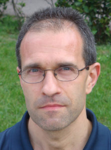 Massimo Del Fabbro合作论文数Department of Biomedical, Surgical & Dental Sciences, University of Milan;Section of Oral Physiopathology, Galeazzi Orthopedic Institute5
 Claudio Stacchi合作论文数University of Trieste, University of Trieste4
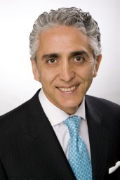 Zadeh Homayoun H合作论文数Ostrow Sch Dent, Univ Southern Calif4
 Giovanni Felisati合作论文数San Paolo Hospital, University of Milan4
 Christoph Hämmerle合作论文数Medizinische Fakultät, Universität Zürich;Center of Dental Medicine, University of Zurich;Dental Campus4Ole T Jensen合作论文数3Lorenzo Pignataro合作论文数3
 Luca Cordaro合作论文数Department of Periodontology and Prosthodontics, “George Eastman” Dental Hospital University Policlinic “Umberto I”3Artzi Zvi合作论文数3Rocchesani Marcel合作论文数3Albertini Jacques F合作论文数3
 Gustavo Avila-Ortiz合作论文数Division of Periodontology, Department of Oral Medicine, Infection and Immunity, Harvard School of Dental Medicine;Departamento de Especialidades Clínicas Odontológicas - Periodoncia, Complutense University of Madrid;Atelier Dental Madrid3
 Raffaele Vinci合作论文数3Wallace Stephen S合作论文数3
 Matteo Deflorian合作论文数3Michael A. Pikos合作论文数3
 Bach Le合作论文数3
 Hom-Lay Wang (王鴻烈)合作论文数Department of Periodontics & Oral Medicine, School of Dentistry, University of Michigan3
 Danilo Di Stefano合作论文数3
 Roberto Pistilli合作论文数3Jose Manuel Navarro合作论文数2Blas Noguerol合作论文数2Turker Ornekol合作论文数2Tommaso Clauser合作论文数2
 Antonio Rapani合作论文数2
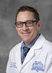 John R Craig合作论文数Dept Otolaryngol Head & Neck Surg, Henry Ford Hosp2Alberto Sicilia合作论文数2
 Hsun-Liang Chan合作论文数Department of Periodontics & Oral Medicine, School of Dentistry, University of Michigan-Ann Arbor2Mario Mantovani合作论文数2
 Mariano Sanz Alonso合作论文数Departamento de Especialidades Clínicas Odontológicas, Facultad de Odontología, Universidad Complutense Madrid2Lozada Jaime L合作论文数2
 Francesco Azzola合作论文数2
 Henning Schliephake合作论文数Abteilung fur Mund-, Kiefer- und Gesichtschirurgie der Georg-August-Universitat Gottingen2Klinge B合作论文数2
 Federico Mandelli合作论文数2
 Ignacio Sanz Sanchez合作论文数Section of Graduate Periodontology, University Complutense2
 Gil Alcoforado合作论文数2
 Teresa Lombardi合作论文数2
 Massimo Simion合作论文数2Sara Torretta合作论文数2
 Lisa J. A. Heitz-Mayfield合作论文数School of Health and Clinical Sciences, The University of Western Australia;The University of Sydney;University of Bern;University of Shanghai2
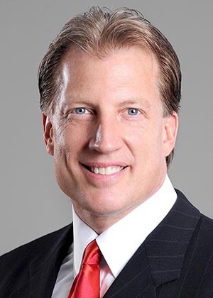 Craig Martin Misch合作论文数Departments of Periodontics & Oral Medicine, School of Dentistry, University of Michigan;Departments of Periodontics and Prosthodontics, University of Florida;Departments of Periodontics and Prosthodontics, University of Alabama2
 Carlotta Pipolo合作论文数2
 Ronald E. Jung合作论文数Clinic of Reconstructive Dentistry, Center of Dental Medicine, University of Zurich2




