Epilepsy is increasingly recognized as a disorder of brain networks rather than a solely focal disease. Beyond the seizure focus, connected brain networks can exert inhibitory control over seizure initiation and propagation resulting in antiseizure effects. Here, we present data from animal and human studies on antiseizure networks and integrate recent results from lesion-, stimulation- and neurophysiological-experiments. Consistent with these results, we present the Interictal Suppression Hypothesis that posits that strong inward connectivity to the seizure focus maintains seizure freedom between events, while collapse of this connectivity accompanies seizure spread and generalization. Workshop discussions highlighted future directions, including mechanistic stimulation studies to interrogate the role of antiseizure networks seizure control and design of therapeutic studies towards network-guided neuromodulation for epilepsy. A deeper mechanistic understanding of antiseizure networks may enable development of precision therapies that target endogenous seizure control rather than simply target the seizure focus.
Introduction: Temporal lobe epilepsy (TLE) is clinically relevant due to its severity and the high percentage of patients resistant to available medications [...]
OBJECTIVE:Current pharmacotherapy for temporal lobe epilepsy (TLE) is limited to symptomatic treatment and leaves approximately one third of patients with inadequate seizure control. Discovering disease-modifying targets is an unmet clinical need. We have previously identified senescent cells (SCs) as one such target. Many drugs that eliminate SCs (senolytics) interfere with apoptotic resistance proteins, potentially resulting in broad cytotoxicity and numerous side effects. Newer, more targeted therapies, like selective senescence killing compound 1 (SSK1), a gemcitabine prodrug that is selectively activated in SCs, offer the possibility to reduce off-target effects, but SSK1 has yet to be investigated in any preclinical epilepsy model. METHODS:We used pilocarpine to induce status epilepticus (SE) in 3- to 4-month-old mice. Immediately following SE, mice were randomly assigned to receive either SSK1 treatment or vehicle for the remainder of the study. We assessed behavioral performance on memory tasks, seizure burden by EEG, and histological markers of SCs. RESULTS:SE robustly increased hippocampal and thalamic expression of SC marker p16 by over 100% compared to saline controls. SSK1 treatment reduced p16+ cells by ~45%, without any apparent neurotoxicity. In addition, SSK1 treatment normalized spatial memory impairments and reduced spontaneous seizure burden, completely protecting a majority (60%) of animals from seizures. SC burden in the hippocampus, but not the thalamus, correlated with seizure burden in vehicle-treated animals. SIGNIFICANCE:These findings lend further credence to the viability of targeting SCs to treat TLE. As with other genetic and pharmacologic SC ablation strategies, SSK1 produced a similar reduction in p16+ cells and normalization of spatial memory. SSK1, however, displays a stronger protective effect against seizures. In short, SSK1 is a compelling, translationally viable option for senolysis in TLE.
Despite a century of development of antiseizure medications, up to a third of people with epilepsy do not achieve seizure freedom with drug therapy. Deep brain stimulation is of growing use, but just as with pharmacotherapy, is not universally effective. Identifying new targets for deep brain stimulation-and in particular sites that are effective against a range of seizure types-may close this gap. Engagement of the basal ganglia experimental seizures was first observed almost 75 y ago. However, the role of the basal ganglia's input nucleus, the striatum, in seizure control is relatively understudied. To address this gap, we used an optogenetic approach to activate and inactivate neurons in the dorsal striatum of rats submitted to the gamma-butyrolactone (GBL) model of absence epilepsy, amygdala kindling model of temporal lobe epilepsy, and pilocarpine-induced Status Epilepticus (SE). Open-loop (continuous light delivery) optogenetic activation of dorsal striatal neurons robustly suppressed seizures in all models. By contrast, open-loop optogenetic silencing increased absence seizure expression and facilitated SE onset but had no effect on kindled seizures. In the GBL model, we also tested the effects of closed-loop modulation (light delivery in response to seizure detection). Closed-loop activation reduced duration of spike-wave discharges (SWDs), while closed-loop inhibition increased SWD duration. These results demonstrated previously unrecognized antiabsence effects associated with striatal neuromodulation. These findings demonstrate a robust, bidirectional role of the dorsal striatum in the control of multiple seizure types, suggesting that the striatum is a site that can exert broad-spectrum control of seizures.
Rapidly approaching visual stimuli (i.e. looming objects) are known to evoke unconditioned defense responses across species. In rodents, this threat reactivity repertoire includes freezing and fleeing behavior. Although components of the circuitry underlying unconditioned response to a looming threat have been elucidated, both a temporal characterization and drug effects on the freezing response have not yet been reported. Here, we describe a modified version of a looming threat task in which no escape route is available. In this task, we observed unconditioned freezing prior to, during, and after exposure to a looming threat stimulus. In Long Evans (LE) and Sprague-Dawley (SD) rats, we report looming stimulus-specific freezing response. We further explored the specificity and pharmacosensitivity of this response in male and female LE rats. Administration of a GABA-A receptor negative allosteric modulator (FG-7142) did not re-establish freezing in habituated animals; however, administration of a GABA-A receptor positive allosteric modulator (diazepam) in naïve LEs significantly reduced freezing during the post-looming period in a sex-dependent manner. Presentation of an unescapable looming stimulus results in freezing that extends beyond the acute threat exposure. Because freezing responses outlast the initial threat, and display only modest sensitivity to conventional anxiolytic therapy, this may represent a platform for screening agents in treatment-refractory anxiety.
OBJECTIVE:For over four decades, the substantia nigra pars reticulata (SNr) has been recognized as a critical structure in the modulation of seizure activity. Pharmacological and optogenetic inhibition of the SNr produces robust seizure suppression in a range of seizure models. These findings have given rise to a longstanding, yet unresolved question: do seizures involve a failure of inhibition within the SNr? METHODS:We recorded single-unit activity in the SNr during spike-and-wave discharges (SWDs) in male and female WAG/Rij rats, a model of genetic absence epilepsy. We monitored extracellular γ-aminobutyric acid (GABA) levels using intensity-based GABA sensing fluorescence reporter (iGABASnFR). To emphasize the multi-modal efficacy of SNr inhibition on seizure suppression, we optogenetically inhibited the SNr. RESULTS:Fifty percent of recorded neurons exhibited a marked increase in firing at SWD onset, with activity returning to baseline at SWD termination. Extracellular GABA levels revealed a decrease in fluorescence during SWDs, consistent with reduced GABAergic transmission. Optogenetic inhibition of SNr neurons using continuous (open-loop) inhibition, but not closed-loop (responsive) inhibition, significantly reduced SWD incidence. SIGNIFICANCE:These data suggest that a loss of GABAergic input to the SNr is associated with increased neuronal activity. Optogenetically restoring inhibition effectively reduced seizure burden. Together, these findings address a long-standing gap in the literature and provide compelling evidence that impaired inhibition within the SNr contributes to seizure expression.
Rigorous and transparent procedures in preclinical epilepsy research studies are important to permit assessing the reproducibility of their findings and derisk their translation into the clinic. The General Pharmacology Working Group of the ILAE/AES Task Force (TASK3-WG1A) developed common data elements (CDEs) addressing rigor and transparency and organized the CDEs into a case report form (CRF) to provide guidance on study planning, conduct, analysis, and reporting. CDEs specifying the type of study, use of inclusion-exclusion criteria, quantitative methods, randomization, blinding, and masking were developed and defined. This companion paper provides additional information and interpretation on the use of the rigor and transparency CDEs to assist preclinical investigators. Adoption of these CDEs and CRF will enhance data quality and transparent reporting to improve the reuse of preclinical data sets and the successful translation of preclinical epilepsy research. The materials provided in the form of CDEs, CRF, and this companion paper can be used in the research community for training purposes and to promote more rigorous conduct and transparent reporting of basic and translational epilepsy research. PLAIN LANGUAGE SUMMARY: The purpose of the Common Data Elements described in this companion paper is to improve the rigor, reproducibility, and transparency of basic and translational epilepsy research studies. Enhancing these important elements of preclinical epilepsy research will facilitate data sharing and comparisons between research laboratories and potentially the reliability of translating preclinical research findings to clinical studies that reduce the burden of disease for individuals with epilepsy.
OBJECTIVE:The pharmacological treatment of temporal lobe epilepsy (TLE), a disorder characterized by recurrent seizures and cognitive dysfunction, is limited to symptomatic control. Identifying novel targets to modify disease progression is of great clinical and translational interest. Cellular senescence has been recently implicated in the development and progression of other neurodegenerative diseases, but its role in TLE is unstudied. METHODS:We first investigated cellular senescence markers in resected hippocampi from patients with medically intractable TLE through multiplexed immunofluorescence. We next used a mouse model of TLE (pilocarpine induced status epilepticus [SE]) for a combination of immunohistochemistry, behavioral testing, and electroencephalogram (EEG) monitoring. We implemented 2 strategies for removal of senescent cells (SCs), a genetic mouse model allowing for targeted senolysis, and a pharmacological approach using dasatinib and quercetin. RESULTS:We found a 5-fold elevation of senescent glia in human TLE cases as compared with controls. In mice, we found increases in senescence markers at both the transcript and protein level and predominantly expressed in microglia, which developed within 2 weeks following SE. Senolytic treatment produced a 50% reduction in SCs, rescued long-term potentiation deficits, normalized spatial memory impairments, reduced seizures, and protected a third of animals from epilepsy. INTERPRETATION:Our data demonstrate that SCs accumulate in both human TLE and in a mouse model of TLE and suggest that clearing SCs may be a viable strategy to reduce seizures and associated cognitive comorbidities. ANN NEUROL 2026;99:1059-1075.
The genetically epilepsy-prone rat (GEPR) is a long-standing animal model for inherited epilepsy, with seizures triggered by high-intensity acoustic stimulation (audiogenic seizures), leading to generalized tonic-clonic seizures, which propagate through a well-characterized brainstem network and are known as brainstem seizures. Repeated exposure to acoustic stimuli can lead to the development of brainstem-triggered limbic seizures accompanied by cortical epileptiform activity, a process called audiogenic kindling (AK). Although the GEPR-3 strain has been in use for over 50 years, the influences of age and sex on the development of AK in GEPR-3s remain unknown; thus, this study aims to address this knowledge gap. Male and female GEPR-3s, aged 1, 2, 4, and 6 months, were exposed to acoustically evoked seizures once daily for 30 consecutive days. Seizure latency, duration, and severity were analyzed in relation to age and number of stimulations. The findings revealed that AK was age-dependent, with limbic motor seizures occurring exclusively in 4- and 6-month-old GEPR-3s, while younger GEPR-3s showed no limbic seizures. Female GEPR-3s developed AK more rapidly and experienced more severe seizures than males. Furthermore, as AK developed, brainstem seizures became more severe in the GEPR-3s. A subset of 4- and 6-month-old male and female GEPR-3s exhibited forelimb and hindlimb extensions or spontaneous limbic seizures. The results indicate that age and sex impact AK development in GEPR-3s, highlighting the need for further studies to understand their significance in advancing our understanding of epileptogenesis and developing more effective treatments.
Decades of studies robustly support a critical role for the hippocampus in spatial memory across a wide range of species. Hippocampal damage produces clear and consistent deficits in allocentric spatial memory that requires navigating through space in rodents, non-human primates, and humans. By contrast, damage to the hippocampus spares performance in most non-navigational spatial memory tasks-which can typically be resolved using egocentric cues. We previously found that transient inactivation of the hippocampus impairs performance in the Hamilton Search Task (HST), a self-ordered non-navigational spatial search task. A key question, however, still needs to be addressed. Acute, reversible inactivation of the hippocampus may have resulted in an impairment in the HST because this approach does not allow for neuroplastic compensation, may prevent the development of an alternative learning strategy, and/or may produce network-based effects that disrupt performance. We compared learning and performance on the HST in male rhesus macaques (six unoperated control animals and six animals that underwent excitotoxic lesions of the hippocampus). We found a significant impairment in animals with hippocampal lesions. While control animals improved in performance over the course of 45 days of training, performance in animals with hippocampal lesions remained at chance levels. The HST thus represents a sensitive assay for probing the integrity of the hippocampus in non-human primates. These data provide evidence demonstrating that the hippocampus is critical for this type of non-navigational spatial memory, and help to reconcile the many null findings previously reported.
Extinction of conditioned fear is considered a fundamental process in the recovery from posttraumatic stress disorder and anxiety disorders. Sleep, especially rapid-eye-movement (REM) sleep, has been implicated in promoting extinction memory. The orexin system contributes to the regulation of sleep and wakefulness and emotional behaviors. In rodents, administrations of an orexin receptor antagonist following fear extinction training enhanced consolidation of extinction memory. Although orexin antagonists increase sleep, including REM sleep, the possible contribution of sleep to the effects of orexin antagonists on extinction memory has not been examined. Therefore, this study examined the effects of suvorexant, a dual orexin receptor antagonist, on extinction memory and sleep and their associations in mice. C57BL/6 mice underwent sleep recording for 24 h before and after contextual fear conditioning with footshocks and extinction learning during the early light phase or early dark phase. Mice were systemically injected with either 25 mg/kg of suvorexant or vehicle immediately after the extinction session. We found that suvorexant neither altered sleep nor improved extinction memory recall compared with vehicle. The higher percentages of REM sleep during the post-extinction dark phase were associated with lower extinction memory recall and greater freezing responses to the fear context. Results also indicate that animals did not reach complete extinction of fear with the fear extinction training protocol used in this study. These findings suggest that promoting REM sleep may not enhance fear extinction memory when extinction of fear is incomplete.
IntroductionExposure to a range of anti-seizure medications (ASMs) during early brain development adversely impacts neurodevelopmental outcomes in both animal models and in clinical studies. Many ASMs, including phenobarbital, phenytoin, valproate (VPA), and benzodiazepines, are associated with acute neurotoxicity (cell death), impaired synaptic development, and long-term behavioral changes following gestational or neonatal exposure in animals. This is mirrored in clinical studies which show lasting neurodevelopmental deficits following early-life or gestational exposure to these drugs. Brivaracetam (BRV) and perampanel (PER) are two newer generation anti-seizure medications and are of interest based on their mechanisms of action (SV2A modulator, AMPA antagonist, respectively), as other drugs with these mechanisms of action do not trigger acute neurotoxicity. Both BRV and PER show anti-seizure efficacy in developing animals, but potential neurotoxicity of these drugs is unexplored.MethodsTo address this gap, we treated postnatal day (P)7 Sprague-Dawley rats with BRV (20, 40, 80 mg/kg) and PER (0.1, 0.9, 2.7 mg/kg), and assessed the induction of cell death across a range of vulnerable brain regions 24 h after exposure. Cell death was assessed using pathogreen staining.ResultsIn each of the regions examined (dorsal striatum, nucleus accumbens, motor cortex, cingulate cortex, lateral thalamus, septum, hippocampus), VPA, which served as a positive control, significantly increased cell death as measured by the numer of pathogreen positive cells. By contrast, neither BRV nor PER increased the number of pathogreen positive cells in any region examined.DiscussionOur results suggest that BRV and PER may have a positive safety profile–at least with respect to acute induction of cell death - and therefore may offer a safer option for the treatment of early life seizures.
The nucleus accumbens (NAc) is a central component of the brain circuitry that mediates motivated behavior, including reward processing. Since the rewarding properties of social stimuli have a vital role in guiding behavior (both in humans and nonhuman animals), the NAc is likely to contribute to the brain circuitry controlling social behavior. In rodents, prior studies have found that focal pharmacological inhibition of NAc and/or elevation of dopamine in NAc increases social interactions. However, the role of the NAc in social behavior in nonhuman primates remains unknown. We measured the social behavior of eight dyads of male macaques following (1) pharmacological inhibition of the NAc using the GABAAagonist muscimol and (2) focal application of quinpirole, an agonist at the D2 family of dopamine receptors. Transient inhibition of the NAc with muscimol increased social behavior when drug was infused in submissive, but not dominant partners of the dyad. Focal application of quinpirole was without effect on social behavior when infused into the NAc of either dominant or submissive subjects. Our data demonstrate that the NAc contributes to social interactions in nonhuman primates.
Introduction: Phenobarbital (PB) and levetiracetam (LEV) are the first-line therapies for neonates with diagnosed seizures, however, a growing body of evidence shows that these drugs given during critical developmental windows trigger lasting molecular changes in the brain. While the targets and mechanism of action of these drugs are well understood-what is not known is how these drugs alter the transcriptomic landscape, and therefore molecular profile/gene expression during these critical windows of neurodevelopment. PB is associated with a range of neurotoxic effects in developing animals, from cell death to altered synaptic development to lasting behavioral impairment. LEV does not produce these effects.Methods: Here we evaluated the effects of PB and Lev on the hippocampal transcriptome by RNA sequencing. Neonatal rat pups were given a single dose of PB, Lev or vehicle and sacrificed 72 h later-at time at which drug is expected to be cleared.Results: We found PB induces broad changes in the transcriptomic profile (124 differentially expressed transcripts), as compared to relatively small changes in LEV-treated animals (15 transcripts). PB exposure decreased GABAergic and oligodendrocyte markers pvalb and opalin, and increased the marker of activated microglia, cd68 and the astrocyte- associated gene vegfa. These data are consistent with the existing literature showing developmental neurotoxicity associated with PB, but not LEV.Discussion: The widespread change in gene expression after PB, which affected transcripts reflective of multiple cell types, may provide a link between acute drug administration and lasting drug toxicity.
Padsevonil (PSL) is a rationally designed anti-seizure medication (ASM) which has overlapping mechanisms of action with the two most common ASMs used for neonatal seizures, phenobarbital (PB) and levetiracetam (LEV). Here we evaluated the anti-seizure properties of PSL across the neonatal and adolescent period in rats in the pentlyenetetrazole (PTZ) induced seizures model. Postnatal day (P)7, P14 and P21 Sprague-Dawley rat pups were pre-treated with PSL (1–30 mg/kg), and assessed for seizure latency and severity 30 min later following injection of PTZ. A separate cohort of P7 pups were treated with neonatal ASMs and euthanized 24 h later (on P8) to assess induction of cell death, a feature common to many ASMs when given to P7 rodents. This effect has been extensively reported with PB, but not with LEV. Cell death was assessed by PathoGreen staining. PSL suppressed PTZ-evoked seizures across multiple age groups, particularly at higher doses, without producing increased cell death compared to vehicle. The effects of PSL were particularly notable at suppressing tonic-clonic seizure manifestations (82
Increased vulnerability to seizures in aging has been well documented both clinically and in various models of aging in epilepsy. Seizures can exacerbate cognitive decline that is already prominent in aging. Senescent cells are thought to contribute to cognitive impairment in aging and clearing senescent cells with senolytic drugs improves cognitive function in animal models. It remains unclear whether senescent cells render the aged brain vulnerable to seizures. Here, we demonstrate that prophylactic senolytic treatment with Dasatinib and Quercetin (D&Q) reduced both seizure severity and mortality in aged C57BL/6J mice. We subjected the D&Q and VEH-treated aged mice to spatial memory testing before and after an acute seizure insult, Status Epilepticus [SE], which leads to epilepsy development. We found that senolytic therapy improved spatial memory before injury, however, spatial memory was not rescued after SE. Senescence-related proteins p16 and senescence-associated β-galactosidase were reduced in D&Q-treated aged mice. Our findings indicate that senescent cells increase seizure susceptibility in aging. Thus, prophylactically targeting senescent cells may prevent age-related seizure vulnerability.
Abstract ID 101688Poster Board 135While there are dozens of anti-seizure medications available for patients with epilepsy, up to one third of patients experience seizures refractory to treatment. Identifying treatments that prevent epileptogenesis is an unmet need. Epileptogenesis involves DNA damage, apoptosis, and inflammation, features also relevant to the cellular senescence program. Senescent cells (SCs) arise in response to extreme stress or injury, leading to a decline in normal functioning and increased inflammation. Ongoing studies from our group show that both genetic and pharmacologic ablation of SCs normalizes spatial memory and reduces seizure burden in the status epilepticus mouse model. However, no studies have looked at the contribution of SCs in the development of post-traumatic epilepsy (PTE), where recurrent seizures develop following a traumatic brain injury. (TBI) This model is translationally compelling because the inciting epileptogenic event (a TBI) is often brought to clinical attention, making prophylactic anti-epileptogenic drugs a clinically viable strategy to prevent PTE. Here, we investigate the effects of dasatinib and quercetin (DQ) combination therapy on seizure burden and behavioral comorbidities in the PTE mouse model.3-4mo old p16-TdTomato reporter mice on a C57Bl/6 background were given a controlled cortical impact (CCI) or sham surgery with the following parameters: 5.25m/s velocity, 2mm depth, 0.1sec dwell time, 3.5mm diameter impounder tip. Mice were then randomly assigned to receive either DQ or a vehicle injection IP once per week for the duration of the study. 2 months after injury, mice were tested in a battery of behavioral tests in order to assay object and spatial memory and anxiety, common behavioral phenotypes associated with PTE. After behavioral testing, at 3.5 months after injury, telemeters were implanted in the mice, and EEGs were recorded continuously for 2 weeks with synchronous video. Following EEG recording, mice were tested with a chemoconvulsant challenge (pentylenetetrazole-PTZ, a GABA-a antagonist) as a secondary measure of seizure-protective effects. Mice were then euthanized, and their brains were fixed for histology to confirm SC ablation and assess neurodegeneration. A similar number of animals were used from each sex. Analysis of behavior, EEGs, and histology were all performed blinded.Mice given a CCI displayed a robust seizure phenotype, with a majority of animals exhibiting electrographic seizures. DQ-treated CCI mice have significantly fewer SCs compared to their vehicle-treated counterparts. SC ablation appears to reduce behavioral deficits in the open field test, elevated plus maze, and novel location test, although at the time of writing we lack the power to claim so conclusively. Finally, DQ-treated mice have a significantly longer latency to reach motor seizures and tonic-clonic seizures when challenged with PTZ.This study assesses the use of senolytic therapy in a translationally-compelling context, PTE. While DQ does reduce SC burden in the PTE mouse model, more work is needed to understand the mechanisms of epileptogenesis in PTE and the role SCs play therein. Therefore, further work will explore the effect of DQ on rescuing CCI-mediated deficits in long-term potentiation and the effect of DQ on the transcriptome of CCI-injured mice.R21NS125552-01, NIH NIGMS T32 GM142520
Amygdala is proposed as site of action for serotonin drugs ameliorating anxiety. Contrary to this, intra-amygdala infusion of SSRIs did not affect social behavior. Similarly, 5-HT1A, 5-HT2A, and 5-HT3 agonism/antagonism in amygdala had no effect. Thus, serotonin effects on social behavior are likely not modulated by amygdala. Serotonin signaling plays critical roles in social and emotional behaviors. Likewise, decades of research demonstrate that the amygdala is a prime modulator of social behavior. Permanent excitotoxic lesions and transient amygdala inactivation consistently increase social behaviors in non-human primates. In rodents, acute systemic administration of drugs that increase serotonin signaling is associated with decreased social interactions. However, in primates, the direct involvement of serotonin signaling in the amygdala, particularly in affiliative social interaction, remains unexplored. Here, we examined the effects of serotonin manipulations within the amygdala on social behavior in eight pairs of familiar male macaques. We microinfused drugs targeting the serotonin system into either the basolateral (BLA) or central (CeA) amygdala and measured changes in social behavior. Surprisingly, the results demonstrated no significant differences in social behavior following the infusion of a selective serotonin reuptake inhibitor, 5-HT1A agonist or antagonist, 5-HT2A agonist or antagonist, or 5-HT3 agonist or antagonist into either the BLA or CeA. These findings suggest that serotonin signaling in the amygdala does not directly contribute to the regulation of social behavior between familiar conspecifics. Future research should explore alternative mechanisms and potential interactions with other brain regions to gain a comprehensive understanding of the complex neural circuitry governing social behavior.
Engagement of the striatum (caudate/putamen) and other basal ganglia nuclei during seizures was first observed over 75 years ago. Basal ganglia output nuclei, and the substantia nigra pars reticulata, in particular, have well-established anti-seizure effects across a large array of experimental models. However, striatal control of seizures is understudied. To address this gap, we used optogenetic approaches to activate and inactivate neurons in the dorsal striatum of Sprague-Dawley rats submitted to the gamma-butyrolactone (GBL) model of absence epilepsy, amygdala kindling model of temporal lobe epilepsy, and pilocarpine-induced Status Epilepticus (SE). All tests were performed on a within-subject basis. Animals were tested in two different light frequencies (5 Hz and 100 Hz). Open-loop (continuous light delivery) optogenetic activation of the dorsal striatal neurons robustly suppressed seizures in all models. On the other hand, optogenetic silencing of the dorsal striatal neurons increased absence seizure expression and facilitated SE onset but had no effect on kindled limbic seizures. In the GBL model, we also verified if the closed- loop strategy (light delivery in response to seizure detection) would be enough to induce antiseizure effects. On-demand light delivery in ChR2-expressing animals reduced SWD duration, while the same approach in ArchT-expressing animals increased SWD duration. These results demonstrated previously unrecognized anti-absence effects associated with striatal continuous and on-demand neuromodulation. Together, these findings document a robust, bidirectional role of the dorsal striatum in the control of seizure generation and propagation in a variety of seizure models, including focal seizure onset and generalized seizures.
Current therapies for the epilepsies only treat the symptoms, but do not prevent epileptogenesis (the process in which epilepsy develops). Many cellular responses during epileptogenesis are also common hallmarks of cellular senescence , which halts proliferation of damaged cells. Clearing senescent cells (SCs) restores function in several age-associated and neurodegenerative disease models. It is unknown whether SC accumulation contributes to epileptogenesis and associated cognitive impairments. To address this question, we used a mouse model of temporal lobe epilepsy (TLE) and characterized the senescence phenotype throughout epileptogenesis. SCs accumulated 2 weeks after SE and were predominantly microglia. We ablated SCs and reduced (and in some cases prevented) the emergence of spontaneous seizures and normalized cognitive function in mice. Suggesting that this is a translationally-relevant target we also found SC accumulation in resected hippocampi from patients with TLE. These findings indicate that SC ablation after an epileptogenic insult is a potential anti-epileptogenic therapy.
 Ludise Malkova合作论文数Laboratory of Neuropsychology, NIMH31Karen Gale合作论文数26
 Stefano Vicini合作论文数Department of Pharmacology and Physiology, School of Medicine, Georgetown University12Alexei Kondratyev合作论文数12Evan Wicker合作论文数11
 Carolina Campos-Rodriguez合作论文数10Hannah F Waguespack合作论文数9Prosper N'Gouemo合作论文数Department of Pharmacology, Southern Illinois University School of Medicine9
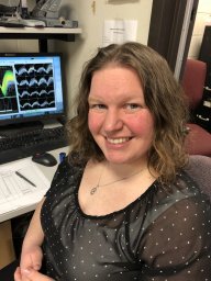 Elizabeth A West合作论文数Graduate School of Biomedical Sciences, Rowan University School of Osteopathic Medicine7
 Brittany Aguilar合作论文数Department of Pharmacology and Physiology, Georgetown University7Safwan K Hyder合作论文数6Eric Witherspoon合作论文数5Megan N Huizenga合作论文数5
 Tahiyana Khan合作论文数5Sean Quinlan合作论文数Royal Coll Surg Ireland, Univ Med & Hlth Sci5
 Laurie L Wellman合作论文数5
 Colin Kulick-Soper合作论文数5
 Kulick Catherine V合作论文数Department of Pharmacology and Physiology, Georgetown University5
 Samuel Gutherz合作论文数School of Medicine, Georgetown University5
 Rafael S. Maior合作论文数Institute of Biology, University of Brasilia5
 Anjik Ghosh合作论文数4Jessica Jacobs合作论文数4Elyssa M LaFlamme合作论文数4Catherine Elorette合作论文数Icahn School of Medicine at Mount Sinai4
 Alberto Sepulveda Rodriguez合作论文数Department of Pharmacology and Physiology, Georgetown University4
 DesJardin Jacqueline T合作论文数Department of Pharmacology and Physiology, Georgetown University Medical Center4
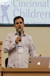 Victor Rodrigues Santos合作论文数Department of Anesthesia, Cincinnati Children's Hospital Medical Center4Murnen Alice合作论文数4Holmes Angela L合作论文数4
 D. T. S. Pak合作论文数4Judith F. Rubinson合作论文数4Anthony D. Kammerich合作论文数4Veronica C Beck合作论文数3Devin Palmer合作论文数3Robert Hammack合作论文数3Bevan Main合作论文数Georgetown Univ3
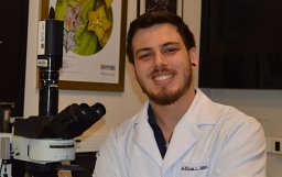 Willian Lazarini-Lopes合作论文数Ribeirão Preto School of Medicine, University of São Paulo3
 Al-Muhtasib Nour合作论文数Department of Pharmacology and Physiology, Georgetown University School of Medicine3Dybdal David合作论文数3
 Lauren E Ullrich合作论文数Interdisciplinary Program in Neuroscience, Georgetown University;Department of Neurology, Georgetown University;Georgetown University, Georgetown University3
 Italo Mocchetti合作论文数Department of Neuroscience, Georgetown University Medical Center3Kenneth J. Kellar合作论文数3
 Norberto Garcia-Cairasco合作论文数Psychobiology Post-Graduation Program, University of São Paulo3Cameron Sweeney合作论文数3
 katherine Conant合作论文数National Institute of Neurological Disorders and Stroke, National Institutes of Health3
 Sonya Dumanis合作论文数3
 Mohamad Z. Koubeissi合作论文数Department of Neurology, Johns Hopkins University3K GALE合作论文数2Mark F. Dubach合作论文数2




