Abstract Schistosoma mansoni and its schistosome relatives are parasitic worms that impose a substantial disease burden on human populations and livestock. On the rationale that calcium signalling is a critical process in multicellular organisms, we have examined wildtype and engineered S. mansoni STIM and ORAI— orthologues of STIM and ORAI known in mammals and other species for their central role in cellular calcium signalling— by imaging their localization, interactions, and contribution to ion currents and calcium influx in living cells. The ER membrane protein S. mansoni STIM recapitulates the essential functions of mammalian STIM1, namely, calcium-sensing by its ER-luminal domain, targeting to ER-plasma membrane junctions through interactions with the plasma membrane and with plasma membrane S. mansoni ORAI channels, and an ability to gate the S. mansoni ORAI channel. S. mansoni ORAI is a plasma membrane calcium channel that exhibits striking parallels with mammalian ORAI1 in its pore architecture and gating mechanism. The schistosome and human proteins are not completely interchangeable, however, and schistosome-human ORAI chimeras point to a special role of the ORAI N terminus in channel gating. Importantly, we demonstrate pharmacological differences between the schistosome and human channels that may offer an opportunity for selective therapeutic targeting of schistosome STIM-ORAI-dependent calcium entry. Author Summary Calcium channels represent potential targets to parasitic helminths. We investigated Schistosoma mansoni CRAC channel activation through the expression of its proteins. We have established that the fundamental protein conformational changes and protein-protein interactions underlying STIM-ORAI signaling are shared between humans and schistosome proteins. Importantly, a key finding is that evolutionary divergence in residues that are not implicated in the basic mechanisms of STIM-ORAI activation appears to offer a window for pharmacological inhibitors that would be selective for the schistosome ORAI channel. We identified pharmacological differences for two compounds tested. These differences open avenues for the development of selective drugs that can target the S. mansoni CRAC channel without affecting human physiology, thus offering the prospect of new treatments for schistosomiasis.
Introduction:T regulatory cells (Treg cells) express the transcription factor FOXP3 and maintain immune homeostasis by attenuating effector responses. Treg cells are prone to lose FOXP3 and convert to pathological 'ex-Treg' cells under conditions of strong or chronic inflammation. One mechanism for loss of FOXP3 expression involves increased DNA methylation of intronic enhancers CNS1 and CNS2 in the Foxp3 locus; these enhancers are maintained in a demethylated state by TET enzymes, 5-methylcytosine (5mC) dioxygenases that generate 5-hydroxymethylcytosine (5hmC) and other oxidized methylcytosines that are essential intermediates in all pathways of DNA demethylation. We previously showed that FOXP3+ Treg cells from Tet2/3-deficient (Tet2/3 DKO) mice displayed increased methylation of CNS1 and CNS2 and converted to FOXP3-negative ex-Treg cells considerably more efficiently than wild-type (WT) Treg cells. Method:We extend our previous analysis of Foxp3-Cre Tet2/3 fl/fl mice. Results and discussion:We classified the mice as DKO-moderate or DKO-severe based on the total number of leukocytes in the spleen and peripheral lymph nodes and investigated the phenotypic and molecular basis for the progressive inflammation occurring in these mice. RNA-seq as well as histological and immunocytochemical analyses showed a striking expansion of T follicular helper (Tfh) cells and plasma cells in Tet2/3 DKO-severe mice. And single-cell (sc) RNA-seq analyses suggested that this was due to skewed differentiation of both Tet2/3 DKO FOXP3+ Treg cells and Tet2/3 DKO FOXP3 - ex-Treg cells into Tfh-like cells. Base-resolution "6-base" sequencing showed the expected loss of 5hmC and increased 5mC in Tfh cells purified from Tet2/3 DKO-severe mice, and suggested that the observed bias in gene expression patterns could arise either from a direct increase in methylation of essential enhancers due to TET deficiency, or from interference with binding of methylation-sensitive transcriptional repressors including CCCTC-bindingfactor (CTCF).
Two prominent mechanisms by which tumors fend off immune control are by constraining the ability of T cells and CAR T cells to survive and expand in the tumor, and by restraining their ability to sustain full cytotoxic capacity. We identified IκBδ, encoded by Nfkbid, a poorly characterized IκB family member, as a molecular lever that overcomes both of these constraints on anti-tumor CD8+ tumor-infiltrating lymphocytes (TILs). Nfkbid is an NFAT target gene that is expressed in CD8+ effector T cells and, at modest levels, in CD8+ TILs. We found that Nfkbid depletion impaired TIL accumulation, exacerbating the growth of solid tumors. On the other hand, ectopic IκBδ overexpression enhanced TIL expansion, reduced the expression of exhaustion-associated transcription factors and inhibitory receptors, and elevated cytotoxic molecule production, leading to enhanced tumor control. IκBδ has a shorter protein isoform that is identical in a core region spanning the ankyrin-repeat domain known to interact with NFκB proteins, but that lacks the ~150-residue N-terminal region. We showed that the shared core region is sufficient to drive T cell accumulation, whereas the N-terminal peptide region is required for robust effector function and to counter exhaustion, underscoring that tumor-infiltrating CD8+ T cell accumulation and effector differentiation are separable programs. Our current study provides evidence that IκBδ, an atypical member of the NFκB family, is a lever to overcome two cardinal deficits that limit CD8+ TIL anti-tumor efficacy: impaired accumulation in the tumor and diminished effector function.
The conformational change in STIM1 that communicates sensing of ER calcium-store depletion from the STIM ER-luminal domain to the STIM cytoplasmic region and ultimately to ORAI channels in the plasma membrane is broadly understood. However, the structural basis for the STIM luminal-domain dimerization that drives the conformational change has proven elusive. A recently published study has approached this question via molecular dynamics simulations. The report pinpoints STIM residues that may be part of a luminal-domain dimerization interface, and provides unexpected insight into how torsional movements of the STIM luminal domains might trigger release of the cytoplasmic SOAR/CAD domain from its resting tethers to the STIM CC1 segments.
Ten-eleven translocation (TET) proteins are iron-dependent and α-ketoglutarate-dependent dioxygenases that sequentially oxidize the methyl group of 5-methylcytosine (5mC) to 5-hydroxymethylcytosine (5hmC), 5-formylcytosine (5fC) and 5-carboxylcytosine (5caC). All three epigenetic modifications are intermediates in DNA demethylation. TET proteins are recruited by transcription factors and by RNA polymerase II to modify 5mC at enhancers and gene bodies, thereby regulating gene expression during development, cell lineage specification, and cell activation. It is not yet clear, however, how the established biochemical activities of TET enzymes in oxidizing 5mC and mediating DNA demethylation relate to the known association of TET deficiency with inflammation, clonal hematopoiesis, and cancer. There are hints that the ability of TET deficiency to promote cell proliferation in a signal-dependent manner may be harnessed for cancer immunotherapy. In this review, we draw upon recent findings in cells of the immune system to illustrate established as well as emerging ideas of how TET proteins influence cellular function.
A Perspective on 'A Reappraisal of the Effects of L-type Ca(2+) Channel Blockers on Store-Operated Ca(2+) Entry and Heart Failure' Over a decade ago, we showed that differentiated, contractile arterial myocytes do not express functional Ca 2+ release-activated Ca 2+ (CRAC) channels that mediate store-operated Ca 2+ entry (SOCE).However, CRAC currents emerge in dedifferentiated fibroproliferative non-contractile arterial myocytes because of increased expression of STIM1 and Orai1 1,2 .Dedifferentiated myocytes (called synthetic) are a hallmark of neointimal hyperplasia in diseases such as hypertension, restenosis and atherosclerosis.The emergence of CRAC channels is only one aspect of synthetic myocytes.Synthetic myocytes remodel their ion channel repertoire to promote hyperplasia and migration at the expense of excitability and contractility.Synthetic myocytes downregulate the major orchestrators of excitation-contraction coupling: dihydropyridinesensitive voltage-gated L-type Ca 2+ channels (Ca v 1.2) and ryanodine receptors (for review 3 ).Recently, we examined the effects of Ca V 1.2 channel antagonists on synthetic arterial myocytes (Johnson et al 4 ).We reported that Ca V 1.2 channels blockers at concentrations ranging from 0.5-20 µM increased intracellular Ca 2+ in synthetic arterial myocytes.Our data support the idea that Ca V 1.2 channel blockers, including amlodipine, nifedipine, verapamil, and diltiazem activate STIM proteins and CRAC channels independently of store depletion 4 .We further showed that 0.5 µM amlodipine synergizes with sub-optimal concentrations of the platelet-derived growth factor (0.5 ng/mL PDGF) in a STIM1-dependent manner to induce arterial myocyte proliferation and migration to levels approaching those produced by maximal PDGF stimulation (10 ng/mL).Bird et al 5 challenged our study and contended that our findings represent an artifact of fluorescence Ca 2+ measurements because "amlodipine is strongly fluorescent in the cytoplasm with an excitation spectrum that overlaps with that of fura-2".They reported Ca 2+ imaging experiments using the single-excitation/single-emission dye Cal520 but did not attempt to replicate our key biological observations, most notably the synergistic effects of 0.5 M amlodipine on PDGF-triggered arterial myocyte proliferation and migration, and their dependence on STIM1 (Figure 1H-K; Figure 7A-G; Figure
A recent study by Jain et al. published in Nature followed up on evidence suggesting that depletion of 5-methylcytosine dioxygenase TET2 in chimeric antigen receptor CAR T cells could enhance their expansion, persistence, and antitumor efficacy. Their findings are cautionary, but offer hope of a path forward.
The first detailed investigation of CD8 + tumor-infiltrating T cell differentiation in the hours after cells enter a tumor has yielded an unexpected twist. Naive T cells veer away from effector fate and enter the path towards exhaustion much earlier than expected.
The protein phosphatase calcineurin has long been familiar to the calcium community, but the definition of its physiological substrates has been far from complete. A new study rectifies this deficiency and sets the stage for new insights into the role of calcineurin in diverse cellular processes.
Chronic antigen stimulation leads to CD8(+) T cell exhaustion, which is mediated by persistent activation of the transcription factor NFAT in the absence of AP-1. Seo, Gonzalez-Avalos and colleagues show that overexpressed BATF cooperates with IRF4 to counteract NFAT-induced exhaustion and promote better tumor control by CAR T cells in mouse models.The transcription factors nuclear factor of activated T cells (NFAT) and activator protein 1 (AP-1; Fos-Jun) cooperate to promote the effector functions of T cells, but NFAT in the absence of AP-1 imposes a negative feedback program of T cell hyporesponsiveness (exhaustion). Here, we show that basic leucine zipper ATF-like transcription factor (BATF) and interferon regulatory factor 4 (IRF4) cooperate to counter T cell exhaustion in mouse tumor models. Overexpression of BATF in CD8(+) T cells expressing a chimeric antigen receptor (CAR) promoted the survival and expansion of tumor-infiltrating CAR T cells, increased the production of effector cytokines, decreased the expression of inhibitory receptors and the exhaustion-associated transcription factor TOX and supported the generation of long-lived memory T cells that controlled tumor recurrence. These responses were dependent on BATF-IRF interaction, since cells expressing a BATF variant unable to interact with IRF4 did not survive in tumors and did not effectively delay tumor growth. BATF may improve the antitumor responses of CAR T cells by skewing their phenotypes and transcriptional profiles away from exhaustion and towards increased effector function.
How natural or innate-like lymphocytes generate the capacity to produce IL-4 and other cytokines characteristic of type 2 immunity remains unknown. Invariant natural killer T (iNKT) cells differentiate in the thymus into NKT1, NKT2, and NKT17 subsets, similar to mature, peripheral CD4+ T helper cells. The mechanism for this differentiation was not fully understood. Here, we show that NKT2 cells required higher and prolonged calcium (Ca2+ ) signals and continuing activity of the calcium release-activated calcium (CRAC) channel, than their NKT1 counterparts. The sustained Ca2+ entry via CRAC pathway in NKT2 cells was apparently mediated by ORAI and controlled in part by the large mitochondrial Ca2+ uptake. Unique properties of mitochondria in NKT2 cells, including high activity of oxidative phosphorylation, may regulate mitochondrial Ca2+ buffering in NKT2 cells. In addition, the low Ca2+ extrusion rate may also contribute to the higher Ca2+ level in NKT2 cells. Altogether, we identified ORAI-dependent Ca2+ signaling connected with mitochondria and cellular metabolism, as a central regulatory pathway for the differentiation of NKT2 cells.
TET enzymes facilitate DNA demethylation by oxidizing 5-methylcytosine to 5-hydroxymethylcytosine and other oxidation products in DNA. Loss-of-function mutations in TET enzymes are associated with increased proliferation and increased stem cell function in many cellular lineages. Moreover, there are several hints that inhibition of TET enzymes in tumor-infiltrating T cells might enhance anti tumor immune responses in solid tumors. For instance, TET2 deficiency in mouse T cells increased the number of CD8+ central memory T cells, and TET deficiency human CD8 chimeric antigen receptor (CAR) T cells increased their antigen-specific proliferation and capacity to reject tumor cells in a patient with chronic lymphocytic leukemia. Furthermore, we previously showed that TET deficiency in regulatory T cells decreased the stability of Foxp3 expression; hence TET deficiency would be expected to counter the ability of regulatory T cells to suppress anti-tumor responses. In this study, we found that TET loss of function in CD8 TILs indeed slowed tumor growth and promoted anti tumor responses in a mouse CAR T cell model. The function of TET proteins and other Fe(II) and a-ketoglutarate dependent dioxygenases is inhibited by L-2-Hydroxyglutarate (L2HG), which is normally kept at low levels by the enzyme L-2HG dehydrogenase (L2HGDH). We found that when CAR T cells were rendered low in TET activity in any one of three ways: (i) deficiency of the TET enzymes, (ii) depletion of L2HGDH and (iii) pretreatment of L2HG, they showed decreased expression of inhibitory receptors as well as increased secretion of inflammatory cytokines. Based on these results, we suggest that TET inhibition in T cells might be a promising general approach in cancer immunotherapy.
Voltage-gated L-type Ca2+ channel (Cav1.2) blockers (LCCBs) are major drugs for treating hypertension, the preeminent risk factor for heart failure. Vascular smooth muscle cell (VSMC) remodeling is a pathological hallmark of chronic hypertension. VSMC remodeling is characterized by molecular rewiring of the cellular Ca2+ signaling machinery, including down-regulation of Cav1.2 channels and up-regulation of the endoplasmic reticulum (ER) stromal-interacting molecule (STIM) Ca2+ sensor proteins and the plasma membrane ORAI Ca2+ channels. STIM/ORAI proteins mediate store-operated Ca2+ entry (SOCE) and drive fibro-proliferative gene programs during cardiovascular remodeling. SOCE is activated by agonists that induce depletion of ER Ca2+, causing STIM to activate ORAI. Here, we show that the three major classes of LCCBs activate STIM/ORAI-mediated Ca2+ entry in VSMCs. LCCBs act on the STIM N terminus to cause STIM relocalization to junctions and subsequent ORAI activation in a Cav1.2-independent and store depletion-independent manner. LCCB-induced promotion of VSMC remodeling requires STIM1, which is up-regulated in VSMCs from hypertensive rats. Epidemiology showed that LCCBs are more associated with heart failure than other antihypertensive drugs in patients. Our findings unravel a mechanism of LCCBs action on Ca2+ signaling and demonstrate that LCCBs promote vascular remodeling through STIM-mediated activation of ORAI. Our data indicate caution against the use of LCCBs in elderly patients or patients with advanced hypertension and/or onset of cardiovascular remodeling, where levels of STIM and ORAI are elevated.
'T cell exhaustion' is a broad term that has been used to describe the response of T cells to chronic antigen stimulation, first in the setting of chronic viral infection but more recently in response to tumours. Understanding the features of and pathways to exhaustion has crucial implications for the success of checkpoint blockade and adoptive T cell transfer therapies. In this Viewpoint article, 18 experts in the field tell us what exhaustion means to them, ranging from complete lack of effector function to altered functionality to prevent immunopathology, with potential differences between cancer and chronic infection. Their responses highlight the dichotomy between terminally differentiated exhausted T cells that are TCF1- and the self-renewing TCF1+ population from which they derive. These TCF1+ cells are considered by some to have stem cell-like properties akin to memory T cell populations, but the developmental relationships are unclear at present. Recent studies have also highlighted an important role for the transcriptional regulator TOX in driving the epigenetic enforcement of exhaustion, but key questions remain about the potential to reverse the epigenetic programme of exhaustion and how this might affect the persistence of T cell populations.
STUDY OBJECTIVE:Methicillin-resistant Staphylococcus aureus (MRSA) transmission dynamics in the emergency department (ED) are not well defined; environmental surfaces may serve as reservoirs for transmission. This study investigates the effect of patients with a history of MRSA colonization or infection on subsequent MRSA contamination of the ED environment. METHODS:Adult ED patients with evidence of an MRSA-positive surveillance result or clinical microbiologic culture in the year preceding their current ED visit were enrolled. Cultures from 5 anatomic sites were obtained to detect active MRSA colonization. After patients' discharge and before environmental disinfection, up to 16 prespecified surfaces in their ED rooms were cultured. Strain typing was performed by repetitive-sequence polymerase chain reaction on all recovered MRSA isolates to determine concordance with the corresponding patient strain. RESULTS:Of 42 patients enrolled, 25 (60%) remained colonized with MRSA. Nineteen of the 25 ED rooms (76%) occupied by MRSA-colonized patients contained greater than or equal to 1 MRSA-contaminated environmental surface on patient discharge. Surfaces were more likely to be contaminated when rooms were occupied by patients colonized with MRSA at 1 body site (odds ratio 11.7; 95% confidence interval 1.5 to 91.5) and greater than or equal to 2 body sites (odds ratio 16.3; 95% confidence interval 3.1 to 86.8) compared with noncolonized patients. In 16 of the 19 ED rooms (84%) where MRSA was recovered, all environmental strains were concordant with the corresponding patient strain. CONCLUSION:Contamination of the ED environment with MRSA from actively colonized patients is common. Improved environmental surface disinfection may help reduce transmission of MRSA to ED health care professionals and patients during emergency care.
T cells expressing chimeric antigen receptors (CAR T cells) have shown impressive therapeutic efficacy against leukemias and lymphomas. However, they have not been as effective against solid tumors because they become hyporesponsive ("exhausted" or "dysfunctional") within the tumor microenvironment, with decreased cytokine production and increased expression of several inhibitory surface receptors. Here we define a transcriptional network that mediates CD8+ T cell exhaustion. We show that the high-mobility group (HMG)-box transcription factors TOX and TOX2, as well as members of the NR4A family of nuclear receptors, are targets of the calcium/calcineurin-regulated transcription factor NFAT, even in the absence of its partner AP-1 (FOS-JUN). Using a previously established CAR T cell model, we show that TOX and TOX2 are highly induced in CD8+ CAR+ PD-1high TIM3high ("exhausted") tumor-infiltrating lymphocytes (CAR TILs), and CAR TILs deficient in both TOX and TOX2 (Tox DKO) are more effective than wild-type (WT), TOX-deficient, or TOX2-deficient CAR TILs in suppressing tumor growth and prolonging survival of tumor-bearing mice. Like NR4A-deficient CAR TILs, Tox DKO CAR TILs show increased cytokine expression, decreased expression of inhibitory receptors, and increased accessibility of regions enriched for motifs that bind activation-associated nuclear factor κB (NFκB) and basic region-leucine zipper (bZIP) transcription factors. These data indicate that Tox and Nr4a transcription factors are critical for the transcriptional program of CD8+ T cell exhaustion downstream of NFAT. We provide evidence for positive regulation of NR4A by TOX and of TOX by NR4A, and suggest that disruption of TOX and NR4A expression or activity could be promising strategies for cancer immunotherapy.
The transcription factor nuclear factor of activated T cells (NFAT) has a key role in both T cell activation and tolerance and has emerged as an important target of immune modulation. NFAT directs the effector arm of the immune response in the presence of activator protein-1 (AP-1), and T cell anergy/exhaustion in the absence of AP-1. Envisioning a strategy for selective modulation of the immune response, we designed a FRET-based high-throughput screen to identify compounds that disrupt the NFAT:AP-1:DNA complex. We screened ∼202,000 small organic compounds and identified 337 candidate inhibitors. We focus here on one compound,N-(3-acetamidophenyl)-2-[5-(1H-benzimidazol-2-yl)pyridin-2-yl]sulfanylacetamide (Compound 10), which disrupts the NFAT:AP-1 interaction at the composite antigen-receptor response element-2 site without affecting the binding of NFAT or AP-1 alone to DNA. Compound 10 binds to DNA in a sequence-selective manner and inhibits the transcription of theIl2gene and several other cyclosporin A-sensitive cytokine genes important for the effector immune response. This study provides proof-of-concept that small molecules can inhibit the assembly of specific DNA–protein complexes, and opens a potential new approach to treat human diseases where known transcription factors are deregulated.
ORAI1 Ca2+ channels in the plasma membrane (PM) are gated by STIM1 at endoplasmic reticulum (ER)-PM junctions to effect store-dependent Ca2+ entry into cells, but little is known about how local STIM-ORAI signalling at junctions is coordinated with overall cellular architecture. Filamentous septins can specify cytoskeletal rearrangements and have been found recently to modulate STIM-ORAI signalling. Here we show by super-resolution imaging of ORAI1, STIM1, and septin 4 in living cells that septins facilitate Ca2+ signalling indirectly. Septin 4 does not colocalize preferentially with ORAI1 in resting or stimulated cells, assemble stably at ER-PM junctions, or specify a boundary that directs or confines ORAI1 to junctions. Rather, ORAI1 is recruited to junctions solely through interaction with STIM proteins, while septins regulate the number of ER-PM junctions and enhance STIM1-ORAI1 interactions within junctions. Thus septins communicate with STIM1 and ORAI1 through protein or lipid intermediaries, and are favorably positioned to coordinate Ca2+ signalling with rearrangements in cellular architecture.
 Anjana Rao合作论文数Department of Pharmacology, School of Medicine, University of California, San Diego;Division of Signaling and Gene Expression, La Jolla Institute for Immunology54
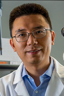 Yubin Zhou合作论文数Department of Translational Medical Sciences, College of Medicine, Texas A&M University;Center for Translational Cancer Research, Institute of Biosciences and Technology, Texas A&M University Health Science Center18
 Gudlur Aparna合作论文数17Ariel Quintana合作论文数12
 Stefan Feske合作论文数Department of Pathology, Grossman School of Medicine, New York University11
 Stephanie Fritz合作论文数9
 Sonia Sharma合作论文数La Jolla Inst Allergy & Immunol9
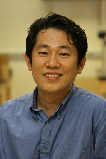 Yousang Gwack合作论文数Molecular Biology Institute, University of California, Los Angeles8Huiming Li合作论文数8
 Sonal Srikanth合作论文数Department of physiology, University of California, Los Angeles7
 Youjun Wang (王友军)合作论文数Beijing Key Laboratory of Gene Resource and Molecular Development, College of Life Sciences, Beijing Normal University7Masatsugu Oh-Hora合作论文数7
 Ana Zeraik合作论文数Chemistry Institute of São Carlos, University of São Paulo6Ma Guolin合作论文数6Hirve Nupura合作论文数6Paul Meraner合作论文数6Yun Huang (黄韵)合作论文数5Carol E. Muenks合作论文数5Edahí González-Avalos合作论文数Div Signaling & Gene Express, La Jolla Inst Immunol5
 Carey-Ann D. Burnham合作论文数Department of Pathology & Immunology, School of Medicine, Washington University in St. Louis;Pattern Bioscience5Green M K合作论文数5Ji Jing合作论文数5
 Lian He (何涟)合作论文数Department of Pharmacology, School of Medicine, Southern University of Science and Technology5
 Rajanikanth V合作论文数Centre for Cellular and Molecular Biology (CCMB),, Council of Scientific and Industrial Research (CSIR),5Wade Zhang合作论文数4Hyungseok Seo合作论文数4Melanie Sullivan合作论文数Dept Pediat, Washington Univ4
 Murali Prakriya合作论文数Allergy and Immunology, Department of Medicine, Feinberg School of Medicine, Northwestern University4
 Zhang Chen合作论文数4
 Thompson Ryley M合作论文数Department of Pediatrics, Washington University School of Medicine in St Louis4Edward D. Lamperti合作论文数4
 Barsha Dash合作论文数Department of Pediatrics, University of California at San Diego3
 Elizabeth Komives合作论文数Department of Chemistry and Biochemistry, Division of Physical Sciences, University of California, San Diego3
 Aomin Sun合作论文数3
 John Morelli合作论文数Department of Pediatrics, Washington University School of Medicine3
 Daniela Samaniego-Castruita合作论文数La Jolla Inst Immunol, Div Signaling & Gene Express, La Jolla, CA 92037 USA3Lillemeier Björn F合作论文数3
 Shenyuan L Zhang合作论文数3
 Srimoyee Ghosh合作论文数3
 Gregory M. Findlay合作论文数Immune Disease Institute and Program in Cellular and Molecular Medicine, Harvard Medical School;Immune Disease Institute and Program in Cellular and Molecular Medicine, Harvard Medical School3Atsushi Onodera合作论文数Departments of Immunology,3
 Katz Zachary合作论文数Department of Anatomy and Structural Biology, Albert Einstein College of Medicine3
 Tan Peng合作论文数Institute of Biosciences and Technology, The Methodist Hospital Research Institute;Health Science Center, M University;The Methodist Hospital Research Institute, Texas A&M University Health Science Center3Boyle Mary合作论文数3
 Yuehe Ding合作论文数National Institute of Biological Sciences, National Institute of Biological Sciences3
 López-Moyado Isaac F合作论文数3
 Stephen C. Harrison合作论文数Center for Molecular and Cellular Dynamics, Harvard Medical School;Howard Hughes Medical Institute3
 Liangyi Chen (陈良怡)合作论文数Institute of Molecular Medicine, College of Future Technology, Peking University3
 Bogdan Tanasa合作论文数Supplemental Experimental Procedures;Cell Culture and Transfection;Supplemental Experimental Procedures|Cell Culture and Transfection3




