Endoscopic endonasal approaches (EEA) have become more prevalent for minimally invasive skull base and sinus surgeries. However, rigid scopes and tools significantly decrease the surgeon's ability to operate in tight anatomical spaces and avoid critical structures such as the internal carotid artery and cranial nerves. This paper proposes a novel tendon-actuated concentric tube endonasal robot (TACTER) design in which two tendon-actuated robots are concentric to each other, resulting in an outer and inner robot that can bend independently. The outer robot is a unidirectionally asymmetric notch (UAN) nickel-titanium robot, and the inner robot is a 3D-printed bidirectional robot, with a nickel-titanium bending member. In addition, the inner robot can translate axially within the outer robot, allowing the tool to traverse through structures while bending, thereby executing follow-the-leader motion. A Cosserat-rod based mechanical model is proposed that uses tendon tension of both tendon-actuated robots and the relative translation between the robots as inputs and predicts the TACTER tip position for varying input parameters. The model is validated with experiments, and a human cadaver experiment is presented to demonstrate maneuverability from the nostril to the sphenoid sinus. This work presents the first tendon-actuated concentric tube (TACT) dexterous robotic tool capable of performing follow-the-leader motion within natural nasal orifices to cover workspaces typically required for a successful EEA.
Intraoperative neuromonitoring of the facial nerve (cranial nerve VII) is critical during skull base surgery due to the nerve's proximity to surgical pathologies and the significant morbidity associated with a facial nerve injury. Objective: This systematic review aims to summarize and describe the numerous intraoperative neuromonitoring techniques available to the skull base surgeon for identification, preservation, and prognostication of facial nerve function during skull base surgery. A systematic review was performed in accordance with the Preferred Reporting Items for Systematic reviews and Meta-Analysis (PRISMA) checklist. Terms related to related to intraoperative neuromonitoring of the facial nerve and the skull base were searched on PubMed/MEDLINE, Embase via Elsevier, and the Web of Science Core Collection via Clarivate databases. Title/abstract and full-text screening was performed by two reviewers. Data on monitoring type, pathology, and surgical approach were collected. The review identified commonly utilized neuromonitoring methods including free-running and stimulated facial nerve electromyography (FEMG), facial corticobulbar motor evoked potentials (FCoMEP), abnormal muscle responses (AMR), and the blink reflex. Less frequently discussed methods were also identified such as the Z-L response. FEMG was the most commonly reported modality, described in 80.50 % of studies, followed by FCoMEPs (25.86 %) and AMR (21.26 %). Each technique demonstrated unique strengths and limitations regarding specificity, sensitivity, and integration into surgical workflow. This systematic review underscores the importance of a multidisciplinary and standardized approach in facial nerve IONM, emphasizing continued collaboration among surgeons, neurophysiologists, and anesthesiologists to enhance patient safety and surgical outcomes.
OBJECTIVE:To optimize neurosurgical tumor resection, tissue types and borders must be appropriately identified. Authors of this study established the use of a nondestructive laser-based endogenous fluorescence spectroscopy device, "TumorID," to almost immediately classify a specimen as glioma, meningioma, pituitary adenoma, or nonneoplastic tissue in the operating room, utilizing a machine learning algorithm. METHODS:TumorID requires only 0.5 seconds to collect data, without the need for any dyes or tissue manipulation, and utilizes a 100-mW, 405-nm laser that does not damage the tissue. The system was used in the operating room to scan ex vivo specimens from 46 patients (mean age 52 years) with glioma (8 patients), meningioma (10 patients), pituitary adenoma (23 patients), and nonneoplastic tissue resected during an epilepsy operation (5 patients). A support vector machine algorithm was trained to distinguish between these lesions and classify them in near real time. Statistical significance was determined through a generalized estimating equation on the area under the known fluorophore emission regions for free reduced nicotinamide adenine dinucleotide (NADH), bound NADH, flavin adenine dinucleotide, and neutral porphyrins. RESULTS:Ultimately, the machine learning model showed a high degree of classification power with a multiclass area under the receiver operating characteristic curve of 0.809 ± 0.002. The areas under the curve for neutral porphyrins were found to be statistically significant (p < 0.001) and to have the largest impact on model output. CONCLUSIONS:This initial ex vivo clinical study demonstrated the ability of TumorID to rapidly differentiate and classify various pathologies and surrounding brain in a configuration that can be easily translated to scan in vivo. This classification power could allow TumorID to augment surgical decision-making by enabling rapid intraoperative tissue diagnostics and border delineation, potentially improving patient outcomes by allowing for a more informed and complete resection.
Laser-based surgical ablation relies heavily on surgeon involvement, restricting precision to the limits of human error and perception. The interaction between laser and tissue is governed by various laser parameters that control the laser irradiance on the tissue, including the power, distance, spot size, orientation, and exposure time. This complex interaction lends itself to robotic automation, allowing the surgeon to focus on high-level tasks, such as choosing the region and method of ablation, while the lower-level ablation plan can be handled autonomously. This paper describes a sampling-based model predictive control (MPC) scheme to plan ablation sequences for arbitrary tissue volumes. Using a steady-state point ablation model to simulate a single laser-tissue interaction, a random search technique explores the reachable state space while preserving sensitive tissue regions. The sampled MPC strategy provides an ablation sequence that accounts for parameter uncertainty without violating constraints, such as avoiding nerve bundles.
Robot-assisted neurological surgery is receiving growing interest due to the improved dexterity, precision, and control of surgical tools, which results in better patient outcomes. However, such systems often limit surgeons' natural sensory feedback, which is crucial in identifying tissues – particularly in oncological procedures where distinguishing between healthy and tumorous tissue is vital. While imaging and force sensing have addressed the lack of sensory feedback, limited research has explored multimodal sensing options for accurate tissue boundary delineation. We present a user-friendly, modular test bench designed to evaluate and integrate complementary multimodal sensors for tissue identification. Our proposed system first uses vision-based guidance to estimate boundary locations with visual cues, which are then refined using data acquired by contact microphones and a force sensor. Real-time data acquisition and visualization are supported via an interactive graphical interface. Experimental results demonstrate that multimodal fusion significantly improves material classification accuracy. The platform provides a scalable hardware-software solution for exploring sensor fusion in surgical applications and demonstrates the potential of multimodal approaches in real-time tissue boundary delineation.
Laser scalpels are precise, dexterous, and efficient tools for soft tissue surgeries. However, surgical lasers are hard to control manually, require experience, and are often incompatible with conventional intraoperative imaging sensors. Integrating compatible sensing technology with laser scalpels for precise soft tissue surgery opens vital avenues for widespread adoption and previously unrealized automation. This paper proposes a dual-sensor strategy to generate high-resolution surgical scene visualization based on surgeon feedback for robotic laser surgery. The proposed method uses a coarse depth sensor to localize the tissue of interest in the surgical scene, and a fine optical coherence tomography (OCT) sensor to create a detailed (< 30 mu m lateral resolution) tissue representation. The method achieves RMSE error in the range of 0.0878mm to 0.102mm in large-area tissue reconstruction and 0.050mm to 0.427mm in pattern-based laser ablation using user feedback over various fiducial samples. The findings demonstrate the proposed system's capability in large-area tissue imaging for precise laser-based surgery.
OBJECTIVE Cranial nerve (CN) preservation remains a challenge for skull base neurosurgeons, and neurophysiological intraoperative monitoring presents many methods for CN identification and mapping. The blink reflex, which is the electrophysiological representation of the corneal reflex, can be used to test both trigeminal and facial nerve function. The objective of this study was to present a method for obtaining a reliable blink reflex response and maintaining it during the course of a procedure. METHODS A method for robust blink reflex recording is presented. Electrode placement, recording parameters, stimulation parameters, anesthetic considerations, and reliability troubleshooting are described. RESULTS This method has been iteratively developed at the authors' institution across multiple sites for more than 5 years. The blink reflex was monitored in multiple cranial approaches and for various pathologies. The most common cases monitored were vestibular schwannoma resections and microvascular decompressions. The most common cranial approaches were the translabyrinthine, retrosigmoid/suboccipital, and middle cranial fossa approaches. CONCLUSIONS To gain a more comprehensive understanding of the clinical utility of the blink reflex in surgical decision-making and outcome prediction, prospective studies involving larger patient cohorts are warranted. This report outlines a reproducible methodology and invites validation and constructive input from the broader neurosurgical and neuromonitoring communities.
Introduction: Surgical training has long relied on the apprenticeship model of education (McDougall 2007). However, with changing ACGME work-hour requirements and the increasing scope of endoscopic endonasal surgery, a thoughtful and graduated approach to endoscopic endonasal surgery is essential. There has been little research looking into the competence and confidence of neurosurgical trainees in endoscopic endonasal surgery. Our own preliminary data suggests that endoscopic endonasal skills may be more emphasized in otolaryngology residents compared to neurosurgery residents ([Figs. 1] and [2]). However, besides subjective assessments of resident autonomy and case minimums, there are no objective methods for determining resident involvement in a case. We plan to use instrument tracking to determine neurosurgical resident involvement in endoscopic endonasal cases and to identify transitional moments where control of instruments is given over to the attendings.
Introduction: WHO pathologic grade serves as a key element in driving clinical management of skull base meningiomas, with grade 2 and 3 lesions requiring different surgical, radiation, and surveillance strategies compared to grade 1 lesions. We currently have limited ability to predict meningioma grading preoperatively. Here, we apply machine learning to a 3D, MRI-based analysis of tumors’ topologic and geometric features to predict pathologic grade of skull base meningiomas using imaging alone.
There is a need for precision pathological sensing, imaging, and tissue manipulation in neurosurgical procedures, such as brain tumor resection. Precise tumor margin identification and resection can prevent further growth and protect critical structures. Surgical lasers with small laser diameters and steering capabilities can allow for new minimally invasive procedures by traversing through complex anatomy, then providing energy to sense, visualize, and affect tissue. In this paper, we present the design of a small-scale tendon-actuated galvanometer (TAG) that can serve as an end-effector tool for a steerable surgical laser. The galvanometer sensor design, fabrication, and kinematic modeling are presented and derived. It can accurately rotate up to 30.14 +/- 0.90 degrees (or a laser reflection angle of 60.28 degrees). A kinematic mapping of input tendon stroke to output galvanometer angle change and a forward-kinematics model relating the end of the continuum joint to the laser end-point are derived and validated.
BackgroundPeritumoral edema alters diffusion anisotropy, resulting in false negatives in tractography reconstructions negatively impacting surgical decision-making. With supratotal resections tied to survival benefit in glioma patients, advanced diffusion modeling is critical to visualize fibers within the peritumoral zone to prevent eloquent fiber transection thereafter. A preoperative assessment paradigm is therefore warranted to systematically evaluate multi-subject tractograms along clinically meaningful parameters. We propose a novel noninvasive surgically-focused survey to evaluate the benefits of a tractography algorithm for preoperative planning, subsequently applied to Synaptive Medical’s free-water correction algorithm developed for clinically feasible single-shell DTI data.MethodsTen neurosurgeons participated in the study and were presented with patient datasets containing histological lesions of varying degrees of edema. They were asked to compare standard (uncorrected) tractography reconstructions overlaid onto anatomical images with enhanced (corrected) reconstructions. The raters assessed the datasets in terms of overall data quality, tract alteration patterns, and the impact of the correction on lesion definition, brain-tumor interface, and optimal surgical pathway. Inter-rater reliability coefficients were calculated, and statistical comparisons were made.ResultsStandard tractography was perceived as problematic in areas proximal to the lesion, presenting with significant tract reduction that challenged assessment of the brain-tumor interface and of tract infiltration. With correction applied, significant reduction in false negatives were reported along with additional insight into tract infiltration. Significant positive correlations were shown between favorable responses to the correction algorithm and the lesion-to-edema ratio, such that the correction offered further clarification in increasingly edematous and malignant lesions. Lastly, the correction was perceived to introduce false tracts in CSF spaces and - to a lesser degree - the grey-white matter interface, highlighting the need for noise mitigation. As a result, the algorithm was modified by free-water-parameterizing the tractography dataset and introducing a novel adaptive thresholding tool for customizable correction guided by the surgeon’s discretion.ConclusionHere we translate surgeon insights into a clinically deployable software implementation capable of recovering peritumoral tracts in edematous zones while mitigating artifacts through the introduction of a novel and adaptive case-specific correction tool. Together, these advances maximize tractography’s clinical potential to personalize surgical decisions when faced with complex pathologies.
Introduction: The WHO pathologic grade of meningiomas have significant prognostic and management implications, including influencing extent of surgical resection, need for postoperative radiation and frequency of postoperative surveillance imaging. Gold standard for this grading is via a time consuming, effort-intensive process driven by pathologists. We have developed a new technology, “TumorID,” which enables immediate/real-time, intraoperative tissue analysis based on laser-induced endogenous fluorescence spectroscopy ([Fig. 1]). Our aim is to apply machine learning (ML) methods to spectral emission data collected by TumorID to rapidly differentiate Grade 1 vs. Grade 2 meningiomas.
Manual surgical resection of soft tissue sarcoma tissue can involve many challenges, including the critical need for precise determination of tumor boundary with normal tissue and limitations of current surgical instrumentation, in addition to standard risks of infection or tissue healing difficulty. Substantial research has been conducted in the biomedical sensing landscape for development of non-human contact sensing devices. One such point-of-care platform, previously devised by our group, utilizes autofluorescence-based spectroscopic signatures to highlight important physiological differences in tumorous and healthy tissue. The following study builds on this work, implementing classification algorithms, including Artificial Neural Network, Support Vector Machine, Logistic Regression, and K-Nearest Neighbors, to diagnose freshly resected murine tissue as sarcoma or healthy. Classification accuracies of over 93% are achieved with Logistic Regression, and Area Under the Curve scores over 94% are achieved with Support Vector Machines, delineating a clear way to automate photonic diagnosis of ambiguous tissue in assistance of surgeons. These interpretable algorithms can also be linked to important physiological diagnostic indicators, unlike the black-box ANN architecture. This is the first known study to use machine learning to interpret data from a non-contact autofluorescence sensing device on sarcoma tissue, and has direct applications in rapid intraoperative sensing.
In-vivo tissue stiffness identification can be useful in pulmonary fibrosis diagnostics and minimally invasive tumor identification, among many other applications. In this work, we propose a palpation-based method for tissue stiffness estimation that uses a sensorized beam buckled onto the surface of a tissue. Fiber Bragg Gratings (FBGs) are used in our sensor as a shape-estimation modality to get realtime beam shape, even while the device is not visually monitored. A mechanical model is developed to predict the behavior of a buckling beam and is validated using finite element analysis and bench-top testing with phantom tissue samples (made of PDMS and PA-Gel). Bench-top estimations were conducted and the results were compared with the actual stiffness values. Mean RMSE and standard deviation (from the actual stiffnesses) values of 413.86 KPa and 313.82 KPa were obtained. Estimations for softer samples were relatively closer to the actual values. Ultimately, we used the stiffness sensor within a mock concentric tube robot as a demonstration of in-vivo sensor feasibility. Bench-top trials with and without the robot demonstrate the effectiveness of this unique sensing modality in in-vivo applications.
INTRODUCTION: Phenotype is the detectable expression of genotype. Despite this, little is known about how the three-dimensional structure of brain tumors correlates with their genetic biomarkers. METHODS: The UCSF-PDGM dataset was filtered for histopathologically-proven gliomas. Segmentation masks of the enhancing tumor, non-enhancing/necrotic tumor, and surrounding FLAIR abnormality were obtained from each patient’s preoperative brain MRI. All tumors were evaluated for IDH mutations and 1p/19q codeletion; all grade III and IV tumors were tested for MGMT methylation status. The segmentation masks were processed to create topological features describing the tumor’s 3D shape. These features, without other clinical variables, were used in a custom machine learning pipeline to predict the presence of IDH mutations, 1p/19q codeletion, and MGMT methylation. RESULTS: After filtration, 103 of 494 gliomas tested had an IDH mutation, 29 of 494 had a 1p/19q codeletion, and 247 of 409 had methylation of MGMT. On the blinded test-subset, the machine learning model, using only features describing shape, rendered an AUROC of 0.902 (95% CI: 0.875-0.929), specificity of 90.3% (83.9%-96.6%), and sensitivity of 75.7% (68.0%-83.3%) for IDH mutation. For 1p/19q codeletion, we found an AUROC of 0.949 (0.908-0.990), specificity of 94.5% (84.3%-100%), and sensitivity of 86.6% (79.6%-93.6%). For MGMT methylation, the performance was poor with an AUROC of 0.445 (0.385-0.504). CONCLUSIONS: The three-dimensional shape of a glioma may be used to predict the presence of some key underlying genetic mutations before biopsy. Additional research is needed to validate this, improve its fidelity, and generalize it to other biomarkers.
 Ralph Abi Hachem合作论文数Department of Head and Neck Surgery and Communication Sciences, Duke University School of Medicine12
 Jihad Abdelgadir合作论文数Department of Neurosurgery, Duke University Medical Center11
 Ali R. Zomorodi合作论文数Department of Neurosurgery, Duke University School of Medicine11
 Adil Syed M合作论文数11
 Weston Ross合作论文数8
 David W Jang合作论文数Department of Head and Neck Surgery and Communication Sciences, Duke University School of Medicine7
 Peter Edward Fecci合作论文数Center for Brain and Spine Metastasis, Duke University;Clinical Science Departments, Pratt School of Engineering, Duke University;Department of Pathology, Pratt School of Engineering, Duke University;Duke Cancer Institute7
 Tanner Zachem合作论文数6C. Rory Goodwin合作论文数Department of Neurosurgery, Johns Hopkins Hospital6Guangshen Ma合作论文数Duke Univ, Durham, NC 27708 USA5
 Kent K. Yamamoto合作论文数4Pranav Warman合作论文数Division of Global Neurosurgery and Neurology, Duke University Medical Center4
 Matthew Tucker合作论文数4
 Andreas Seas合作论文数1Duke Global Neurosurgery and Neurology, Duke University4
 Jordan Komisarow合作论文数Department of Neurosurgery, Duke University School of Medicine4
 Timothy Dunn合作论文数Department of Neurosurgery, Duke University School of Medicine4
 Benjamin Wissel合作论文数Medical Center, Cincinnati Children's Hospital4Anoop Patel合作论文数3Kevin Choi合作论文数3Siobhan R. Oca合作论文数Duke University3
 Shivanand Lad合作论文数Thomas Lord Department of Mechanical Engineering & Materials Science, Pratt School of Engineering, Duke University3
 Yash Chitalia合作论文数Georgia Institute of Technology3
 Ackall Feras Y合作论文数3
 Gerald Arthur Grant合作论文数Department of Neurosurgery, School of Medicine, Stanford University;Department of Neurosurgery, Duke University3
 Evan Calabrese合作论文数3Behnam Moradkhani合作论文数2Kymberly K. Levine合作论文数2Ravi Prakash合作论文数2
 Vadim Tsvankin合作论文数2
 Daniel John Sexton合作论文数Department of Medicine, School of Medicine, Duke University2
 Matthew Koch合作论文数2Steven Cook合作论文数2Kimberly B Hoang合作论文数2Tomer Anor合作论文数2
 daniel m buckland合作论文数beth israel deaconess medical center2Lyndon Chan合作论文数Div Rhinol & Endoscop Skull Base Surg, Duke Univ2
 Aatif Mairaj Husain合作论文数Department of Neurology, Duke University School of Medicine;Duke Clinical Research Institute2
 Leila Bridgeman合作论文数Thomas Lord Department of Mechanical Engineering & Materials Science, Pratt School of Engineering, Duke University2
 Coumans Jean-Valery合作论文数Massachusetts Gen Hosp, Dept Neurosurg, Boston, MA 02114 USA2
 Pankaj K. Agarwalla合作论文数Department of Neurosurgery, New Jersey Medical School2
 Rahimpour Shervin合作论文数Surgical Neurology Branch, National Institute of Neurological Disorders and Stroke2
 Allan Howard Friedman合作论文数Department of Neurosurgery, School of Medicine, Duke University2
 Pierre E. Dupont合作论文数Boston Children's Hospital2
 David N. Louis合作论文数Department of Pathology, Massachusetts General Hospital;Mass General Research Institute, Massachusetts General Hospital;Department of Pathology, Brigham and Women's Hospital;Harvard Medical School2
 Rebecca Betensky合作论文数Department of Biostatistics, School of Global Public Health, New York University2
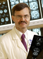 Allan H. Friedman合作论文数Duke University Medical Center2
 Mike Stratton合作论文数Wellcome Sanger Institute, Genome Research Limited2
 Henry Seth Friedman合作论文数Department of Medicine, School of Medicine, Duke University2
 Joseph Madsen合作论文数Department of Neurosurgery, Boston Children's Hospital;Harvard Medical School2




