INTRODUCTION: Laser interstitial thermal therapy (LITT) allows for confirmatory tissue diagnosis, surgical cytoreduction, and faster return to systemic therapies for patients with brain tumors or radiation necrosis. Ablated tissue remains in situ following LITT, with post-LITT edema potentially associated with transient clinical worsening and complicating subsequent response assessment. METHODS: All patients receiving LITT at a single center for tumors or radiation necrosis from 2015 – 2023 with = 9 months of MRI follow-up were retrospectively reviewed. A 3D U-net segmentation model implemented in nnU-Net was developed for automated segmentation of Contrast-enhancing Lesion Volume (CeLV) of LITT-treated lesions on T1-weighted post-contrast MR images. CeLVs were analyzed to perform volumetric post-LITT response assessments. RESULTS: Sixty-one LITT-treated lesions and 6 control cases of medically-managed radiation necrosis were analyzed across 384 unique MRI exams. Automated segmentation was qualitatively accurate in 367/384 (95.6%) images. CeLV increased to a median of 68.3% (IQR 35.1 – 109.2%) from baseline at 1 – 3 months from LITT (P = 0.0012) and returned to baseline thereafter. Median overall survival (mOS) for LITT-treated patients was 39.1 (9.2 – 93.4) months. In accordance with previously established volumetric thresholds, volumetric disease progression was defined as CeLV increase = 40% from volumetric nadir or baseline. Twenty-one of 56 (37.5%) patients experienced volumetric progression with a progression-free survival of 21.4 (6.0 – 93.4) months. Volumetric progression was associated with lower mOS (17.3 vs 62.1 months, P = 0.0015). CONCLUSIONS: CeLV increased to nearly 70% from baseline at 1 - 3 months following LITT, resolving within 3 - 6 months of the procedure. To facilitate inclusion of LITT patients in clinical trials, we developed and demonstrated feasibility of response assessment criteria to account for this transient post-ablation CeLV expansion. Automated lesion segmentation may increase the rate of adoption for volumetric response assessment in clinical practice.
BackgroundPeritumoral edema alters diffusion anisotropy, resulting in false negatives in tractography reconstructions negatively impacting surgical decision-making. With supratotal resections tied to survival benefit in glioma patients, advanced diffusion modeling is critical to visualize fibers within the peritumoral zone to prevent eloquent fiber transection thereafter. A preoperative assessment paradigm is therefore warranted to systematically evaluate multi-subject tractograms along clinically meaningful parameters. We propose a novel noninvasive surgically-focused survey to evaluate the benefits of a tractography algorithm for preoperative planning, subsequently applied to Synaptive Medical’s free-water correction algorithm developed for clinically feasible single-shell DTI data.MethodsTen neurosurgeons participated in the study and were presented with patient datasets containing histological lesions of varying degrees of edema. They were asked to compare standard (uncorrected) tractography reconstructions overlaid onto anatomical images with enhanced (corrected) reconstructions. The raters assessed the datasets in terms of overall data quality, tract alteration patterns, and the impact of the correction on lesion definition, brain-tumor interface, and optimal surgical pathway. Inter-rater reliability coefficients were calculated, and statistical comparisons were made.ResultsStandard tractography was perceived as problematic in areas proximal to the lesion, presenting with significant tract reduction that challenged assessment of the brain-tumor interface and of tract infiltration. With correction applied, significant reduction in false negatives were reported along with additional insight into tract infiltration. Significant positive correlations were shown between favorable responses to the correction algorithm and the lesion-to-edema ratio, such that the correction offered further clarification in increasingly edematous and malignant lesions. Lastly, the correction was perceived to introduce false tracts in CSF spaces and - to a lesser degree - the grey-white matter interface, highlighting the need for noise mitigation. As a result, the algorithm was modified by free-water-parameterizing the tractography dataset and introducing a novel adaptive thresholding tool for customizable correction guided by the surgeon’s discretion.ConclusionHere we translate surgeon insights into a clinically deployable software implementation capable of recovering peritumoral tracts in edematous zones while mitigating artifacts through the introduction of a novel and adaptive case-specific correction tool. Together, these advances maximize tractography’s clinical potential to personalize surgical decisions when faced with complex pathologies.
Due to improvements in population health, systemic cancer therapies and screening tools, the incidence of brain cancer metastases has continued to rise. The constituent cells possess unique characteristics that allow them to penetrate the blood–brain barrier, colonize the central nervous system, and co-opt their surroundings to thrive while evading surveillance by the immune system. This presents a unique challenge both to the multidisciplinary teams that care for these patients and the investigators striving to leverage these tumors' distinctive attributes into novel treatments. In this chapter, we outline the pathways and mechanisms underlying the development and survival of brain metastases, and how they inform current and emerging treatment strategies.
Abstract BACKGROUND Laser interstitial thermal therapy (LITT) is a minimally invasive option for tissue diagnosis, cytoreduction, and rapid post-operative return to systemic therapies for patients with brain tumors or radiation necrosis. As ablated tissue remains in situ, post-LITT edema may be associated with apparent increased lesion size and transient clinical worsening, complicating assessment of progression. METHODS All patients receiving LITT at a single center for tumors or radiation necrosis from 2015 – 2023 with ≥ 9 months of MRI follow-up were retrospectively reviewed. A 3D U-net segmentation model implemented in nnU-Net was developed for automated segmentation of Contrast-enhancing Lesion Volume (CeLV) of LITT-treated lesions on T1-weighted post-contrast MR images. CeLVs were analyzed to establish volumetric post-LITT response assessments. RESULTS Across 384 unique MRI exams, 61LITT-treated lesions and 6 control cases of medically-managed radiation necrosis were analyzed. Automated segmentation was qualitatively accurate in 367/384 (95.6%) images. CeLV increased to a median of 68.3% (IQR 35.1 – 109.2%) from baseline at 1 – 3 months from LITT (P = 0.0012) and returned to baseline thereafter. Based on established criteria, volumetric disease progression was defined as lesion expansion 40% from volumetric nadir or baseline. Twenty-one of 56 (37.5%) patients experienced volumetric progression with a progression-free survival of 21.4 (6.0 – 93.4) months. Patients experiencing volumetric progression had lower mOS (17.3 vs 62.1 months, P = 0.0015). CONCLUSIONS We observed a stereotyped increase in CeLV at 1 – 3 months post-LITT, which resolved within 6 months of the procedure. Post-LITT disease response can be reliably and feasibly assessed with criteria that account for this transient CeLV expansion. Automated lesion segmentation may speed adoption of volumetric response assessment criteria to clinical practice.
Abstract BACKGROUND Laser interstitial thermal therapy (LITT) allows for definitive tissue diagnosis, surgical cytoreduction, and faster post-operative return to systemic therapies for patients with brain tumors or radiation necrosis. Ablated tissue remains in situ following LITT, resulting in characteristic post-LITT lesion expansion. Post-LITT edema may be associated with transient clinical worsening and complicates subsequent response assessment. METHODS All patients receiving LITT at a single center for tumors or radiation necrosis from 2015 – 2023 with ≥ 9 months of MRI follow-up were retrospectively reviewed. A 3D U-net segmentation model implemented in nnU-Net was developed for automated segmentation of Contrast-enhancing Lesion Volume (CeLV) of LITT-treated lesions on T1-weighted post-contrast MR images. CeLVs were analyzed to establish volumetric post-LITT response assessments. RESULTS Sixty-one LITT-treated lesions and 6 control cases of medically-managed radiation necrosis were analyzed across 384 unique MRI exams. Automated segmentation was qualitatively accurate in 367/384 (95.6%) images. CeLV increased to a median of 68.3% (IQR 35.1 – 109.2%) from baseline at 1 – 3 months from LITT (P = 0.0012) and subsequently returned to baseline. Median overall survival (mOS) for LITT-treated patients was 39.1 (9.2 – 93.4) months. Using previously established volumetric thresholds, volumetric disease progression was defined as lesion expansion ≥ 40% from volumetric nadir or baseline. Twenty-one of 56 (37.5%) patients experienced volumetric progression with a progression-free survival of 21.4 (6.0 – 93.4) months. Patients with volumetric progression had lower mOS (17.3 vs 62.1 months, P = 0.0015). CONCLUSION We observed a nearly 70% increase in CeLV at 1 – 3 months post-LITT, which characteristically resolved within 6 months of the procedure. Development of response assessment criteria that account for transient post-LITT lesion expansion is feasible and should be considered for clinical trials. Automated lesion segmentation may facilitate adoption of volumetric response assessment into clinical practice.
BACKGROUND:Laser interstitial thermal therapy (LITT) of intracranial tumors or radiation necrosis enables tissue diagnosis, cytoreduction, and rapid return to systemic therapies. Ablated tissue remains in situ, resulting in characteristic post-LITT edema associated with transient clinical worsening and complicating post-LITT response assessment. METHODS:All patients receiving LITT at a single center for tumors or radiation necrosis from 2015 to 2023 with ≥9 months of MRI follow-up were included. An nnU-Net segmentation model was trained to automatically segment contrast-enhancing lesion volume (CeLV) of LITT-treated lesions on T1-weighted images. Response assessment was performed using volumetric measurements. RESULTS:Three hundred and eighty four unique MRI exams of 61 LITT-treated lesions and 6 control cases of medically managed radiation necrosis were analyzed. Automated segmentation was accurate in 367/384 (95.6%) images. CeLV increased to a median of 68.3% (IQR 35.1-109.2%) from baseline at 1-3 months from LITT (P = 0.0012) and returned to baseline thereafter. Overall survival (OS) for LITT-treated patients was 39.1 (9.2-93.4) months. Lesion expansion above 40% from volumetric nadir or baseline was considered volumetric progression. Twenty-one of 56 (37.5%) patients experienced progression for a volumetric progression-free survival of 21.4 (6.0-93.4) months. Patients with volumetric progression had worse OS (17.3 vs 62.1 months, P = 0.0015). CONCLUSIONS:Post-LITT CeLV expansion is quantifiable and resolves within 6 months of LITT. Development of response assessment criteria for LITT-treated lesions is feasible and should be considered for clinical trials. Automated lesion segmentation could speed the adoption of volumetric response criteria in clinical practice.
Glioma is one of the most common primary malignant brain tumors. Despite progress in therapeutic approaches, the median survival of patients with glioma remains less than 2 years, generating the need for new therapeutic approaches. Ultrasound (US) is widely used in medical fields and is used as a therapeutic tool mainly for improving the performance of therapeutic entities. In this study, we examined a novel approach using low frequency US (20 kHz) (LFUS) as an independent treatment tool for malignant glioma, since primary studies showed that cancer cells are more susceptible to LFUS than healthy cells. LFUS safety and efficacy were examined in a 9L gliosarcoma-bearing female Fischer 344 rats. Two LFUS protocols were examined: a one-time treatment (US1X), and two treatments 24 h apart (US2X). For safety evaluation, rats were monitored for weight change and pain measurements. For efficacy, tumor volume was measured as a function of time and the tumor structural chances were examined histopathologically. LFUS treatment showed rapid inhibition of tumor growth, seen as soon as 12 h after US application. In addition, LFUS was found to affect the tumor structure, which was more extensive (>60% of tumor area) in smaller tumors. In US2X, the tumor tissue was completely destroyed, and an extensive immune response was observed. Importantly, the treatment was highly selective, keeping the healthy tissue surrounding the tumor unharmed. We developed a highly efficient and selective therapeutic protocol for treating malignant glioma with minimal side effects based solely on LFUS.
INTRODUCTION:Increasingly, physicians find themselves in demanding leadership positions. However, leadership education for medical trainees remains lacking with most physicians reporting that they are ill-equipped to tackle the challenges of leadership. Here, we set out to describe the Feagin Leadership Program (FLP) and assess its reception and impact on trainees over the past 12 years. MATERIALS AND METHODS:During the 1-year FLP, selected scholars from Duke University, Wake Forest University, and the University of North Carolina participate in five leadership sessions, individual coaching, a leadership forum, and a multidisciplinary team-based capstone project. A 28-question survey with six optional free-response questions was distributed to the Feagin Alumni Network, and descriptive statistics were assessed. RESULTS:Since its founding, 212 scholars have graduated from the FLP and 117 (55%) alumni have gone on to surgical specialties. A survey was distributed among all Feagin alumni. A total of 56 (26%) surveys were completed. Forty-three percent (n = 24) had held at least one leadership position since completing the FLP. When asked about the impact of their experience, 96% (n = 54) said that the program encouraged them to pursue a position of leadership within their field, 95% (n = 53) stated that it prepared them for such a position, and 93% (n = 52) stated that the program positively influenced their decision to be involved with current or future positions of leadership. CONCLUSIONS:Over the last 12 years, the FLP has demonstrated a high perceived impact on personal growth, leadership proficiency, and the decision to pursue leadership positions in medicine. The current dearth of leadership education for surgical trainees can best be addressed with models such as the FLP, with adoption benefiting medical trainees, the medical community, and patients they serve.
BACKGROUND:Medulloblastoma is the most common pediatric malignant brain tumor, consisting of four molecular subgroups (WNT, SHH, Group 3, Group 4) and 12 subtypes. Expression of the cell surface poliovirus receptor (PVR), CD155, is necessary for entry of the viral immunotherapeutic agent, PVSRIPO, a polio:rhinovirus chimera. CD155, physiologically expressed in the mononuclear phagocytic system, is widely expressed ectopically in solid tumors. The objective of this study is to elucidate CD155 expression as both a receptor for PVSRIPO and a therapeutic target in medulloblastoma.METHODS:PVR mRNA expression was determined in several patient cohorts and human medulloblastoma cell lines. Patient samples were also analyzed for CD155 expression using immunohistochemistry and cell lines were analyzed using Western Blots. CD155 was blocked using a monoclonal antibody and cell viability, invasion, and migration were assessed.RESULTS AND DISCUSSION:PVR mRNA expression was highest in the WNT subgroup and lowest in Group 4. PVR expression in the subgroups of medulloblastoma were similar to other pediatric brain and non-brain tumors. PVR expression was largely not associated with subgroup or subtype. Neither PVR protein expression intensity nor frequency were associated with overall survival. PVR expression was elevated in Group 3 patients with metastases but there was no difference in paired primary and metastatic medulloblastoma. Blocking PVR resulted in dose-dependent cell death, decreased invasion in vitro, and modestly inhibited cell migration.CONCLUSIONS:CD155 is expressed across medulloblastoma subgroups and subtypes. Blocking CD155 results in cell death and decreased cellular invasion. This study provides rationale for CD155-targeting agents including PVSRIPO and antibody-mediated blockade of CD155.
Cells were first gated on size and singularity by forward scatter and side scatter. Nonviable cells were excluded by live/dead gating. Live cells were gated on CD11c and CD11b, then on CD45 and F4/80 to identify macrophage and dendritic cell phenotypes. Finally myeloid cells were gated for expression of TIM-3. Final populations were (A) F4/80+CD45hiCD11c-CD11bhi (B) F4/80+CD45dimCD11bhi (C) F4/80+CD45hiCD11c+CD11b+ and (D) F4/80-CD45dimCD11c+CD11b-.
BACKGROUND: The accuracy of pedicle screws placed with instrument tracking and robotic navigation are indi-vidually comparable or superior to placement using stan-dard fluoroscopy, however head-to-head comparisons between these adjuncts in a similar surgical population have yet to be performed.METHODS: Consecutive patients undergoing percuta-neous thoracic and lumbosacral spinal instrumentation were retrospectively enrolled. Instrumentation was per-formed using either fluoroscopy-based instrument tracking system (TrackX, TrackX Technologies) or robotic-navigation (ExcelsiusGPS, Globus Medical). Postinstrumentation computed tomography scans were graded for breach ac-cording to the Gertzbein-Robbins scale, with "acceptable" screws deemed as Grade A or B and "unacceptable" screws deemed as Grades C through E. Accuracy data was compared between both instrumentation modalities.RESULTS: Fifty-three patients, comprising a total of 250 screws (167 robot, 83 instrument tracking) were included. The overall accuracy between both modalities was similar, with 96.4% and 97.6% of screws with acceptable accuracy between instrument tracking and robotic navigation, respectively (I-squared 0.30, df = 1, P = 0.58). Between instrument tracking and robotic navigation, 92.8% and 95.8% of screws received Grade A, 3.6% and 1.8% a Grade B, 1.2% and 1.2% a Grade C, 1.2% and 0.6% a Grade D, and 1.2% and 0.6% a Grade E, respectively. The robot was abandoned intraoperatively in 2 cases due to unrecoverable registration inaccuracy or software failure, leading to abandonment of 8 potential screws (4.8%). -CONCLUSIONS: In a similar patient population, there is a similarly high degree of instrumentation accuracy be-tween fluoroscopy-based instrument tracking and robotic navigation. There is a rare chance for screw breach with either surgical adjunct.
Each mouse was individually imaged with cone beam computed tomography (CBCT) using the SARRP with a 65 kVp and 0.7 mA beam. Using the treatment planning system from the SARRP system (Muriplan) and the CBCT, the target was exactly placed 3mm below the skull's burr hole. The planning system calculated the x-ray beam's (220kVp and 13mA) time of exposure according to the prescribed dose and moved the motorized couch to its target location, after which a 3-mm beam centered on the burr hole and underlying tumor was used to administer a total of 10 Gy radiation per animal at a rate of 1.9 Gy/min (18). The isodose distribution is shown on the figure provided. The dose to adjacent organs or rest of the brain is insignificant as the beam's penumbra demonstrates complete drop off on the edge of the field (17, 50, 51)
Gating strategy to assess for surface expression of TIM-3 on (A) CD4+ and (B) CD8+ T cells isolated from peripheral lymph nodes, lungs, livers, spleens, and brains.
INTRODUCTION:Intraoperative neuromonitoring (IONM) is commonly used during surgery of the spine and spinal cord for early surveillance of iatrogenic injury to the central and peripheral nervous system. However, for infants and young children under 3 years of age, the use of IONM is challenging due to incomplete central and peripheral myelination.CASE PRESENTATION:We report a case of a T4-T6 dermal sinus tract (DST) that was resected on day of life 23, with the successful use of IONM.CONCLUSION:To our knowledge, this is the youngest reported case of the use of IONM in the surgical correction of a DST in a neonatal patient. This case demonstrates the potential efficacy of IONM in neonatal spine surgery and the techniques used to adapt the technology to an immature nervous system.
Prone transpsoas fusion (PTP) is a minimally invasive technique that maximizes the benefit of lateral access interbody surgery and the prone positioning for surgically significant adjacent segment disease. The authors describe the feasibility, reproducibility and radiographic efficacy of PTP when performed for cases of lumbar ASD. Adult patients undergoing PTP for treatment of lumbar ASD at three institutions were retrospectively enrolled. Demographic information was recorded, as was operative data such as adjacent segment levels, operative time, blood loss, laterality of approach, open versus percutaneous pedicle screw instrumentation and need for primary decompression. Radiographic measurements including segmental and global lumbar lordosis, pelvic incidence, pelvic tilt, sacral slope and sagittal vertical axis were recorded both pre- and immediately post-operatively. Twenty-four patients met criteria for inclusion. Average age was 60.4 ± 10.4 years and average BMI was 31.6 ± 5.0 kg/m2. Total operative time was 204.7 ± 83.3 min with blood loss of 187.9 ± 211 mL. Twenty-one patients had pedicle screw instrumentation exchanged percutaneously and 3 patients had open pedicle screw exchange. Two patients suffered pulmonary embolism that was treated medically with no long-term sequelae. One patient had transient lumbar radicular pain and all patients were discharged home with an average length of stay of 3.0 days (range 1–6). Radiographically, global lumbar lordosis improved by an average of 10.3 ± 9.0 degrees, segmental lordosis by 10.1 ± 13.3 degrees and sagittal vertical axis by 3.2 ± 3.2 cm. Single-position prone transpsoas lumbar interbody fusion is a clinically reproducible minimally invasive technique that can effectively treat lumbar adjacent segment disease.
BACKGROUND: Radiation necrosis (RN) after stereotactic radiosurgery (SRS) for brain metastases (BM) can result in significant morbidity, compounded by the effects of extended steroid therapy. Laser interstitial thermal therapy (LITT) is a minimally invasive procedure that can offer definitive treatment for RN while potentially obviating the need for prolonged steroid use. OBJECTIVE: To compare LITT vs medical management (MM) in the treatment of RN. METHODS: A multicenter, retrospective study was performed of SRS-treated patients with BM who developed biopsy-proven RN and were treated with LITT or MM. Clinical outcome data were compared by treatment modality. RESULTS: Seventy-two patients met criteria with a median follow-up of 10.0 months (4.2-25.1), and 57 patients (79%) underwent LITT. Four MM (27%) and 3 LITT patients (5%) demonstrated radiographic progression ( P = .031) at a median of 5.3 and 4.0 months ( P = .40). There was no significant difference in overall survival (LITT median of 15.2 vs 11.6 months, P = .60) or freedom from local progression (13.6 vs 7.06 months, P = .40). Patients stopped steroid therapy earlier in the LITT cohort at a median of 37 days compared with 245 days ( P < .001). When controlled for follow-up duration, patients treated with LITT were 3 times more likely to be weaned off steroids before the study end point ( P = .003). CONCLUSION: These data suggest that LITT for treatment of biopsy-proven RN after SRS for BM significantly decreases time to steroid independence. Prospective trials should be designed to further validate the utility of LITT for RN and its impact on steroid-induced morbidity.
Background Improved survival for patients with brain metastases has been accompanied by a rise in tumor recurrence after stereotactic radiotherapy (SRT). Laser interstitial thermal therapy (LITT) has emerged as an effective treatment for SRT failures as an alternative to open resection or repeat SRT. We aimed to evaluate the efficacy of LITT followed by SRT (LITT+SRT) in recurrent brain metastases. Methods A multicenter, retrospective study was performed of patients who underwent treatment for biopsy-proven brain metastasis recurrence after SRT at an academic medical center. Patients were stratified by "planned LITT+SRT" versus "LITT alone" versus "repeat SRT alone." Index lesion progression was determined by modified Response Assessment in Neuro-Oncology Brain Metastases (RANO-BM) criteria. Results Fifty-five patients met inclusion criteria, with a median follow-up of 7.3 months (range: 1.0-30.5), age of 60 years (range: 37-86), Karnofsky Performance Status (KPS) of 80 (range: 60-100), and pre-LITT/biopsy contrast-enhancing volume of 5.7 cc (range: 0.7-19.4). Thirty-eight percent of patients underwent LITT+SRT, 45% LITT alone, and 16% SRT alone. Median time to index lesion progression (29.8, 7.5, and 3.7 months [P = .022]) was significantly improved with LITT+SRT. When controlling for age in a multivariate analysis, patients treated with LITT+SRT remained significantly less likely to have index lesion progression (P = .004). Conclusions These data suggest that LITT+SRT is superior to LITT or repeat SRT alone for treatment of biopsy-proven brain metastasis recurrence after SRT failure. Prospective trials are warranted to validate the efficacy of using combination LITT+SRT for treatment of recurrent brain metastases.
Abstract INTRODUCTION Laser interstitial thermal therapy (LITT) is an effective minimally-invasive treatment option for intracranial tumors. Our group produced plasmonics-active gold nanostars (GNS) designed to preferentially accumulate within intracranial tumors and amplify the ablative capacity of LITT while better conforming to tumor boundaries and protecting surrounding tissue. MATERIALS AND METHODS The 12nm GNS were synthesized using reduced HAuCl4 with Na3C6H5O7 seeds, mixed with AgNO3, C6H8O6, and HAuCL4, and coated with polyethylene glycol then functionalized with methoxy PEG thiol. CT-2A glioma cells were intracranially implanted into mice, followed 18 days later by IV injection of GNS. PET-CT was performed at 10-minutes, 24-, and 72-hours post-GNS administration, with autoradiography (AR) and histopathology (HP) on sacrifice after the last scan. To test the impact of GNS on LITT coverage capacity in appropriately sized ex vivo models, we utilized agarose gel-based phantoms incorporating control and GNS-infused central “tumors” in multiple shapes. LITT was administered with the NeuroBlate System. RESULTS In vivo, GNS preferentially accumulated within intracranial tumors on PET-CT at the 24- and 72-hour timepoints. AR and HP confirmed high GNS accumulation within tumor. Ex vivo, in cuboid tumor phantoms, the GNS-infused phantom heated 5.5x faster than the control, rising 0.49°C per minute compared to 0.09°C. In a split-cylinder tumor phantom with half containing GNS, the GNS-infused border heated 2x faster and the surrounding area was exposed to 30% lower temperatures. In a GNS-infused star-shaped phantom, the heat spread contoured along phantom boundaries. CONCLUSIONS Our results provide evidence for use of GNS to improve the specificity, efficiency, and potentially safety of LITT. The in vivo data support selective accumulation within intracranial tumors, and the GNS-infused phantom experiments demonstrate increased rates of heating within the tumor model, heat contouring to tumor borders, and decreased heating of surrounding regions representing normal structures.
 C. Rory Goodwin合作论文数Department of Neurosurgery, Johns Hopkins Hospital50Liu Ann合作论文数27
 Ignacio Jusue Torres合作论文数Loyola University Medical Center22
 Daniel M. Sciubba合作论文数Johns Hopkins University22
 Daniele Rigamonti合作论文数Johns Hopkins University;KHDR Consulting22
 Benjamin D Elder合作论文数21
 Peter Edward Fecci合作论文数Center for Brain and Spine Metastasis, Duke University;Clinical Science Departments, Pratt School of Engineering, Duke University;Department of Pathology, Pratt School of Engineering, Duke University;Duke Cancer Institute20
 Vikram A Mehta合作论文数Duke Canc Inst, Duke Univ18
 Timothy Y. Wang合作论文数18
 Henry Brem合作论文数Department of Neurosurgery, School of Medicine, The Johns Hopkins University17
 Muhammad Abd-El-Barr合作论文数Department of Neurosurgery, Duke University School of Medicine15
 Betty Tyler合作论文数Departments of Neurology and Neurosurgery, School of Medicine, Biomedical Engineering15Antonella Mangraviti合作论文数Sant'Andrea University Hospital;Department of Neuroscience, Mental Health and Sense Organs, Sapienza University of Rome15Jennifer Lu合作论文数14
 Isaac O. Karikari合作论文数Departments of Neurosurgery, College of Medicine, The University of Oklahoma14
 Srinivasan Ethan合作论文数the Life Sciences Institute, University of Michigan14Grabowski Matthew M合作论文数12
 Ziya L. Gokaslan合作论文数Department of Neurosurgery, Warren Alpert Medical School, Brown University11
 Debebe Theodros合作论文数Department of Neurosurgery, School of Medicine, Stanford University10
 Nancy Abu-Bonsrah合作论文数Research Department, Association of Future African Neurosurgeons9
 Elizabeth P. Howell合作论文数9
 Patel Mira A合作论文数Johns Hopkins Hospital, Johns Hopkins University School of Medicine9Thomas Kosztowski合作论文数9Alessandro Olivi合作论文数8
 Yuan Wang合作论文数Tangdu Hosp, Fourth Mil Med Univ8Khoi Than合作论文数Department of Neurosurgery, Duke University School of Medicine8
 Jean-Paul Wolinsky合作论文数Department of Neurological Surgery, Feinberg School of Medicine, Northwestern University8
 Ali Bydon合作论文数Department of Otolaryngology-Head and Neck Surgery, School of Medicine, Johns Hopkins University8
 Witham Timothy F合作论文数Johns Hopkins University School of Medicine8
 Michael Lim合作论文数School of Medicine, Johns Hopkins University;School of Medicine, Stanford University8
 Vadim Tsvankin合作论文数7
 Ari Blitz合作论文数Division of Neuroradiology, Department of Radiology and Radiological Science, School of Medicine, Johns Hopkins University;Case Western Reserve University7Hoffberger Jamie合作论文数7
 Gene Barnett合作论文数Department of Neurological Surgery, Cleveland Clinic;Brain Tumor and Neuro-Oncology Center Cleveland Clinic;Health System Gamma Knife Center, Cleveland Clinic;Case Comprehensive Cancer Center, Case Western Reserve University;Department of Neurological Surgery, School of Medicine, Case Western Reserve University7Jamie Robison合作论文数6
 Balint Otvos合作论文数6Eileen Kim合作论文数6
 Christina Jackson合作论文数Dept Neurosurg, Johns Hopkins Univ6
 Tito Vivas-Buitrago合作论文数Dept Neurosurg, Mayo Clin6
 Christopher Ignatius Shaffrey合作论文数Department of Neurosurgery, School of Medicine, Duke University6
 Phuoc T. Tran合作论文数Department of Radiation Oncology, School of Medicine, University of Maryland, Baltimore6
 Xiaobu Ye合作论文数Departments of Neurology and Neurosurgery, School of Medicine, Biomedical Engineering6Garzon-Muvdi Tomas合作论文数Department of †Neurosurgery;‡Oncology Center-Chemical Therapeutics, The Johns Hopkins University School of Medicine;§Technion Faculty of Medicine, Technion-Israel Institute of Technology;Technion-Israel Institute of Technology, The Johns Hopkins University School of Medicine6
 Griffin Andrew S合作论文数6
 Pakawat Chongsathidkiet合作论文数Department of Surgery, Duke University Medical Center6
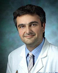 Mathios Dimitrios合作论文数6Esteban Velarde合作论文数School of Medicine, Johns Hopkins University6
 Drew Mark Pardoll合作论文数Department of Oncology, School of Medicine, Johns Hopkins University;Bloomberg~Kimmel Institute for Cancer Immunotherapy, School of Medicine, Johns Hopkins University;Sidney Kimmel Comprehensive Cancer Center, School of Medicine, Johns Hopkins University6
 Peter Burger合作论文数Division of Neuropathology, Department of Pathology, School of Medicine, The Johns Hopkins University6




