The liver contains a diverse repertoire of leukocytes, with liver B cells representing a significant population of hepatic immune cells in both newborns and adults. Despite their importance, these cells remain largely unexplored. In this study, we comprehensively characterized liver B cells from newborns through adulthood in both humans and mice applying a combination of in vivo imaging, immunophenotyping, RNA sequencing, and cell transfer protocols to characterize hepatic B cell subtypes from birth to adulthood. We found that newborn liver B cell population are transcriptionally and phenotypically distinct from those in adults, with significant alterations in genes related to immunoglobulin processing, release, and major histocompatibility complex class I and II functions. Photoconversion-based tracking revealed that liver-resident B cells actively migrate to distant lymphoid organs, including the spleen and bone marrow, where they integrate into the mature immune cell pool. Our findings establish the newborn liver as a crucial niche for B cell maturation and migration, highlighting its role in seeding secondary lymphoid organs and contributing to immune function throughout life. These insights provide new understanding of liver-derived B cells in the postnatal development of the immune system.
The peritoneal cavity contains a large population of GATA6-expressing large peritoneal macrophages (LPMs), known to support healing of intraabdominal organs. In this study, we aimed to explore their full sphere of influence by examining their ability to perform wound healing at distant sites outside the cavity. In a mouse model combining a remote skin injury with peritoneal stimulation we observed a significant acceleration of skin wound healing in response to LPM activation. Tracking GATA6-expressing LPMs, we demonstrated that LPMs do not migrate to distant wound sites following peritoneal activation. Using parabiosis experiments and administration of activated peritoneal contents indicated an important role of molecules secreted by LPMs in remote skin wound healing. More specifically, proteomic and transcriptomic analyses identified fibronectin as a key factor produced by activated LPMs. In fact, depletion of LPMs or genetic knockout of fibronectin in myeloid cells eliminated the enhanced healing effect. These findings highlight the endocrine function of LPMs in systemic tissue repair, challenging the traditional perspective of plasma fibronectin being exclusively liver derived. Our results suggest that LPMs, strategically positioned in the peritoneal cavity, serve as a source of circulating fibronectin, promoting matrix formation and accelerating wound healing at distant sites.
Abstract Protective immunity against many infectious diseases develops following primary infection, called infection induced immunity (III), and provides a blueprint for vaccination. However, many vaccination strategies have failed. In the parasitic Leishmania major model of self-healing cutaneous disease, control of secondary challenge infection relies on pre-existing T helper (Th)1-dependent activation of skin-infiltrating monocytes for elimination of intracellular parasites. To better understand immune-evasion of pre-existing Th1 immunity by pathogens, we investigated the pathogen-niche established following non-healing challenge infection with the L. amazonensis parasite in a setting of pre-existing III. Following secondary challenge, pre-existing Th1 III initially controlled infection but ultimately failed. Loss of protection was not overtly STAT6- or IL-10-mediated. Rather, monocyte-lineage tracing revealed inflammatory monocyte-derived PD-L1 + PD-L2 + macrophages provide an intracellular pathogen-niche and facilitate evasion of pre-existing Th1 immunity. Anti-PD-1 immune checkpoint blockade enhanced uninfected, but not infected, monocyte-derived cell activation and depletion of monocyte-derived precursors improved parasite control. These observations suggest that evasion of pre-existing Th1 immunity in this setting is not due to a failure of the Th1 response, but rather due to infected-cell intrinsic defects in activation.
Abstract Introduction Every organ in the body is innervated and contains populations of tissue-resident macrophages. Studies show communications between peripheral nerves and immunity in various organs; however, this is seldom explored in the liver. The liver is a firewall against bloodstream infection — a function made possible by Kupffer cells (KCs), the liver resident macrophage. Being uniquely situated in liver capillaries, KCs clear up to 95% of bacteria in the blood. Despite the liver being innervated, connections between hepatic nerves and KC are unclear. Here, we explore the interplay between hepatic nerves and KCs, hypothesizing that hepatic innervation is critical for KC identity, phenotype, and function. Methods We used tissue clearing to visualize the nerves in the liver. Intravital microscopy for assessing bacteria catching by KCs. Results We show that hepatic nerve fibers are primarily sympathetic nerves which line the portal vein of the liver but terminate before reaching the capillaries where KCs reside. This suggests that there is no direct physical interaction between KCs and hepatic sympathetic nerves. Despite this, sympathetic denervation reduced expression of genes associated with KC identity and led to reduced levels of receptors critical for KC function, like CRIg — a receptor required for catching bacteria. Sympathetic denervation significantly decreased bacteria catching by KCs, resulting in increased bacterial burden in the circulation and worse survival following bloodstream infection. Mechanistically, we show that liver-specific nerves release neurotransmitters and activate β-adrenergic receptors to maintain KC function and phenotype. Specific ablation of the β2-adrenergic receptor on KCs reduced bacteria catching suggesting that KCs directly sense neurotransmitters. Lastly, reintroducing norepinephrine into denervated mice rescued bacteria catching by KCs. Conclusion Overall, this study suggests that KC identity and function depend on norepinephrine released by hepatic sympathetic nerves. Funding Source Cancer Research Institute CRI4653 Topic Categories Neuroimmunology (NEUR)
Neutrophils are intrinsically antimicrobial innate immune cells, but they have recently been observed to play diverse roles across various conditions. The concept of neutrophil heterogeneity has thus emerged, yet a fundamental understanding of the mechanisms and processes underlying neutrophil diversity in vivo is lacking. Here, we interrogated the native propensities of neutrophils to diversify by studying them in the setting of host defense to barrier challenge. Using both high-parameter, high throughput methods and intravital microscopy, we tracked cellular dynamics, identity, and behaviors and asked how these aligned with critical host defensive functions. We found that neutrophil diversity was underscored by both turnover and plasticity of cells within an infection site, and that environment-specific adaptation occurred rapidly upon extravasation and swarm initiation. The population-level phenotypic landscape shifted considerably through phases of inflammation initiation, peak, and resolution; and adaptation varied substantially between infections and aseptic wounds. Like mature neutrophils, immature neutrophils were capable of phenotypic adaptation in infection sites but were deficient in host defense functions. We unexpectedly identified a novel population of CD101-low, mature neutrophils, expressing elevated levels of PD-L1 and ICAM-1, that was associated with key antimicrobial effector functions and most pronounced during the neutrophil response to large or recalcitrant challenge. Our findings highlight that neutrophil specification is rapid and environment-dependent, and that specific phenotypes are linked to critical host defensive behaviors and functions in vivo. ### Competing Interest Statement The authors have declared no competing interest. Damon Runyon Cancer Research Foundation, DRG-2494-23 Canadian Institutes of Health Research, https://ror.org/01gavpb45, PJT-178265, PJT-186272 Snyder Chair in Critical Care Medicine, END611952
Staphylococcus aureus forms biofilms on biotic and abiotic surfaces, enabling persistent infections that evade immune clearance. Although many S. aureus strains can produce the biofilm-associated exopolysaccharide, poly-β-1,6-N-acetyl-D-glucosamine (PNAG), they often form protein-dominated biofilm matrices in vitro, leaving a role for PNAG unclear. Using intravital imaging in a foreign-body infection model, we found that PNAG in biofilms hinders neutrophil access and delays bacterial clearance. Neutrophil elastase was crucial for eventual biofilm clearance. In vivo PNAG labeling revealed that the exopolysaccharide forms a physical barrier that prevents neutrophils from reaching bacterial clusters. In contrast, PNAG-deficient strains permitted greater neutrophil infiltration and were cleared more rapidly than wild-type bacteria. Enzymatic degradation of PNAG with the glycoside hydrolases PgaB or dispersin B (DspB) disrupted the biofilm, restored neutrophil access, and enhanced bacterial clearance. Together, these findings identify PNAG as a key structural barrier protecting S. aureus from innate immunity and suggest that targeting PNAG with glycoside hydrolases may offer a promising therapeutic strategy for biofilm-associated S. aureus infections.IMPORTANCEThe biofilm-associated exopolysaccharide PNAG is frequently expressed in Staphylococcus aureus clinical isolates but is often reduced during laboratory passage, with expression highly dependent on growth conditions. While in vitro analyses have revealed that PNAG is not a dominant matrix component, our intravital imaging of community-acquired methicillin-resistant S. aureus (CA-MRSA) skin infections demonstrates that PNAG is robustly produced in vivo and plays a central role in immune evasion. These findings highlight how PNAG function in tissue environments may be non-obvious in vitro and underscore the need for in vivo models to understand biofilm pathogenesis. By revealing PNAG as a key barrier to neutrophil-mediated clearance, this work positions PNAG and PNAG-targeting glycoside hydrolases as compelling therapeutic candidates for treating antibiotic-resistant S. aureus biofilm infections, a major cause of morbidity in both healthcare and community settings.
Abstract Introduction The fatality rate for metastatic cancers remains high, with the lungs being a common and deadly site of metastasis. Before cancer cells arrive, host tissues undergo changes that prepare them for tumour growth. This process forms the pre-metastatic niche, making the lung environment more receptive to colonization. Neutrophils populate the pre-metastatic niche, but their phenotype and whether they support metastasis at this stage remain unclear. Methods A model of E0771-GFP breast cancer cells that metastasize to the lungs was established, where the lung pre-metastatic niche and arrival of cancer cells can be captured using intravital microscopy. Using this model, we observed that not only does neutrophil behaviour change in the pre-metastatic niche from homeostasis, but their responsiveness to arriving cancer cells is compromised. Results The Kubes Lab has previously demonstrated that under healthy conditions, the majority of resident lung neutrophils crawl along the alveolar capillary wall seeking perturbations. Behavioural analysis of neutrophils imaged in pre-metastatic lungs showed reduced crawling and increased adhesion to the endothelium. Further, pre-metastatic neutrophils were imaged in the presence of both intravenously injected and spontaneously arrived cancer cells. In both cases, neutrophils exhibited limited interactions with cancer cells. In contrast, in healthy lungs, numerous neutrophils maintained prolonged contact with intravenously injected cancer cells. Super-resolution microscopy of healthy resident lung neutrophils showed acquisition of GFP+ particles following interaction with GFP+ cancer cells. Conclusion Our findings reveal that neutrophils in the lung pre-metastatic niche adopt a distinct behavioural state compared to healthy resident neutrophils. Such behaviour may contribute to the permissive environment that enables metastatic seeding in the lungs. These findings lay the foundation for therapeutic targeting of neutrophils to prevent metastatic seeding. Funding Source Canadian Research Excellence Chair (CERC), Infrastructure Operating Fund (IOF), Canadian Foundation for Innovation (CFI), and Bruce Mitchell Research Program Topic Categories Tumor Immunology: Cellular Responses and Tumor Microevironment (TIME)
Background: Antimicrobial resistance (AMR) is a major global health challenge, worsened by the lack of new antibiotics. This study aimed to design a redox-responsive bola amphiphile derived from an antimicrobial peptide (AMP) to enhance vancomycin (VCM) activity against both Gram-positive and Gram-negative bacteria. Methods: The bola amphiphile was synthesized via SN2 substitution, incorporating a thioether bond, and used to create self-assembling peptidosomes (VCM-AMP-PEP). Biocompatibility tests were done via red blood cell hemolytic and non-cytotoxic assays. Drug release was evaluated in vitro via the dialysis method. Antimicrobial activity was evaluated via the microbroth dilution method against methicillin-resistant Staphylococcus aureus (MRSA) and Gram-negative bacteria, Escherichia coli and Pseudomonas aeruginosa. Bactericidal activity of was evaluated via time killing and FACS (Fluorescence-Activated Cell Sorting) assays. The in vivo efficacy was evaluated Balb/c mice model, and intravital experiments were conducted on adult C57BL/6 mice. Results: Characterization showed an average particle size of 230.2 f 2.19 nm, a polydispersity index (PDI) of 0.223 f 0.028, zeta potential (ZP) of 13.1 f 1.25 mV, and entrapment efficiency (EE) of 51.4 f 1.08%. The particle size increased from 241.8 f 14.93 nm to 2906.0 f 191.5 nm upon exposure to reactive oxygen species (ROS), confirming redox responsiveness. Biocompatibility tests showed the peptidosomes were non-hemolytic and non-cytotoxic, while drug release studies demonstrated redox-triggered release in hydrogen peroxide and sustained release under physiological conditions. In vitro, VCM-AMP-PEP exhibited a modest increase in minimum inhibitory concentration (MIC) against methicillin-resistant Staphylococcus aureus (MRSA) (10.41 mu g/mL vs. VCM, 7.8 mu g/mL) but significantly enhanced activity against Gram-negative bacteria, reducing MICs by 12-fold against Escherichia coli and 3-fold against Pseudomonas aeruginosa. Flow cytometry revealed enhanced bacterial cell death with 99.77% MRSA and 85.79% Pseudomonas aeruginosa elimination at MICs, compared to VCM alone. In vivo, VCM-AMP-PEP demonstrated a 2.7-fold, 13-fold, and 10.9-fold reduction in bacterial load in blood, liver, and kidneys, respectively. Intravital imaging showed complete uptake of VCM-AMP-PEP by the liver via Kupffer cells. Conclusions: These findings suggest redox-responsive peptidosomes as a promising strategy to enhance vancomycin's efficacy against AMR and polymicrobial infections.
The liver, a key metabolic organ, has a central role in maintaining systemic homeostasis but is vulnerable to numerous diseases. Its metabolic functions are mainly carried out by hepatocytes; however, the liver also harbours diverse non-parenchymal cell populations, including immune cells. Among these, Kupffer cells, the resident macrophages of the liver, are critical modulators of liver function and immunity. Emerging research highlights their dynamic roles throughout life, from maintaining tissue homeostasis to shaping the balance between immune tolerance and activation in adulthood. Kupffer cells are located in liver sinusoids, where they act as frontline defenders, clearing pathogens and cellular debris from the circulation. Beyond their established phagocytic and immune regulatory functions, Kupffer cells influence metabolic processes, tissue repair and oncogenesis. Moreover, they shape the response of the liver to metabolic disorders such as metabolic dysfunction-associated steatohepatitis, infections and malignancies, including hepatocellular carcinoma. Here we explore Kupffer cell biology, focusing on the development, heterogeneity and multifaceted roles of these cells in liver health and disease. We further discuss how advances in imaging, transcriptomics and macrophage-targeted therapies can inform future strategies to combat liver-associated health challenges.
Abstract Introduction Neutrophils are powerful antimicrobial innate immune cells, without which a host is highly susceptible to morbidity and death caused by infections. However, in recent years, their role in noninfectious contexts has been highlighted, including in cardiac stress, autoimmunity, and cancer. Across settings, the concept of neutrophil heterogeneity has emerged, yet the basis and functional implications of this diversity remain largely unknown. Many devoted studies have examined contexts of cancer or other lifestyle diseases–arenas largely departed from the selective pressures that shaped neutrophil biology. Therefore, we sought to study neutrophil diversity in an evolutionarily powerful setting: infection and acute injury to the skin. Methods Using spectral flow cytometry, along with intravital microscopy and transgenic mice that enable tracking neutrophils over time, we assessed neutrophil dynamics and plasticity, as well as relationships between environment, phenotype, function, and behavior in vivo. Results We found that neutrophils rapidly specify their phenotype upon extravasating to a site of challenge. During response to a biofilm infection, neutrophil phenotypes shifted over time, reflecting both plasticity and turnover. Several phenotypic changes seen during the infection response were absent in a noninfectious wound, highlighting the challenge-specific nature of neutrophil phenotypic adaptation. Notably, we identified an unexpected population of CD101-low mature neutrophils, expressing elevated levels of PD-L1 and ICAM-1, associated with the infection response. This population was characteristic of a highly structured neutrophil swarm. Conclusion This work provides fundamental insights into the evolved capacity of neutrophils to diversify in vivo, revealing that neutrophil phenotypes are dynamic even in acute settings of inflammation, and tightly linked to a cell’s environment. It further aligns observations of phenotypic adaptation with specific host defensive behaviors. Funding Source Damon Runyon Postdoctoral Fellowship Award Topic Categories Innate Immune Responses and Host Defense: Cellular Mechanisms (INC)
Large peritoneal macrophages (LPMs), located in the peritoneal cavity, are essential for local tissue repair. However, their contribution to tissue repair at distant sites remains unclear. This study investigates the role of LPM activation in skin wound healing at remote sites A novel mouse model combining peritoneal stimulation (PS) with a skin injury was used to assess LPM-mediated effects on remote skin wound healing. Dual Cre and Flippase fate-mapping tracked GATA6-expressing LPMs to evaluate their migratory behavior. Parabiosis experiments and administration of activated peritoneal fluid were conducted to identify the role of circulating factors. Proteomic and transcriptomic analyses were used to characterize LPM-secreted molecules. Fluorescently labelled fibronectin was tracked in plasma and skin wounds. Activation of LPMs trough PS significantly accelerated healing of remote skin wounds. Removing LPMs or using mice lacking LPMs abolished the beneficial effect. Adoptive transfer of LPMs but not B-cells after LPM depletion was sufficient to rescue the phenotype. Fate-mapping demonstrated that LPMs did not migrate to distant wounds after activation. Parabiosis and peritoneal fluid transfer experiments indicated the role of LPM-derived circulating signaling molecules in remote skin wound healing. Proteomic and transcriptomic analyses identified fibronectin as critical mediator, as adoptive transfer of LyzMcre Fnflox peritoneal cells failed to rescue the impaired remote wound healing phenotype in LPM deficient mice. Protein-coding fibronectin isoforms transcribed by LPMs, correspond to the soluble plasma. Fluorescently labelled fibronectin was detected in the plasma and incorporated into skin wounds following adoptive transfer of Fngfp/gfp peritoneal cells and PS. LPMs act as a source of circulating fibronectin, facilitating extracellular matrix formation and promoting wound healing at remote sites. These findings reveal a novel endocrine role for LPMs in systemic tissue repair, and challenge the traditional perspective, that plasma fibronectin is exclusively liver derived.
PURPOSE OF REVIEW:Kupffer cells (KCs), the resident liver macrophages, are absolutely critical in immune surveillance and intravascular pathogen eradication. This mini-review highlights KCs' contributions to host protection of intravascular infections. RECENT FINDINGS:KCs, uniquely originated and self-renewing, demonstrate remarkable functional plasticity and trained immunity. KCs are frontline responders in infections: they phagocytose microorganisms but can succumb to certain infections. We highlight some of the recent findings in this regard. SUMMARY:Understanding KCs' complex interactions with diverse pathogens is key to improving treatment modalities in infection. Future research, needs to focus on how certain pathogens evade KCs and how we can aid these macrophages in eradicating microbes. A move towards humanized KC models in vivo and in vitro incorporating key environmental factors such as shear flow and unique sinusoidal components will be essential to unravel their comprehensive roles.
OBJECTIVE:To investigate the hepatoprotective effects of mitochondrial transplantation (MTx) in a murine liver ischemia/reperfusion (I/R) model. BACKGROUND:Sequential liver ischemia, followed by reperfusion (I/R), is a pathophysiological process underlying hepatocellular injury in a number of clinical contexts, such as hemorrhagic shock/resuscitation, major elective liver surgery, and organ transplantation. A unifying pathogenic consequence of I/R is mitochondrial dysfunction. Restoration of mitochondria through transplantation (MTx) has emerged as a potential therapeutic in I/R. However, its role in liver I/R and its mechanisms of action remain poorly defined. METHODS:We investigated the hepatoprotective effects of MTx in an in vivo mouse model of liver I/R and used in vivo imaging and various knockout and transgenic mouse models to determine the mechanism of protection. RESULTS:We found that I/R-induced hepatocellular injury was prevented by MTx, as measured by plasma ALT, AST, and liver histology. In addition, I/R-induced pro-inflammatory cytokine release (IL-6, TNFα) was dampened by MTx, and anti-inflammatory IL-10 was enhanced. Moreover, MTx lowered neutrophil infiltration into both the liver sinusoids and lung bronchoalveolar lavage fluid, suggesting a local and distant reduction in inflammation. Using in vivo intravital imaging, we found that I/R-subjected Kupffer cells (KCs), rapidly sequestered transplanted mitochondria, and acidified mitochondria within lysosomal compartments. To specifically interrogate the role of KCs, we depleted KCs using the diphtheria toxin-inducible Clec4f/iDTR transgenic mouse, then induced I/R, and discovered that KCs are necessary for the beneficial effects of MTx. Finally, we induced I/R in the complement receptor of the immunoglobulin (CRIg) superfamily knockout mice and found that CRIg was required for mitochondria capture by KCs and mitochondria-mediated hepatoprotection. CONCLUSIONS:In this study, we demonstrated that CRIg-dependent capture of mitochondria by I/R-subjected KCs is a hepatoprotective mechanism in vivo . These data progress knowledge on the mechanisms of MTx and open new avenues for clinical translation.
Liver regeneration (LR) is essential for recovery from acute trauma, cancer surgery, or transplantation. Neurotransmitters such as acetylcholine (ACh) play a role in LR by stimulating immune cells and augmenting hepatocyte proliferation, but the source of this ACh remains unclear. Here, we demonstrated that B cells expressing choline acetyltransferase (ChAT), which synthesizes ACh, were required for LR. Mice lacking ChAT+ B cells subjected to partial hepatectomy (PHX) displayed greater mortality due to failed LR. Kupffer cells and hepatic CD8+ T cells expressed the α7 nicotinic ACh receptor (nAChR), and LR was disrupted in mice lacking α7 nAChR. Mechanistically, B cell-derived ACh signaled through α7 nAChR to positively regulate the function of regenerative Kupffer cells and to control the activation of hepatic CD8+ T cells to curtail harmful interferon-gamma (IFNγ) production. Our work offers insights into LR mechanisms that may point to therapies for liver damage.
Concussions can cause debilitating symptoms despite no evidence of structural changes on diagnostic imaging. The cellular events occurring in the brain parenchyma following concussion, especially repetitive concussion, are not well elucidated. We developed a concussion model to induce a confined area of injury without causing frank hemorrhage. Using intravital microscopy, we observe activation of the vasculature that supported neutrophil rolling and platelet adhesion but no overt cellular recruitment from blood into brain parenchyma. Activated resident, not monocyte-derived, macrophages relocated to the injury site via Cx3cr1 and phagocytosed dysfunctional/detached astrocytes via scavenger receptors and TLR4, particularly after repetitive concussion. Additionally, microglia sealed areas of blood-brain barrier (BBB) disruption via purinergic pathways. Using a splitCre approach to dissect microglia and perivascular macrophages, we show that microglial invasion into the injury site is key to reducing BBB disruption. Our data suggest that microglia repair the BBB following concussion, but in doing so significantly alter the cellular ultrastructure of the brain milieu.
The liver is a sizeable visceral organ whose primary functions involve nutrient metabolism, clearance of toxins, and energy storage. Besides these critical functions, the liver is also a major immunological site. It is populated by several specialized resident immune cells, including B cells, T Cells, dendritic cells, and several populations of macrophages. It is also the site for the production and release of acute-phase proteins during inflammation. One reason for garrisoning these immune sentinels and effectors in the liver is its relative location in the circulatory system. The liver is the first significant organ downstream of the intestine, where blood originating from the intestine enters the liver through the portal vein. This organization facilitates the liver’s uptake and processing of nutrient-rich blood directly from the intestinal source. However, the intestine is also home to trillions of microbes, many of which are commensals but also represent potential pathogens. As such, the portal blood supply represents an avenue for systemic infection. To sterilize the portal blood, the liver immune system filters pathogens, which is primarily accomplished by liver macrophages. Here, we will discuss the major populations of macrophages resident in the liver, their location, functions, development, and role in maintaining the liver in the face of injury and infection.
 D. Neil Granger合作论文数Department of Molecular and Cellular Physiology, School of Graduate Studies, Louisiana State University Health Sciences Center in Shreveport52
 Craig Jenne合作论文数Cumming School of Medicine, University of Calgary;Alberta Children's Hospital Research Institute;The Calvin, Phoebe and Joan Snyder Institute for Chronic Diseases, University of Calgary;Department of Microbiology, Immunology and Infectious Diseases, University of Calgary45
 Justin F Deniset合作论文数43
 Michael J. Hickey合作论文数Leukocyte Trafficking Group, Centre for Inflammatory Disease Monash Health, Monash University36
 Bas Surewaard合作论文数University of Calgary30Braedon McDonald合作论文数Snyder Institute of Infection, Immunity, and Inflammation, University of Calgary, Alberta, Canada29
 Woo-Yong Lee合作论文数28
 SS Kanwar合作论文数Department of Biotechnology;Himachal Pradesh University;Department of Biotechnology, Himachal Pradesh University28
 Christoph W. Sensen合作论文数Hungarian Centre of Excellence for Molecular Medicine26
 Brent Johnston合作论文数Departments of Microbiology & Immunology and Pediatrics, Dalhousie University25Steven M. Kerfoot合作论文数School of Medicine, Yale University25Rc Woodman合作论文数24Dm Mccafferty合作论文数22
 Bjoern Petri合作论文数Snyder Institute of Infection, Immunity and Inflammation, University of Calgary22
 Bonder Claudine S合作论文数Ctr Canc Biol20Ph Reinhardt合作论文数19G Andonegui合作论文数19
 Moritz Peiseler合作论文数19
 Bryan Heit合作论文数1 Immunology Research Group;Department of Physiology and Biophysics;University of Calgary;1 Immunology Research Group, University of Calgary19
 Connie Hoi Yee Wong合作论文数19
 Rachel M. Kratofil合作论文数Univ Calgary, Cumming Sch Med, Dept Microbiol Immunol & Infect Dis, Snyder Inst Chron Dis, Calgary, AB T2N 4N1, Canada17
 Christopher James Doig合作论文数Department of Critical Care Medicine, Cumming School of Medicine, University of Calgary;Department of Medicine and Community Health Sciences, Cumming School of Medicine, University of Calgary17
 Stephen M Robbins合作论文数Cancer Research Institute;Departments of Oncology and Biochemistry & Molecular Biology;University of Calgary;Cancer Research Institute, University of Calgary17D Payne合作论文数16
 Fernanda Vargas e Silva Castanheira合作论文数Departamento de Microbiologia Imunobiologia e Parasitologia,;Universidade Federal de São Paulo15
 Joel Zindel合作论文数Dept Visceral Surg & Med, Bern Univ Hosp14Zbytnuik Lori合作论文数14Bryan G. Yipp合作论文数14M. Ursula Norman合作论文数14
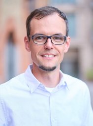 Carsten Deppermann合作论文数University of Würzburg;Department of Experimental Biomedicine;and Rudolf Virchow Center, University Hospital;DFG Research Center for Experimental Biomedicine;Würzburg;University Hospital and Rudolf Virchow Center, University of Würzburg13
 John Lawrence Wallace合作论文数Antibe Therapeutics Inc;Department of Physiology and Pharmacology, Cumming School of Medicine, University of Calgary13XF NIU合作论文数12Erin F. Mcavoy合作论文数12Sarah C. Mullaly合作论文数11
 Zhutian Zeng (曾筑天)合作论文数School of Basic Medical Sciences, University of Science and Technology of China Life Sciences and Medicine11
 Kamala Patel合作论文数Department of Biochemistry & Molecular Biology, Cumming School of Medicine, University of Calgary11
 Alison Fox-Robichaud合作论文数Dept of Medicine, McMaster University11Ajitha Thanabalasuriar合作论文数11
 Pina Colarusso合作论文数Department of Chemistry, University of Waterloo11JEFFREY P. GABOURY合作论文数10
 Caroline Leger合作论文数10McCafferty Donna-Marie合作论文数10
 Derek J Roberts合作论文数10Ostrovsky L合作论文数10
 Bruna A. David合作论文数10Mia Phillipson合作论文数10
 Mark Gordon Swain合作论文数Department of Medicine, Cumming School of Medicine, University of Calgary;The Calvin, Phoebe and Joan Snyder Institute for Chronic Diseases, The University of Calgary;Division of Gastroenterology and Hepatology, The University of Calgary9
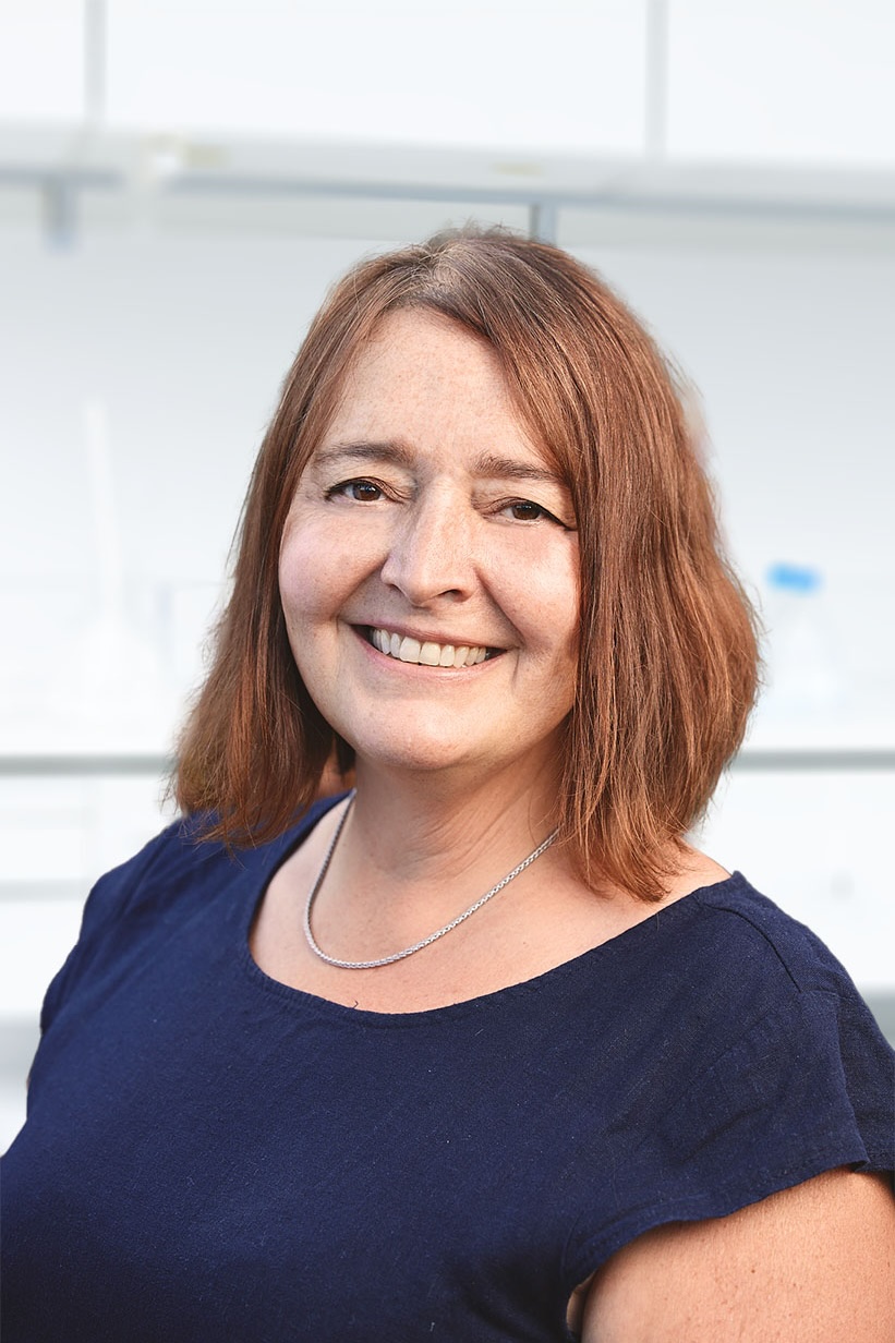 Deborah Stroka合作论文数Department for BioMedical Research, Faculty of Medicine, University of Bern;Department of Visceral Surgery and Medicine, Bern University Hospital9
 Christopher H Mody合作论文数Department of Internal Medicine, University of Calgary9




