Focally evoked calcium waves in astrocyte cultures have been thought to propagate by gap-junction-mediated intercellular passage of chemical signal(s). In contrast to this mechanism we observed isolated astrocytes, which had no physical contact with other astrocytes in the culture, participating in a calcium wave. This observation requires an extracellular route of astrocyte signaling. To directly test for extracellular signaling we made cell-free lanes 10-300 microns wide in confluent cultures by deleting astrocytes with a glass pipette. After 4-8 hr of recovery, regions of confluent astrocytes separated by lanes devoid of cells were easily located. Electrical stimulation was used to initiate calcium waves. Waves crossed narrow (< 120 microns) cell-free lanes in 15 of 36 cases, but failed to cross lanes wider than 120 microns in eight of eight cases. The probability of crossing narrow lanes was not correlated with the distance from the stimulation site, suggesting that cells along the path of the calcium wave release the extracellular messenger(s). Calculated velocity across the acellular lanes was not significantly different from velocity through regions of confluent astrocytes. Focal superfusion altered both the extent and the direction of calcium waves in confluent regions. These data indicate that extracellular signals may play a role in astrocyte-astrocyte communication in situ.
The present study investigates how a neuron's past history of neural activity may alter its responsiveness to subsequent signals. We demonstrate that a depolarizing pulse of extracellular potassium can prime neurons to become responsive to basic fibroblast growth factor (bFGF), even when the pulse is brief and occurs prior to addition of bFGF. Specifically, we subjected cultured embryonic chick ciliary ganglion neurons (E7) to a short pulse of elevated extracellular potassium followed by addition of bFGF and tested the effect of such treatment on neuronal survival. Neurons treated in this manner produced high levels of survival, whereas neurons exposed to either the pulse alone or the continuous presence of bFGF alone failed to promote any significant levels of survival. This priming effect of depolarization on bFGF-induced survival was blocked by calcium channel antagonists. To test the time dependency of this effect, we increased the time interval between termination of the calcium pulse and addition of bFGF. Our results demonstrate that a brief elevation in intracellular calcium has long lasting effects, up to 8 h after cessation of the depolarizing pulse, on neuronal responsiveness to bFGF. These findings suggest how a developing neuron's history of activity can alter its subsequent ability to respond to neurotrophic factors and has significant implications on the mechanisms by which activity may influence neuronal survival.
Communication from astrocytes to neurons has recently been reported by two laboratories, but different mechanisms were though to underlie glial calcium wave activation of associated neurons. Neuronal calcium elevation by glia observed in the present report is similar to that reported previously, where an increase in neuronal calcium was demonstrated in response to glial stimulation. In the present study hippocampal neurons plated on a confluent glial monolayer displayed a transient increase in intracellular calcium following a short delay after the passage of a wave of increased calcium in underlying glia. Activated cells displayed action potentials in response to glial waves and showed antineurofilament immunoreactivity. Finally, the N-methyl-D-aspartate glutamate receptor antagonist DL-2-amino-5-phosphonovaleric acid and the non-NMDA glutamate receptor antagonist 6,7-dinitroquinoxaline-2,3-dione significantly reduced the responsiveness of neurons to glial calcium waves. Our results indicate that hippocampal neurons growing on hippocampal or cortical astrocytes respond to glial calcium waves with elevations in calcium and increased electrical activity. Furthermore, we show that in most cases this communication appears to be mediated by ionotropic glutamate receptor channels.
Proteoglycans have been implicated in neuronal pathfinding during development, yet related second messenger and signaling systems are unknown. We have used the calcium indicator fura-2/AM to monitor cytoplasmic calcium ion concentration ([Ca2+]i) in chick dorsal root ganglion (DRG) neuronal growth cones elongating on laminin during contact with chondroitin sulfate proteoglycan (CSPG): (1) to determine whether there is a change in [Ca2+]i in neurons that contact CSPG, and (2) to determine whether changes in [Ca2+]i are necessary for inhibition of growth cone migration. The majority of DRG neurons responded to CSPG contact with a transient rise in [Ca2+]i (mean Δ[Ca2+]i above resting level was 554 ± 109 nM; P < 0.0001). The effect of CSPG contact was concentration dependent and required the carbohydrate moiety of CSPG. Addition of soluble CSPG did not elevate [Ca2+]. Treatment with reagents that blocked plasma membrane calcium channels, or that perturbed intracellular Ca2+ stores, indicated that extracellular Ca2+ was the major source of the [Ca2+]i elevation, and that Ca2+ entry occurred through non-voltage-gated calcium channels. Although general Ca2+ channel blockers abolished the CSPG-induced [Ca2+]i rise, they did not abolish growth cone avoidance of surface-bound CSPG in these assays. We conclude: (1) that DRG neurons elevate [Ca2+]i in response to CSPG contact to levels that can modify cytoskeletal mechanisms of growth cone migration, and (2) that avoidance of substratum-bound CSPG may not be dependent upon elevated [Ca2+]i.
 Hassinger T D合作论文数3Albrecht H. Kossel合作论文数1Sb Kater合作论文数1Mvl Bennett合作论文数1
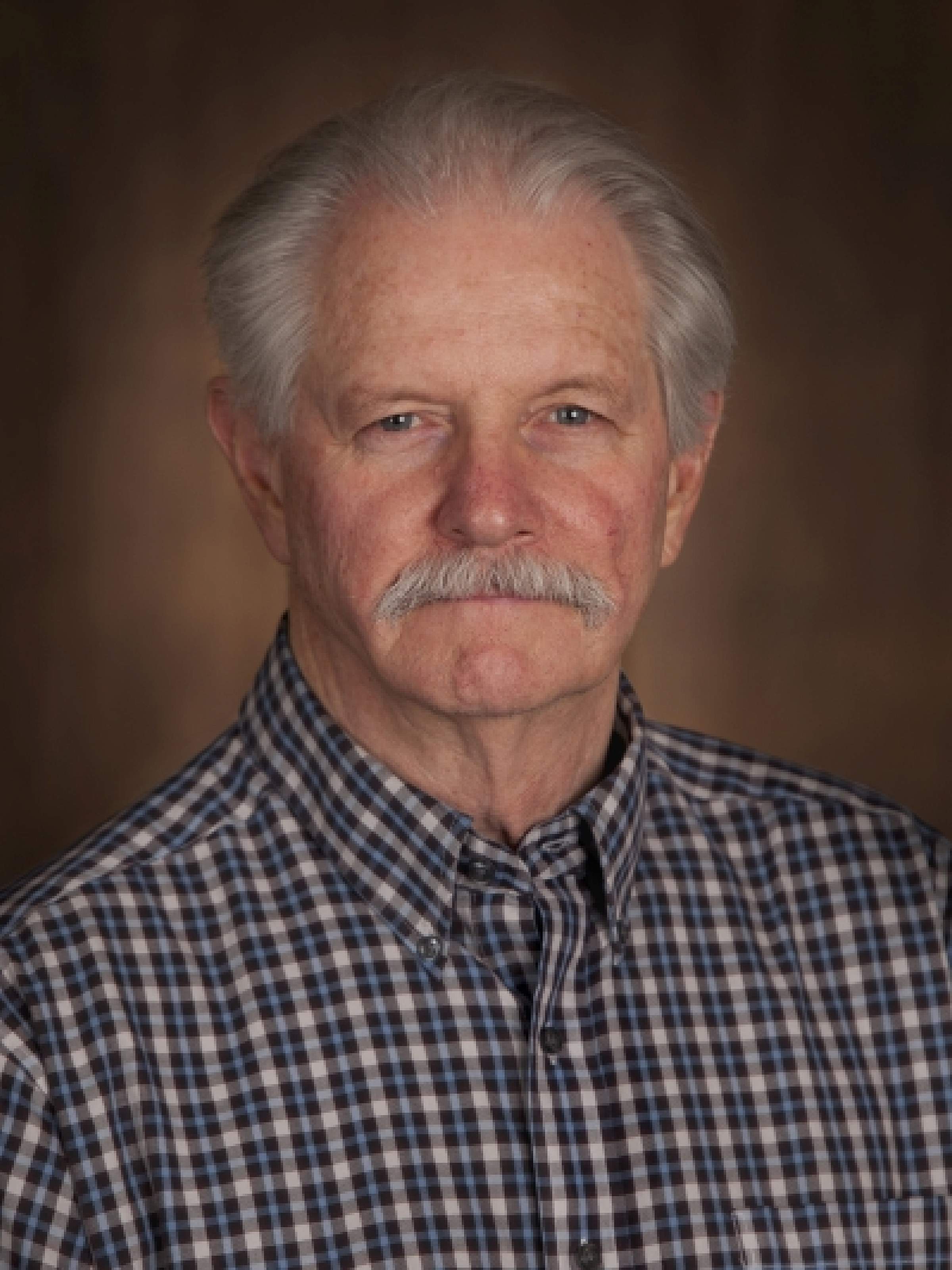 F. Edward Dudek合作论文数Spencer Fox Eccles School of Medicine, University of Utah;Department of Neurosurgery, School of Medicine, University of Utah1Strecker G J合作论文数1Paul C Letourneau合作论文数1S.B. Kater合作论文数1
 Marc Schmidt合作论文数Department of Biology, School of Arts & Sciences, University of Pennsylvania1
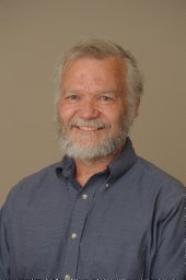 Peter B. Guthrie合作论文数Program in Neuronal Growth and Development;Department of Anatomy and Neurobiology;Colorado State University;Department of Anatomy and Neurobiology, Colorado State University1Diane M. Snow合作论文数1




