Transplantation of skin micrografts in a 1:100 ratio regenerate the epidermis of full-thickness wounds in pigs within 14 days in a wet environment. The aim of the current study was to combine micrografts and commercially available moist dressings. We hypothesized that micrografts regenerate the epidermis when covered with a moist dressing. 5cm×5cm and 10cm×10cm full-thickness wounds were created on the backs of pigs. Wounds were transplanted with 0.8mm×0.8mm micrografts created from a split-thickness skin graft in a 1:100 ratio. 5cm×5cm wounds were treated with wound chambers, moist dressings or dry gauze (non-transplanted control group). 10cm×10cm wounds were compared to non-transplanted wounds, both covered with moist dressings. Reepithelialization was assessed in biopsies from day 10, 14 and 18 post-transplantation. 5cm×5cm transplanted wounds covered with moist dressings showed 69.5±20.6% reepithelialization by day 14 and 90.5±10.4% by day 18, similar to wounds covered with a wound chamber (63.9±16.7 and 86.2±11.9%, respectively). 18 days post-transplantation, 10cm×10cm transplanted wounds covered with moist dressings showed 66.1±10.3% reepithelialization, whereas nontransplanted wounds covered with moist dressings were 40.6±6.6% reepithelialized. We conclude that micrografts combined with clinically available moist dressings regenerate the epidermis of full-thickness wounds.
BACKGROUND: Transplantation of genetically modified keratinocytes has been shown to accelerate wound healing. However, this method is labor-intensive and time-consuming. We have developed a new technique of intraoperative gene delivery to wounds that involves transplantation of transfected minced skin particles (MSPs) derived from harvested partial-thickness skin.STUDY DESIGN: MSPs measuring 0.8 X 0.8 X 0.35 mm were created from a split-thickness skin graft of a pig. In vitro transfection was carried out with adenoviral LacZ (Ad-LacZ) for qualitative and adenoviral vascular endothelial growth factor (Ad-VEGF) for quantitative analysis. Transfected MSPs were transplanted to each of 2.5 X 2.5 cm full-thickness wounds on the dorsum of the pig. Nontransfected MSPs served as controls. Wound chambers were applied and injected with saline to create a wet environment.RESULTS: LacZ expression was detected in migrating cells originating from MSPs both in vitro and in vivo. VEGF expression in the wound fluid of Ad-VEGF-MSP-transplanted wounds on each of days 2 to 4 (mean +/- SEM 6.74 +/- 1.89 ng/mL, day 2; 9.88 +/- 2.27 ng/mL, day 3; 9.87 +/- 1.28 ng/mL, day 4) was significantly higher (p < 0.0001) compared with wounds transplanted with either untransfected MSPs, Ad-LacZ-MSPs, or untransplanted controls. In vitro VEGF expression was significantly higher (p < 0.0001) in Ad-VEGF 1 X 10(10) transfected MSPs compared with either Ad-VEGF 1 X 10(9) transfected MSPs or untransfected MSPs. Wounds transplanted with Ad-VEGF-MSPs showed significantly higher (p < 0.0001) numbers of newly formed blood vessels (12.6 +/- 0.9 vessels/high power field [HPF]) compared with wounds transplanted with either Ad-LacZ-MSPs (4.4 +/- 0.5 vessels/HPF) or untransfected MSPs (5.2 +/- 0.7 vessels/HPF). All MSP-transplanted wounds (Ad-VEGF-MSPs, untransfected MSPs, Ad-LacZ-MSPs) showed significantly higher re-epithelialization compared with untransplanted wounds on days 10 and 14 (p < 0.0001).CONCLUSIONS: We demonstrated successful transfection of MSPs that can be transplanted to wounds as a source of gene-expressing cells. This technique can be used to deliver growth-modulating genes in wound healing. (J Am Coll Surg 2011;212:340-348. (c) 2011 by the American College of Surgeons)
BACKGROUND:Partial facial allotransplantation is an emerging option in reconstruction of central facial defects, providing function and aesthetic appearance. Ethical debate partly stems from uncertainty surrounding identity aspects of the procedure. There is no objective evidence regarding the effect of donors' transplanted facial structures on appearance change of the recipients and its influence on facial recognition of donors and recipients.METHODS:Full-face frontal view color photographs of 100 volunteers were taken at a distance of 150 cm with a digital camera (Nikon/DX80). Photographs were taken in front of a blue background, and with a neutral facial expression. Using image-editing software (Adobe-Photoshop-CS3), central facial transplantation was performed between participants. Twenty observers performed a familiar 'facial recognition task', to identify 40 post-transplant composite faces presented individually on the screen at a viewing distance of 60 cm, with an exposure time of 5s. Each composite face comprised of a familiar and an unfamiliar face to the observers. Trials were done with and without external facial features (head contour, hair and ears). Two variables were defined: 'Appearance Transfer' refers to transfer of donor's appearance to the recipient. 'Appearance Persistence' deals with the extent of recipient's appearance change post-transplantation. A t-test was run to determine if the rates of Appearance Transfer differed from Appearance Persistence.RESULTS:Average Appearance Transfer rate (2.6%) was significantly lower than Appearance Persistence rate (66%) (P<0.001), indicating that donor's appearance transfer to the recipient is negligible, whereas recipients will be identified the majority of the time. External facial features were important in facial recognition of recipients, evidenced by a significant rise in Appearance Persistence from 19% in the absence of external features to 66% when those features were present (P<0.01).CONCLUSION:This study may be helpful in the informed consent process of prospective recipients. It is beneficial for education of donors families and is expected to positively affect their decision to consent for facial tissue donation.
Once belonging to science fiction, face transplantation is a clinical reality now. The controversial first face transplantation in Amiens, France in 2005 was followed by significant ethical debate among physicians, surgeons, scientists and bioethicists. The controversies are being followed by cautious optimism and acceptance. An increasing number of surgical teams are developing protocols of composite tissue transplantation. Twenty five years after the birth of the American Society of Reconstructive Microsurgery (ASRM) and during its 25th anniversary, the founding meeting of American Society of Reconstructive Transplantation (ASRT) took place in 2009 to officially introduce the birth of a new field that combines the two disciplines: transplantation and reconstructive microsurgery. It appears that the field of composite tissue allotransplantation will continue to grow for at least several years before more sophisticated means of replacing the damaged tissues become available. This article takes a brief look at the evolution of organ transplantation and reflects our viewpoint regarding the emerging field of composite tissue allotransplantation (CTA).
Background: Gene therapy is a potentially attractive option in the treatment of nonhealing wounds. It aims at delivering the growth-modulating genes to the wound, enabling over or underexpression of specific molecules hence modifying the healing process. Transplantation of gene-transfected cells (ex vivo gene delivery) is one of the methods of delivering specific genes to the wound. Use of autologous keratinocytes as ex vivo gene delivery vehicles has previously been reported. In addition, successful clinical application of allogenic cultured keratinocyte as a temporary biological dressing for wounds has been described. Combining the two approaches, we evaluated the feasibility of transplantation of allogenic keratinocytes as gene delivery vehicles to accelerate wound healing.The Problem: Prompt skin replacement is critical in the treatment of skin defects to prevent fluid loss and infection. Autografts are the most common technique; however, donor site shortage is a major limiting factor in resurfacing the extensive wounds. Despite the successful clinical application of in vitro-expanded autologous keratinocytes, the long process of preparation is a disadvantage of this approach.Basic/Clinical Science Advances: Allogenic keratinocytes can function as temporary biological dressings, and in combination with gene delivery they can potentially optimize the host skin regenerative capacity before they are eventually rejected by the immune system.Clinical Care Relevance: In a clinical context, cell cultures need to be readily available for treatment. Allogenic cell cultures can be cryopreserved, genetically modified, and used when required.Conclusion: We demonstrated that human epidermal growth factor-transfected allogenic keratinocytes enhance re-epithelialization in porcine full-thickness wounds.
BACKGROUND: Reconstruction of complex abdominal wall defects is challenging. The use of prosthetic mesh can be associated with surgical site infection, fistula formation, and adhesions. This study presents our experience using a non-cross-linked porcine dermal scaffold (NCPDS) in abdominal wall reconstruction.METHODS: Patients undergoing abdominal wall reconstruction with NCPDS between May 2006 and January 2008 underwent a retrospective chart review. Demographics, indications for NCPDS placement, surgical technique, complications, and follow-up data were evaluated.RESULTS: Sixteen patients were identified in whom NCPDS was implanted into complex abdominal wall defects. These included 13 planned and 3 emergency surgeries. Indications for surgery included delayed reconstruction of giant ventral hernia secondary to decompressive laparotomy and open management of abdominal trauma, recurrence of large incisional hernia, temporary coverage of open abdomen secondary to intra-abdominal catastrophes, and open abdominal closure owing to compartment syndrome secondary to necrotizing fasciitis. In all, NCPDS was positioned in a subfascial underlay technique. Forty-four percent required a combination of components separation and NCPDS insertion. At a mean follow-up period of 16.5 months, the majority had desirable outcomes. Complications included seroma (21%), superficial wound dehiscence (7%), recurrence (7%), and infection (7%). Two patients died from multiorgan failure unrelated to NCPDS placement. The material only had to be removed in 1 patient because of wound infection and superficial wound dehiscence.CONCLUSIONS: NCPDS seems to be a safe and effective alternative to prosthetic mesh in the reconstruction of complicated abdominal wall defects. (C) 2010 Elsevier Inc. All rights reserved.
BACKGROUND:Autologous chondrocyte transplantation (ACT) is the most commonly used cell-based surgical procedure for repair of articular cartilage defects. The challenges of this technique include dedifferentiation of chondrocytes following several in vitro passages, invasive means of transplantation, and inadequate cell retention leading to washout of transplanted cells. To overcome these obstacles, we developed a novel technique of transplanting high-density chondrocyte pellets seeded on a prefabricated, resorbable, rigid, 2-dimensional template amenable to minimally invasive implantation. METHODS:Chondrocytes were obtained from the costal cartilage of New Zealand white rabbits and expanded in vitro in monolayer culture. After 2 passages, chondrocyte suspension was centrifuged and a total of 1 x 10(6) cells condensed on the surface of a prefabricated, resorbable template of LactoSorb plate (0.5-mm thick, 4-mm diameter). The construct was incubated for 24 hours in a culture medium before transplantation into circular 4-mm diameter, 0.5-mm deep defects in a non-weight-bearing part of the femoral condyle. Control defects were left empty or implanted with LactoSorb alone. Macroscopic and histological evaluation was performed 4 weeks posttransplantation. RESULTS:Macroscopically, boundaries of all defects were demarcated and distinguishable from adjacent intact cartilage. Regenerative tissue in experimental group appeared white, smooth, and uniform showing more resemblance to hyaline cartilage. Control groups revealed absent cartilaginous tissue and defects were filled with soft, fibrous tissue with an irregular surface. Histologically, the repair tissue in the control groups was fibroinflammatory with irregular surface and no evidence of continuous chondrocytic regeneration. Cartilage regeneration in the experimental defects revealed a continuous, high-density layer of chondrocytes surrounding the LactoSorb plates. Consistently with chondrocyte pellets grown for 4 weeks only, the amount of extracellular matrix deposition in the transplanted group was less than the normal cartilage. CONCLUSION:We have developed a novel approach for ACT, utilizing high-density chondrocyte pellets seeded on a prefabricated, rigid, 2-dimensional resorbable carrier. Our study can serve as a model for further minimally invasive development of this technique and evaluating its potential role as an alternative in ACT.
Face transplantation has opened new horizons in facial reconstructive surgery. It replaces the missing facial tissues or structures with identical ones. Indications include reconstruction of central facial defects, involving all the anatomic layers such as skin, subcutaneous fat, muscles, cartilage or bone. This central part of the face has unique anatomy and complex functions performing mastication, deglutition, expression, verbal communication and integration of these processes.
Objective: The purpose of this study was to determine whether amnion-derived cellular cytokine solution (ACCS) could improve the quality of epithelialization and accelerate closure of dermatome-created partial-thickness wounds in normal and streptozotocin-induced diabetic pigs. Methods: Dermatome-created partial-thickness wounds were sealed with wound chambers in healthy and diabetic pigs and were injected with ACCS. Wound fluid was exchanged daily for total protein concentration, and biopsies were taken on days 6, 8, 10, and 12. Epithelialization, thickness of epidermis, number of epidermal cell layers, and rete ridges were evaluated. Results: The macroscopic appearance of the wounds and speed of healing was similar in all groups at each time point. All wounds were healed by day 6. The epidermis was thicker in the ACCS-treated diabetic wounds than in the controls (140.6 μm vs 82.7 μm on day 12 in diabetic pigs). There were more cell layers (13 vs 7.7) in ACCS-treated diabetic pigs on day 12. The number of rete ridges per 2.5 mm was greater on day 12 in the ACCS-treated diabetic wounds (13 vs 8). There was also a significant increase in the number of rete ridges in ACCS-treated nondiabetic pigs but no difference in epidermal thickness or number of cell layers. Conclusion: In diabetic pigs, we found a significantly thicker epidermis and more cell layers and rete ridges in the ACCS-treated wounds. Healthy pigs showed more rete ridges but no difference in thickness of epidermis or number of cell layers on day 12.
ABSTRACTDecreased inflammatory response seen in wet wound healing may be correlated with diminished scarring. This study seeks to test this hypothesis and to validate a model of scarring in the Yorkshire pig. Four Yorkshire pigs were used to create 36 dorsal wounds per pig (144 wounds total) in the following groups: full‐thickness excisional, partial thickness, meshed split‐thickness skin grafts, sheet split‐thickness skin grafts, minced skin, and incisional wounds. Wounds were randomized into wet and dry groups. Wet wounds were enclosed in polyurethane chambers with 2 mL of normal saline. Dry wounds were covered with regular gauze. Terminal biopsies were performed at 72 hours and day 28. Histology demonstrated significantly less inflammatory infiltrate, thicker neoepidermis, more pronounced rete ridge formation, and decreased scar tissue thickness in wet wounds. The mean macroscopic scar surface area was significantly decreased in full‐thickness excisional wet wounds compared with dry wounds (61.2 mm2 vs. 150.8 mm2, p<0.01). Hydroxyproline content was decreased in full‐thickness wet compared with dry groups (44.81 vs. 62.21 mg/g, p<0.01). Tensile strength was 90% greater in full‐thickness wet compared with dry groups (p<0.01). Healing in the liquid environment significantly reduced scar formation. This model will allow for future investigation of high‐concentration topical scar‐modulating agents in the liquid environment.
Composite tissue transplantation in reconstructing complex facial defects has developed tremendous interest over the recent years, since the first report of partial face transplantation performed in France in 2005. However, the controversy over the ethical, immunological, and psychological issues remains. Recently, we obtained IRB approval to perform partial face transplantation at Brigham & Women's Hospital, Boston. Here we present the rationale and IRB application process of our unique approach to this highly controversial procedure, which focuses on partial face transplantation on patients currently on immunosuppressants due to previous transplanted organ. 'Patient selection criteria', selection process, technical and immunological protocols are discussed. We currently share the concern that life-long immunosuppression associated with facial transplantation may not outweigh its benefits as compared to the alternative reconstructive methods. We asked ourselves the question of which patient population would risk less and overall benefit more from undergoing face transplantation, and identified those currently on immunosuppressive therapy the most suitable candidates. Organ transplant recipients are at increased risk of malignancy, particularly skin cancer commonly located in the facial region, necessitating surgical resection and facial reconstruction. They also have to take immunosuppressants to prevent rejection of their primary transplanted organ, which will minimize the need for additional immunosuppression associated with facial allograft. Being a previous organ recipient also diminishes the difficulty of complying with the strict postoperative immunosuppressive regimen, commonly encountered by organ transplant recipients. This approach could be very beneficial for previously immunosuppressed patients and perhaps take its place in our reconstructive ladder options.
 Elof Eriksson合作论文数8
 Bohdan Pomahac合作论文数Brigham and Women's Hospital, Harvard Medical School8
 Taro Koyama合作论文数6
 Florian Hackl合作论文数5
 Feng Yao合作论文数Brigham and Women’s Hospital, Harvard Medical School5Baraa Zuhaili合作论文数5Juri Bergmann合作论文数5Emily Waisbren合作论文数4Reish Richard G合作论文数3
 Charles Nelson III合作论文数Graduate School of Education, Harvard University;Department of Social and Behavioral Sciences, School of Public Health, Harvard University;Department of Psychiatry, Harvard Medical School, Harvard University;Boston Children’s Hospital2
 Benjamin J Balas合作论文数Department of Brain and Cognitive Sciences;Massachusetts Institute of Technology;Department of Brain and Cognitive Sciences, Massachusetts Institute of Technology2Justin Philip合作论文数1philipp gerner合作论文数1Kapil Verma合作论文数Office of New Drugs Center for Drug Evaluation and Research, US Food and Drug Administration1
 Jose A Canseco合作论文数Rothman Orthopaed Inst, Thomas Jefferson Univ Hosp1
 Anil K. Chandraker合作论文数Harvard Medical School;Brigham and Women's Hospital1Diaz-Siso J Rodrigo合作论文数Brigham and Women's Hospital, Harvard Medical School1Caterson Edward J合作论文数1Vranckx Jan J合作论文数1Pribaz Julian J合作论文数1Sisk Geoffroy C合作论文数1
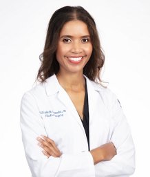 Kiwanuka Elizabeth合作论文数Brigham and Women's Hospital, Harvard Medical School1Johan P.E. Junker合作论文数Center for Tissue Engineering, University Hospital of Linköping1Charlotte A. Smith合作论文数1Martin C. Robson合作论文数1
 Omar Yusef Kudsi合作论文数Department of Surgery, Lankenau Hospital and Institute for Medical Research1
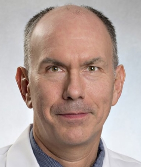 Scott R. Granter合作论文数Harvard Medical School;Dana-Farber Cancer Institute;Brigham and Women's Hospital1Usha Govindarajulu合作论文数Department of Medicine, Brigham & Women's Hospital, 75 Francis Street, Boston, MA 02115, United States1




