Parkinson's disease (PD) is a neurodegenerative disease that is prevalent in the elderly population. At present, the diagnostic method for PD mainly relies on the clinical experience of doctors, which is limited by its locality and subjectivity. Clinical multimodal data can provide different perspectives to understand patients and can ef fectively improve the accuracy of diagnosis. However, noise interference, high-dimensional feature complexity and category imbalance in multimodal data challenge PD diagnosis. Therefore, to solve the above problems, we propose a multimodal flexible subspace learning framework for PD diagnosis and clinical score regression, which learns the key features from multimodal data. Specifically, first, we provide diversified information by fusing PD neuroimaging data(DTI and MRI) and genetic data. Secondly, the self-representation learning method is used to dynamically capture sample similarities, thereby effectively mining the category information between samples. Finally, the l2,0 norm is used for dynamic feature selection, and the relevant features are enhanced by utilizing the similarity between features, while irrelevant and redundant features are excluded. In this way, the intrinsic struc tural properties of the data are learned, and then the classification and regression tasks are performed through Support Vector Machine(SVM). Our extensive experiments on the Parkinson's Progression Markers Initiative (PPMI) dataset demonstrate that this algorithm significantly improves both PD classification(94.07% accuracy for PD vs. NC at baseline) and clinical score regression performance(1.10 Mean Absolute Error for GDS prediction at baseline).
Accurate diagnosis of Parkinson's disease (PD) remains challenging due to heterogeneous symptoms and subtle neuroanatomical changes. Although multimodal Magnetic Resonance Imaging (MRI) provides complementary information, existing methods often fail to effectively model cross-modal interactions and brain anatomical connectivity. We propose a cross-modal graph attention network with self-supervised contrastive pretraining for PD diagnosis. Contrastive learning extracts robust volumetric representations from limited data, while a bidirectional cross-modal attention module enables deep interaction between T1-weighted MRI and quantitative susceptibility mapping features. An anatomically-informed graph attention network further models inter-regional dependencies to enhance diagnostic reliability and interpretability. Experiments on a hospital cohort demonstrate superior performance over representative methods.
Multi-object tracking (MOT) in satellite video is fundamentally limited by low target observability: genuine targets often produce weak and unstable responses, while structured backgrounds can generate persistent target-like interference, leading to missed detections, fragmented trajectories, and identity switches. To address this ambiguity, we propose a weak-observation-aware and reliability-guided framework with a layered two-stage design: the front end enhances weak observations over short temporal windows, whereas the back end controls their use for long-term trajectory association and state updating according to their reliability. The front-end Temporal Prior Module (TPM) constructs a structure-aware temporal prior from motion-aligned historical evidence, strengthening weak-target responses while limiting the propagation of structured-background interference. The back-end Satellite Weak-Observation Reliability-Guided Tracker (SWRT) introduces active-track support into short-window reasoning and combines current, short-term, and long-term evidence into a candidate-reliability score that regulates association eligibility and state-update strength. On VISO, the proposed method achieves a multiple object tracking accuracy (MOTA) of 72.5% and an identity F1 score (IDF1) of 81.1%. On SATMTB-MOT, it achieves the best MOTA and IDF1 for airplanes and vehicles and remains competitive for ships among the compared methods. These results demonstrate that this layered design achieves a better balance between target recovery and identity preservation under low-observability conditions.
Alzheimer's disease (AD) is a neurodegenerative disorder characterized by progressive cognitive decline and brain atrophy, where early diagnosis is critical. But existing multimodal methods integrating structural magnetic resonance imaging (sMRI) and clinical metadata often fail to align heterogeneous semantic levels effectively. To address this, we propose a CLIP-guided cross-modal dual-alignment framework (CGDA) for early AD diagnosis. First, CGDA employs a fine-tuned CLIP model to extract 2D sMRI features and textual embeddings, establishing general semantic alignment, then 3D sMRI encoder captures spatial context, while a text module encodes clinical metadata. Secondly, a dualalignment mechanism refines cross-modal correspondence by performing two critical alignments: 1) spatially aligning 3D anatomical features with metadata, 2) semantically fusing CLIP-derived features with 3D data for medical-specific contextualization. Finally, a dual cross-attention module enables dynamic fusion for robust diagnosis. Experiments on ADNI show CGDA achieves competitive performance in early AD diagnosis, demonstrating its effectiveness in cross-modal medical representation learning.
Alzheimer's disease (AD) is an irreversible neurodegenerative disorder where early diagnosis serves as the only viable window for effective intervention. While Structural MRI (sMRI) is a primary clinical tool for this purpose, manual diagnosis is heavily constrained by clinician expertise and the difficulty of visually quantifying subtle, diffuse anatomical changes. Deep learning has emerged as a powerful solution to capture these complex patterns. However, the inherent black-box opacity of current models creates a trust deficit that severely impedes their clinical adoption. Furthermore, existing interpretability methods predominantly rely on post-hoc visualizations, which frequently suffer from significant background leakage and lack anatomical coherence. To overcome these barriers, we propose a novel multi-level interpretable framework that inherently integrates diagnostic reasoning into the model architecture. The framework operates through a progressive three-stage trajectory to bridge the gap between pixel-level signals and global pathology. First, to eliminate the ambiguity caused by background leakage, we introduce a voxel discovery module implemented via Hierarchical Evidential Masking (HiEM). By leveraging this iterative optimization strategy, the model actively filters out irrelevant background signals to autonomously discover AD-relevant voxel sets. Subsequently, to bridge the semantic gap between discrete voxels and clinical anatomy, these discovered signals are dynamically aggregated into coherent semantic regions. Finally, recognizing that AD pathology propagates through brain networks, we explicitly model the regional interaction among these defined areas using Graph Attention Network (GAT). This voxel-region-network progression allows the model to transparently zoom out from local cues to global interactions. Validated on the Alzheimer's Disease Neuroimaging Initiative (ADNI) and Australian Imaging, Biomarkers and Lifestyle Study (AIBL) datasets, our method achieves performance comparable to state-of-the-art black-box models while autonomously reconstructing established neuropathological trajectories. Crucially, by extracting high-fidelity pathological signals, it offers a trustworthy tool for precise disease diagnosis and potentially serves as a digital biomarker discovery instrument for localizing subtle prodromal lesions. The source code is publicly available at: https://github.com/k-Jayus/HiEM.
Epilepsy is a chronic neurological disorder characterized by recurrent unprovoked seizures, affects over 50 million people worldwide, and poses significant risks, including sudden unexpected death in epilepsy (SUDEP). Conventional unimodal approaches, primarily reliant on electroencephalography (EEG), face several key challenges, including low SNR, nonstationarity, inter- and intrapatient heterogeneity, portability, and real-time applicability in clinical settings. To address these issues, a comprehensive survey highlights the concept of advanced multimodal learning for epileptic seizure detection and prediction (AMLSDP). The survey presents the evolution of epileptic seizure detection (ESD) and prediction (ESP) technologies across different eras. The survey also explores the core challenges of multimodal and non-EEG-based ESD and ESP. To overcome the key challenges of the multimodal system, the survey introduces the advanced processing strategies for efficient AMLSDP. Furthermore, this survey highlights future directions for researchers and practitioners. We believe this work will advance neurotechnology toward wearable and imaging-based solutions for epilepsy monitoring, serving as a valuable resource for future innovations in this domain.
Ventricular septal defect (VSD) can be effectively identified by extracting anatomical structures and blood-flow features through multi-scale analysis of multi-modal ultrasound images. However, existing automated identification methods mainly focus on the interaction of multi-scale features, while ignoring the positive impact of hierarchical image differences on identification performance. Moreover, static multi-scale feature extraction strategies within such methods limit the network’s flexibility in modeling features of different frequencies. In this paper, we propose a hierarchical feature-guided dynamic collaborative learning Transformer model for VSD identification. Specifically, we first propose a hierarchical feature extraction unit that sequentially extracts basic- and detailed-layer features from the multi-modal ultrasound images. Subsequently, a hierarchical feature-guided dynamic collaborative learning Transformer block (HDC_Transformer) is constructed. This block performs linear operations while enhancing local anatomical information by using detailed features to guide low-scale features. Simultaneously, a basic-layer feature is used to guide high-scale features for supplementing global information. Moreover, considering that low-scale and high-scale features mainly capture high-frequency and low-frequency information, respectively, the HDC_Transformer block utilizes a dynamic learning mechanism to achieve an effective balance between high- and low-frequency modeling across different feature extraction layers. Extensive experiments on both public and private datasets demonstrate the superior performance of the proposed method across various identification tasks and offer new insights into VSD identification. Our code is available at: https://github.com/Zhaocheng1/HDC_Transformer.
Alzheimer’s disease (AD) is a prevalent neurodegenerative disorder where early diagnosis is pivotal for effective intervention, yet it is hindered by subtle pathological feature differences among AD subtypes and severe class imbalance in medical imaging datasets. Existing structural magnetic resonance imaging (sMRI) and resting-state functional MRI (rs-fMRI) multimodal fusion methods for AD diagnosis mostly adopt simple concatenation or summation without fine-grained cross-modal alignment and interaction. To address these issues, we propose a Deep Cross-Branch Multi-Modal Fusion Network (DCMFNet) for early AD diagnosis. We first preprocess sMRI and rs-fMRI to extract ROI-based features, then perform dimension unification and normalization to realize cross-modal feature alignment. A novel Deep Cross-branch Multi-modal Feature Fusion (DCMF) module with three parallel branches and a dual-pathway cross-modal branch is designed to fully mine complementary and correlated cross-modal information, and the fused features are input into a Transformer encoder for classification. Moreover, we introduce the Logit Adjustment Cross-Entropy (LACE) loss to mitigate class imbalance by correcting decision boundaries based on class prior probabilities, enhancing the recognition of minor classes. The model is evaluated on a private clinical dataset. Experimental results show that DCMFNet outperforms traditional machine learning methods and state-of-the-art deep learning models in six binary AD subtype classification tasks, with the LACE loss and DCMF module effectively alleviating class imbalance and improving cross-modal feature representation. This work provides a reliable multimodal fusion framework for early AD diagnosis and reduces the diagnostic burden on healthcare professionals.
Recent advancements in attention-based Siamese trackers have led to significant improvements in performance. However, tracking efficiency remains a crucial metric that cannot be overlooked in practical applications. This paper analyses the characteristics of current attention mechanisms and Siamese frameworks. Based on these analyses, we propose a more efficient attention-based tracking algorithm, named MCAT. First, the construction of global feature dependencies, which is crucial for the functionality of attention mechanisms, always involves extensive computation of numerous insignificant dependencies. To address this issue, we propose a progressive attention mechanism LGPA. This method can effectively reduce unnecessary computations by progressively building feature dependencies from local to global levels under the guidance of spatial and channel distribution information. Additionally, despite keypoint-based tracking strategy has demonstrated significant potential in the research of Siamese frameworks, there is still room for improvement in the efficiency of multi-scale information fusion and utilization, which is crucial for improving the robustness of tracking models under complex scenarios. For this issue, we propose a multi-scale pixelwise cross-correlation method and a novel corner head to better mine and integrate multi-scale information. By embedding the proposed methods, we achieve substantial accuracy improvements with minimal computational overhead. With different backbones, we propose two end-to-end Siamese trackers: a fully convolutional tiny version (MCAT) and a transformer-based version (MCAT-swin). Extensive experiments across multiple benchmarks show that our trackers achieve state-of-the-art performance with high efficiency. On the long-term tracking benchmark LaSOT, MCAT-swin achieves an AUC score of 68.1% and a precision score of 72.5%, while running at 140FPS. The running speed of MCAT even exceeds 220FPS.
In neuroimaging-based Alzheimer's Disease (AD) diagnosis, effectively integrating structural MRI (sMRI) and resting-state functional MRI (rs-fMRI) data while preserving clinical interpretability remains a significant challenge. To address this issue, we propose a novel transformer-based framework that unifies heterogeneous imaging features into coherent region-level representations. Our approach uniquely leverages prior anatomical knowledge to guide attention toward AD-relevant regions while employing a learnable mapping mechanism that transforms sMRI spatial features into biologically meaningful regional representations. We implement a consistency constraint to ensure optimal alignment between structural and functional coupling across modalities, followed by a Bayesian fusion strategy to integrate these aligned multi-modal features. Through comprehensive evaluation on the ADNI dataset, our method demonstrates not only superior diagnostic accuracy compared to existing state-of-the-art approaches but also provides clinically interpretable insights into AD-related brain connectivity patterns. This work represents a significant advancement in multi-modal neuroimaging analysis for AD diagnosis, successfully combining enhanced diagnostic performance with clinical interpretability.
Alzheimer’s disease (AD) is a common neurodegenerative disorder. Early and accurate diagnosis of AD is essential for effective treatment. However, due to the class imbalance problem, there is a significant data gap between different categories. Moreover,the data feature differences of AD are relatively small, which poses challenges for its application in the early diagnosis of AD. To tackle these problems, we propose an intelligent early AD diagnosis model based on Transformer. The deep learning diagnosis model utilizes Transformer to integrate image features and non-image features. Furthermore, it incorporates a class imbalance loss function to optimize the performance of early AD diagnosis, thereby enhancing the model’s ability to recognize underrepresented classes. In order to alleviate the problem of class imbalance and test the model performance, we used stratified 5-fold cross validation to verify the model effect.Experimental results demonstrate that our model can significantly improve the accuracy of AD diagnosis, which is markedly better than traditional methods. Additionally, loss function we used more effectively mitigates the problem of class imbalance. We believe this work can effectively reduce the burden on medical staff to diagnose early AD.
Satellite video has revolutionized Earth observation, offering unprecedented capabilities for automated object tracking. However, multiobject tracking (MOT) in satellite videos remains highly challenging due to complex backgrounds, extremely low signal-to-noise ratios (SNR), and subpixel-scale targets. Although existing MOT methods achieve satisfactory performance under conventional conditions, their effectiveness declines significantly when applied to satellite imagery. To address these challenges, we propose a novel MOT framework tailored specifically for remote sensing videos, termed the multiscale collaborative attention network (MCANet), which explicitly incorporates temporal and spatial contextual information to enhance tracking performance. Specifically, we design a collaborative frame enhancement module, which improves small-scale target detection by utilizing cross-frame spatio-temporal feature compensation. This module robustly suppresses background interference and enhances weak target features in low-SNR environments. In addition, we propose a multiscale temporal attention motion capture module that integrates multilayer spatio-temporal attention mechanisms to effectively capture complex motion patterns, significantly improving target association and trajectory continuity under low temporal resolution conditions. Extensive experiments on representative satellite imagery datasets confirm that MCANet substantially outperforms existing state-of-the-art methods, achieving a MOTA score of 69.1% and an IDF1 score of 77.6% on the VISO dataset. These results highlight the effectiveness, robustness, and practical applicability of MCANet in remote sensing MOT scenarios.
Siamese network trackers are a prominent paradigm in visual object tracking due to efficient similarity learning. However, most Siamese trackers are restricted to the bounding box tracking format, which often fails to accurately describe the appearance of non-rigid targets with complex deformations. Additionally, since the bounding box frequently includes excessive background pixels, trackers are sensitive to similar distractors. To address these issues, we propose a novel segmentation-assisted model that learns binary mask representations of targets. This model is generic and can be seamlessly integrated into various Siamese frameworks, enabling pixel-wise segmentation tracking instead of the suboptimal bounding box tracking. Specifically, our model features two core components: (i) a multi-stage precise mask representation module composed of cascaded U-Net decoders, designed to predict segmentation masks of targets, and (ii) a saliency localization head based on the Euclidean model, which extracts spatial position constraints to boost the decoder’s discriminative capability. Extensive experiments on five tracking benchmarks demonstrate that our method effectively improves the performance of both anchor-based and anchor-free Siamese trackers. Notably, on GOT-10k, our method increases the AO scores of the baseline trackers SiamRPN++ (anchor-based) and SiamBAN (anchor-free) by 5.2% and 7.5%, respectively while maintaining speeds exceeding 60 FPS.
Satellite video has revolutionized Earth observation, offering unprecedented capabilities for automated object tracking. However, multi-object tracking (MOT) in satellite videos remains highly challenging due to complex backgrounds, extremely low signal-to-noise ratios (SNR), and sub-pixel-scale targets. Although existing MOT methods achieve satisfactory performance under conventional conditions, their effectiveness declines significantly when applied to satellite imagery. To address these challenges, we propose a novel MOT framework tailored specifically for remote sensing videos, termed the Multi-scale Collaborative Attention Network (MCANet), which explicitly incorporates temporal and spatial contextual information to enhance tracking performance. Specifically, we design a Collaborative Frame Enhancement Module (CFEM), which improves small-scale target detection by utilizing cross-frame spatiotemporal feature compensation. This module robustly suppresses background interference and enhances weak target features in low-SNR environments. Additionally, we propose a Multi-scale Temporal Attention Motion Capture Module (MTAMC) that integrates multi-layer spatiotemporal attention mechanisms to effectively capture complex motion patterns, significantly improving target association and trajectory continuity under low temporal resolution conditions. Extensive experiments on representative satellite imagery datasets confirm that MCANet substantially outperforms existing state-of-the-art methods, achieving a MOTA score of 69.1% and an IDF1 score of 77.6% on the VISO dataset. These results highlight the effectiveness, robustness, and practical applicability of MCANet in remote sensing MOT scenarios.
Alzheimer’s disease (AD) is an incurable, progressive neurodegenerative disease, and its early diagnosis is essential. Previous studies have demonstrated the superiority of utilizing multi-site data to train models for diagnosing AD. However, existing research models mostly use dimensionally reduced image data for model training, which loses the complete spatial information of the image. At the same time, the data from different sites are heterogeneous, and the joint training models often perform poorly when facing data from new sites. Traditional domain adaptation methods require centralized data for training, which results in the leakage of medical privacy. Our study proposes a multi-site federated learning model (FedSADA), which uses a 3D Vision Transformer as the basic framework to learn the spatial information of the complete image fully. We also added a self-attention domain adaptive loss function and a local maximum mean difference loss function to perform domain adaptation on multi-site data. At the same time, a federated learning framework is adopted to protect medical data fully. We conducted experiments on public datasets, demonstrating that our method can effectively improve model generalization and accuracy on new data sites.
As increased nuchal translucency (NT) thickness is notably associated with fetal chromosomal abnormalities, structural defects, and genetic syndromes, accurate measurement of NT thickness is crucial for the screening of fetal abnormalities during the first trimester. We aimed to develop a model for quality assessment of ultrasound images for precise measurement of fetal NT thickness. We collected 2140 ultrasound images of midsagittal sections of the fetal face between 11 and 14 weeks of gestation. Several image segmentation models were trained, and the one exhibiting the highest DSC and HD 95 was chosen to automatically segment the ROI. The radiomics features and deep transfer learning (DTL) features were extracted and selected to construct radiomics and DTL models. Feature screening was conducted using the t-test, Mann-Whitney U-test, Spearman’s rank correlation analysis, and LASSO. We also developed early fusion and late fusion models to integrate the advantages of radiomics and DTL models. The optimal model was compared with junior radiologists. We used SHapley Additive exPlanations (SHAP) to investigate the model’s interpretability. The DeepLabV3 ResNet achieved the best segmentation performance (DSC: 98.07 ± 0.02
Multimodal neuroimaging grounded in standardized brain atlases enables precise decoding of Alzheimer's progression by capturing both structural atrophy and functional decline across neural circuits. Current methods compromise anatomical fidelity in whole-brain modeling while generating biologically inconsistent cross-modal interactions. To address these dual challenges, we develop a graph learning framework that integrates three synergistic components: anatomically constrained feature extraction preserving region-specific biomarkers through spatial priors, channel-wise attention mechanisms for discriminative pattern refinement, and bidirectional cross-modal adaptation governed by alternating attention to enforce neuropathological consistency. This unified architecture processes sMRI and PET data through sequential stages of anatomical feature preservation, noise-robust feature enhancement, and dynamic modality fusion, ultimately mapping neurodegeneration patterns across scales. Evaluated on ADNI, our framework achieves superior classification accuracy while graph topology analysis reveals clinically significant hub reorganization within the default mode network, directly correlating with progressive connectivity deterioration. The method's capacity to reconcile localized biomarker specificity with systemic network dynamics establishes new standards for computational neuropathology.
In studies of Alzheimer's disease (AD), limited sample size considerably hampers the performance of intelligent diagnostic systems. Using multi-site data increases sample size but raises concerns regarding data privacy and inter-site heterogeneity. To address these issues, we developed a knowledge-guided federated graph attention learning network with a diffusion module to facilitate AD diagnosis from multi-site data. We used multiple templates to extract regions-of-interest (ROI)-based volume features from structural magnetic resonance imaging (sMRI) data. These volume features were then combined with previously identified AD features from published studies (prior knowledge) to determine the discriminative features within the images. We then designed an attention-guided diffusion module to synthesize samples by prioritizing these key features. The diffusion module was trained within a federated learning framework, which ensured inter-site data privacy while limiting data heterogeneity. Finally, we designed a federated graph attention learning network as a classifier to capture AD-related deep features and improve the accuracy of diagnosing AD. The efficacy of our approach was validated using three AD datasets. Thus, the classifier developed in this study represents a promising tool for optimizing multi-site neuroimaging data to improving the accuracy of diagnosing AD in the clinic.
Object tracking stands as a cornerstone challenge within computer vision, with blurriness analysis representing a burgeoning field of interest. Among the various forms of blur encountered in natural scenes, defocus blur remains significantly underexplored. To bridge this gap, this article introduces the Defocus Blur Video Object Tracking (DBVOT) dataset, specifically crafted to facilitate research in visual object tracking under defocus blur conditions. We conduct a comprehensive performance analysis of 18 state-of-the-art object tracking methods on this unique dataset. Additionally, we propose a selective deblurring framework based on Deblurring Auxiliary Learning Net (DID-Anet), innovatively designed to tackle the complexities of defocus blur. This framework integrates a novel defocus blurriness metric for the smart deblurring of video frames, thereby enhancing the efficacy of tracking methods in defocus blur scenarios. Our extensive experimental evaluations underscore the significant advancements in tracking accuracy achieved by incorporating our proposed framework with leading tracking technologies.
Recently, leveraging deep neural networks for automated colorectal polyp segmentation has emerged as a hot topic due to the favored advantages in evading the limitations of visual inspection, e.g., overwork and subjectivity. However, most existing methods do not pay enough attention to the uncertain areas of colonoscopy images and often provide unsatisfactory segmentation performance. In this paper, we propose a novel boundary uncertainty aware network (BUNet) for precise and robust colorectal polyp segmentation. Specifically, considering that polyps vary greatly in size and shape, we first adopt a pyramid vision transformer encoder to learn multi-scale feature representations. Then, a simple yet effective boundary exploration module (BEM) is proposed to explore boundary cues from the low-level features. To make the network focus on the ambiguous area where the prediction score is biased to neither the foreground nor the background, we further introduce a boundary uncertainty aware module (BUM) that explores error-prone regions from the high-level features with the assistance of boundary cues provided by the BEM. Through the top-down hybrid deep supervision, our BUNet implements coarse-to-fine polyp segmentation and finally localizes polyp regions precisely. Extensive experiments on five public datasets show that BUNet is superior to thirteen competing methods in terms of both effectiveness and generalization ability.
 Baiying Lei (雷柏英)合作论文数School of Biomedical Engineering, Shenzhen University Medical School14
 Tianfu Wang (汪天富)合作论文数Department of medicine, Shenzhen University12
 Lei Dou合作论文数8
 Qinghui Wang合作论文数5
 Haiyong Chen (陈海永)合作论文数School of Artificial Intelligence, Hebei University of Technology5
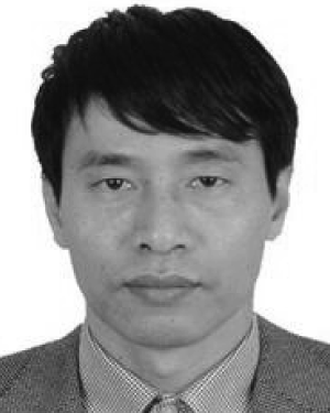 Xiaohua Xiao合作论文数Shenzhen Univ, Affiliated Hosp, Shenzhen, Peoples R China4
 Xuemin (Sherman) Shen (沈学民)合作论文数Department of Electrical and Computer Engineering, University of Waterloo4
 Kun Liu (刘坤)合作论文数School of Artificial Intelligence, Hebei University of Technology4
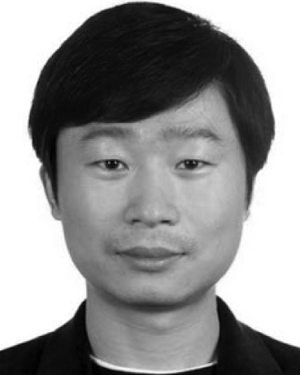 Feng Zhou合作论文数3
 Xuegang Song (宋雪刚)合作论文数School of Biomedical Engineering, Guangzhou Medical University3
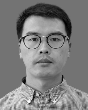 Jie Dou合作论文数3
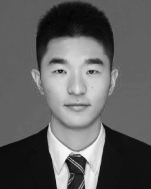 Cheng Zhao合作论文数Shenzhen Univ, Natl Reg Key Technol Engn Lab Med Ultrasound, Sch Biomed Engn,Hlth Sci Ctr, Guangdong Key Lab Biomed Measurements & Ultrasoun, Nanhai Ave 3688, Shenzhen 518060, Guangdong, Peoples R China3
 Wen Wu (吴文)合作论文数School of Electronic and Optical Engineering, Nanjing University of Science and Technology3
 Yongqiang Dong (董永强)合作论文数Key Laboratory of Computer Network and Information Integration, Ministry of Education, China, School of Artificial Intelligence, Southeast University;Future Network Research Center, Southeast University3
 Jun Liang合作论文数School of Engineering, Cardiff University3
 Wen-Long Ming合作论文数3Bin Cao合作论文数2
 Jiali Liu合作论文数2
 Guolin Ma (马国林)合作论文数China-Japan Friendship Hospital;Peking University Health Science Center;Peking Union Medical College;Capital Medical University2
 Guanghui Yue合作论文数2
 Jianwei Zhao (赵健伟)合作论文数School of Chemistry and Chemical Engineering, Nanjing University;Jiaxing University2
 Yu Gu (谷宇)合作论文数School of Biomedical Engineering, Capital Medical University2
 Dapeng Wu (吴大鹏)合作论文数School of Communications and Information Engineering, Chongqing University of Posts and Telecommunications1
 Qiufu Li (李秋富)合作论文数College of Computer Science and Software Engineering, Shenzhen University1
 Yanbiao Sun (孙岩标)合作论文数School of Precision Instrument and Opto-electronics Engineering, Tianjin University1Yuanyuan Gao合作论文数1Xuyuan Kang (康旭源)合作论文数1Cuimei Wei合作论文数1
 Aimei Li合作论文数1HAN Jiang-rui (韩江瑞)合作论文数1for the Alzheimer’s Disease Neuroimaging Initiative and the Alzheimer Disease Metabolomics Consortium合作论文数1Peipei Lv合作论文数1Wang Shilu合作论文数1
 Kai Meng合作论文数1Xi Wen (文熙)合作论文数1
 Jiuling Zhang合作论文数Institute of Software, University of Chinese Academy of Sciences1
 Wang Shuang合作论文数1Li Yanan合作论文数1Jing Qin合作论文数1
 Puning Zhang合作论文数Special Research Centre for Optical Internet and Wireless Information Networks, Chongqing University of Posts and Telecommunications1
 Xiaowei Zhang合作论文数1
 Shushan Si合作论文数1
 Huoyou Hu (胡火有)合作论文数1
 Aocai Yang合作论文数1
 Weiqing Yan (阎维青)合作论文数School of Computer and Control Engineering, Yantai University1Luming Li合作论文数1
 Ingo Lüdtke合作论文数1
 Zhang Gaowei合作论文数Hebei Univ Technol, Sch Artificial Intelligence, Tianjin 300130, Peoples R China1




