Peter Lawrence commemorates the work of developmental biologist Antonio García-Bellido, who revolutionised our understanding of the genetic basis of animal development.
This book is a thriller. And it is written for everyone, not just for boffins. It is especially for those interested in COVID-19, science itself, scientific research, cancer, biotechnology, business, economics and enterprise. And it reaches into some contemporary issues, such as the bureaucrat versus the scientist, regulation versus risk, state sponsorship versus private enterprise, translational versus blue sky research and chauvinism versus internationalism. The narrative itself recounts a scientific adventure story that ranks in excitement with the best of the Bond books but has one signal advantage: it is real. Here, our heroes, with the clock ticking, pitch the power of scientific knowledge and experiments, evidence and argument against the rapidly advancing scythe of death.
In this review we recall a number of important discoveries that took place in Drosophila during the seventies and eighties of the last century. The development of cell lineage methods and of powerful modifications of same, such as the Minute technique, led to the discovery of compartments and provided a clearer picture of the body organization: that came to be seen as a chain of metameric lineage units along the A/P body axis. Further, genetic screens allowed the identification of the genes involved in the establishment of the metameric scaffold — the segmentation genes— and also of Hox genes that are responsible for the specific development of individual body parts. As cloning methods became available, many of the most relevant of these developmental genes were cloned and a molecular analysis of development initiated. The discovery of the homeobox, a molecular mark of Hox and other relevant developmental genes, allowed the finding of Hox genes in animal species, like humans, in which they could not be identified by genetic methods. Analysis of the structure and function of Hox genes provided a general image of the genetic design of the metazoan body.
The slope of a supracellular molecular gradient has long been thought to orient and coordinate planar cell polarity (PCP). Here we demonstrate and measure that gradient. Dachsous (Ds) is a conserved and elemental molecule of PCP; Ds forms intercellular bridges with another cadherin molecule, Fat (Ft), an interaction modulated by the Golgi protein Four-jointed (Fj). Using genetic mosaics and tagged Ds, we measure Ds in vivo in membranes of individual cells over a whole metamere of the Drosophila abdomen. We find as follows. (i) A supracellular gradient rises from head to tail in the anterior compartment (A) and then falls in the posterior compartment (P). (ii) There is more Ds in the front than the rear membranes of all cells in the A compartment, except that compartment's most anterior and most posterior cells. There is more Ds in the rear than in the front membranes of all cells of the P compartment. (iii) The loss of Fj removes intracellular asymmetry anteriorly in the segment and reduces it elsewhere. Additional experiments show that Fj makes PCP more robust. Using Dachs (D) as a molecular indicator of polarity, we confirm that opposing gradients of PCP meet slightly out of register with compartment boundaries.
ABSTRACTWe investigate the mechanisms of planar cell polarity (PCP) in theDrosophilalarva. The epidermis displays an intricate pattern of polarity and is excellent for the study of one system of PCP, the Dachsous/Fat system; partly because the Starry Night/Frizzled system plays no discernable role in the larva. Measurements of the amount of Dachsous reveal a peak near the rear of the anterior compartment. Localisation of Dachs and orientation of ectopic denticles reveal the polarity of every cell in the segment. We discuss how well these findings evidence our gradient model of Dachsous activity. Several groups have proposed that Dachsous and Fat fix the direction of PCP via oriented microtubules that transport PCP proteins to one side of the cell. We test this proposition in the larval cells and find that most microtubules grow perpendicularly to the axis of PCP. We find no meaningful bias in the polarity of those microtubules aligned close to that axis. We also reexamine published data from the pupal abdomen and fail to find evidence supporting the hypothesis that microtubular orientation draws the arrow of PCP.
We investigate the mechanisms of planar cell polarity (PCP) in the Drosophila larva. The epidermis displays an intricate pattern of polarity and is excellent for the study of one system of PCP, the Dachsous/Fat system; partly because the Starry Night/Frizzled system plays no discernable role in the larva. Measurements of the amount of Dachsous reveal a peak near the rear of the anterior compartment. Localisation of Dachs and orientation of ectopic denticles reveal the polarity of every cell in the segment. We discuss how well these findings evidence our gradient model of Dachsous activity. Several groups have proposed that Dachsous and Fat fix the direction of PCP via oriented microtubules that transport PCP proteins to one side of the cell. We test this proposition in the larval cells and find that most microtubules grow perpendicularly to the axis of PCP. We find no meaningful bias in the polarity of those microtubules aligned close to that axis. We also reexamine published data from the pupal abdomen and fail to find evidence supporting the hypothesis that microtubular orientation draws the arrow of PCP.### Competing Interest StatementThe authors have declared no competing interest.
I first met Mike Berridge in Cambridge University’s Zoology Department in 1962 where we were both graduate students of Sir Vincent Wigglesworth (known as VBW). We remained good friends for the next 58 years. He became a great scientist — but why was he so successful? There are many ways to achieve scientific greatness: he was certainly not born great, nor did he have greatness thrust upon him. I don’t think he was unusually lucky, although he did certainly practise a great deal — I remember him going through his lectures again and again, timing, rehearsing — and we know that practice can bring what others call luck. In those salad days we students were free to choose our own project and encouraged to research it independently; if we looked usefully occupied, we were left to get on as best we could. Mike chose to work on urine formation in the cotton stainer bug (Dysdercus), chosen because it was easy to rear and was a pest in his homeland Zimbabwe. Looking back, one can see what an important apprenticeship that was. Later he turned to secretion and used the salivary glands of the blowfly (Calliphora). His collaborator Robin Irvine describes how phenomenally adept he became; he would take 50 or 60 of these glands that he had microdissected so deftly, each undamaged and arrange them nicely in a ring in a petri dish under oil, with the precision of Lalique. Each gland was placed in a drop of bespoke medium, then one end pulled out and pierced so that it released a tiny and growing droplet under the oil. Mike then serially collected these droplets with a micropipette until he had enough saliva for biochemical assays. Mike wasn’t a show-off, nor did he expropriate the work of his students and postdocs. He didn’t go in for any of the standard strategies that help one rise to the top, like hobnobbing with the powerful, or swapping and trading authorships. By their clarity, simplicity and great significance in cellular physiology, two of his papers — in which he revealed and discussed the role of inositol trisphosphate as a second messenger — found themselves in the top five of the citation list for all biomedicine for a whole decade (the 80s). His personality just did not fit with politics: he had integrity, he was modest, diffident and honest, but there was a determination, a persistence and a disciplined perfectionism. I can see VBW in his character and I wonder if Mike consciously or subconsciously modelled himself on his supervisor. But I think Mike’s similarity to VBW in his quiet professionalism and gentility lay in their natures; they were alike intrinsically, genetically if you wish. Mike and I often discussed VBW and what he taught us through his example. VBW rarely had his name on his student’s papers and indeed not on any of ours; his was a bygone era where scientific papers were authored by those who did the work, had the ideas and wrote them. We admired his scientific longevity, his independence and his quiet determination. We learnt from him to believe in the precedence of experiment over theory as well as the value of scholarship. Perhaps Mike did see him as a role model, but I think this whole concept can be dangerous. If one tries to model one’s career on the style of another, or aim to achieve as another has done, then one has to check that characters and abilities match, as with Mike and VBW, or it can be a self-harming ambition. I think the benefit of role models has become oversold; we are all of us so very different. Mike’s career illustrates, as so often with those who make fundamental discoveries, how long in science it takes to learn things well and deep enough to find a way through the hype, fashion and the maze of sidetracks to ask the right questions. Of course it takes even longer to produce concrete impacts on society or medicine. Think how long it has taken for us to begin to benefit fully from the discovery of DNA, its function and its structure. For no good reason, this truth, so endlessly repeated by scientists, does not seem to come home to those who design scientific policy or even grant applications. As Mike and I met on the golf course once a fortnight or so for over 50 years, or when we met in our homes, or when we went together on family holidays, many times to Africa, one issue came up again and again: how fortunate we both were to have had core support for most of our scientific lives and how vital it had been for his discoveries with their important long-term implications for cell physiology and medicine. Robin Irvine remembers this too: oftentimes when he and Mike were confronted by a baffling series of results, Mike would say “it’s a good thing we don’t have to write a grant to do this stuff”. We all expected that Mike would be awarded the Nobel Prize, probably with Yasutomi Nishizuka: would we have viewed his career and achievements differently if he had been? Probably, but actually his achievements would have been the same, almost as well known and just as effective. Mike was a sporting and competitive person, this came out in his fascination with games and his love of keeping records of everything. There was nearly always a twinkle, nearly always a funny side. Every Christmas we received a special compendium of statistics and stick drawings that summarised the year’s events on the golf course (see cartoon). He had a wry sense of observation and a perceptive but friendly sense of humour. VBW must have been very proud of him. Golfing friends: An example of one of Mike’s annual cartoons, this one summarising 2013’s events on the golf course.
We investigate planar cell polarity (PCP) in the Drosophila larval epidermis. The intricate pattern of denticles depends on only one system of PCP, the Dachsous/Fat system. Dachsous molecules in one cell bind to Fat molecules in a neighbour cell to make intercellular bridges. The disposition and orientation of these Dachsous-Fat bridges allows each cell to compare two neighbours and point its denticles towards the neighbour with the most Dachsous. Measurements of the amount of Dachsous reveal a peak at the back of the anterior compartment of each segment. Localization of Dachs and orientation of ectopic denticles help reveal the polarity of every cell. We discuss whether these findings support our gradient model of Dachsous activity. Several groups have proposed that Dachsous and Fat fix the direction of PCP via oriented microtubules that transport PCP proteins to one side of the cell. We test this proposition in the larval cells and find that most microtubules grow perpendicularly to the axis of PCP. We find no meaningful bias in the polarity of microtubules aligned close to that axis. We also reexamine published data from the pupal abdomen and find no evidence supporting the hypothesis that microtubular orientation draws the arrow of PCP.
Summary: Peter Lawrence reflects on the life and work of Sydney Brenner – ‘the Oscar Wilde of science’.
Our aim in this short Primer is to explain the principles of planar cell polarity (PCP) in animal development. The literature in this small field is complex and specialized, but we have extracted a simple and central story from it. We explain our hypothesis that polarity, initially cued by the direction of slope of a multicellular gradient, is interpreted at the cellular level so that each cell becomes molecularly polarised. The mechanism involves a comparison between a cell and its neighbours. To achieve this comparison there are (at least) two disparate and independent molecular systems, each depending on molecular bridges that span between neighbouring cells. Even though the two systems are made up of different molecules, we argue that both systems function in a logically equivalent way.
We respond to a recent report by Abbasi and Marcus who present two main findings: first they argue that there is an organiser and a compartment boundary within the posterior compartment of the butterfly wing. Second, they present evidence for a previously undiscovered lineage boundary near wing vein 5 in Drosophila , a boundary that delineates a "far posterior" compartment. Clones of cells were marked with the yellow mutation and they reported that these clones always fail to cross a line close to vein 5 on the Drosophila wing. In our hands yellow proved an unusable marker for clones in the wing blade and therefore we reexamined the matter. We marked clones of cells with multiple wing hairs or forked and found a substantial proportion of these clones cross the proposed lineage boundary near vein 5, in conflict with their findings and conclusion. As internal controls we showed that these same clones respect the other two well established compartment boundaries: the anteroposterior compartment boundary is always respected. The dorsoventral boundary is mostly respected, and is crossed only by clones that are induced early in development, consistent with many reports. We question the validity of Abbasi and Marcus' conclusions regarding the butterfly wing but present no new data. Arising from: R. Abbasi and J. M. Marcus Sci. Rep. 7, 16337 (2017); https://doi.org/10.1038/s41598-017-16553-5 .
Epithelial cells are polarised within the plane of the epithelium, forming oriented structures that have a coordinated and consistent polarity (planar cell polarity, PCP). In Drosophila, at least two separate molecular systems generate and interpret intercellular polarity signals: Dachsous/Fat, and the 'core' or Starry night/Frizzled system. Here, we study the prickle gene and its protein products Prickle and Spiny leg. Much research on PCP has focused on the asymmetric localisation of core proteins in the cell and as a result prickle was placed in the heart of the Starry night/Frizzled system. We investigate whether this view is correct and how the prickle gene relates to the two systems. We find that prickle can affect, separately, both systems; however, neither Prickle nor Spiny leg are essential components of the Dachsous/Fat or the Starry night/Frizzled system, nor do they act as a functional link between the two systems.
INTRODUCTION The study. of abnormal development is consider.ed by the group to be a method for discovering th~ rules that govern normal development. We divide our topic into several subtopics: (a)the use of genetic methods; ('b) the role of cell lineage in the development of the nervous system; (c) the role of abnormal activity in afferent systems; and· (d) .a comparison of .structural with functional changes that might provide evidence for "suppressed synapses".
Many, if not all, epithelial cells are polarised within the plane of the epithelium and some form oriented structures whose coordinated and consistent polarity (planar cell polarity, PCP) relates to the principal axes of the body or organ. PCP depends on intercellular communication of polarity signals; in Drosophila at least two separate molecular systems generate these signals: Dachsous/Fat, Ds/Ft and the core or Stan system and both are conserved widely (reviewed in Butler and Wallingford, 2017). Here we make a new attempt to understand the PCP gene prickle ( pk ) and its products Pk and Sple. Much research on PCP has asked how and why many PCP proteins, including Pk and Sple, are asymmetrically localised in the cell (Strutt and Strutt, 2009). This question led to the pk gene being placed at the heart of the core or Stan system (Tree et al., 2002b). Here we use direct genetic tests to ask if this view is correct and if and how the pk gene relates to the Stan and the Ds/Ft systems. We conclude that Pk and Sple have been widely misunderstood: we find they can affect, separately, the Ds/Ft system (reversing, or rectifying the polarity of its output) and the core or Stan system (being required for the asymmetrical distribution of its proteins). In the Stan system they appear to work via binding to Vang. Neither Pk nor Sple are essential components of either the Ds/Ft or the Stan systems nor do they act as a bridge between the two systems.
 Jose Casal-Jimenez合作论文数Department of Zoology, University of Cambridge33
 Morata Ginés合作论文数the fruitfly Drosophila19
 P Johnston合作论文数17
 Gary Struhl合作论文数Struhl Lab, Zuckerman Institute, Columbia University;Department of Genetics and Development, Columbia University17
 Alfonso Martinez Arias合作论文数Department of Genetics, University of Cambridge17Jim Smith合作论文数15Lewis Wolpert合作论文数15
 Andrew Lumsden合作论文数MRC Centre for Developmental Neurobiology,King's College London14
 Elizabeth Robertson合作论文数Sir William Dunn School of Pathology, University of Oxford;Oxford Stem Cell Institute, University of Oxford13
 Cheryll Anne Tickle合作论文数University of Bath11
 Jean-Paul Vincent合作论文数Epithelial Cell Interactions Laboratory, Francis Crick Institute9
 Caroline Cecile Gabrielle Fabre合作论文数Department of Zoology, University of Cambridge6Saavedra Pedro合作论文数5Steven M Green合作论文数5Stefano Pietra合作论文数4
 Michael Ashburner合作论文数Department of Genetics, University of Cambridge;Churchill College, University of Cambridge4
 Elliot Meyerowitz合作论文数Division of Biology and Biological Engineering, California Institute of Technology4Cheryll Tickle合作论文数3Amy L. Brittle合作论文数3
 Ada Repiso合作论文数Institut d'Investigacions Biomèdiques August Pi i Sunyer (IDIBAPS),, Universitat de Barcelona3Javier Sampedro Molinuevo合作论文数Universidad Politecnica de Madrid, Univ. Politécnica de Madrid3
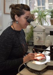 Isabel M. Palacios合作论文数Wellcome/CRC Institute, University of Cambridge3Anne Warner合作论文数2Danny L. Brower合作论文数2Beatriz Ibáñez-Jiménez合作论文数2Rosa Beddington合作论文数2P Simpson合作论文数2Pmj Shelton合作论文数2Jeremy Brockes合作论文数2
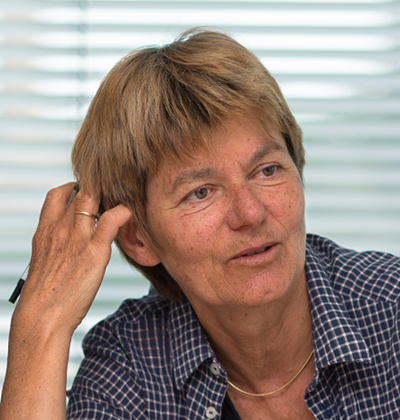 Mariann Bienz合作论文数MRC Laboratory of Molecular Biology2David Strutt合作论文数School of Biosciences, Faculty of Science, The University of Sheffield2
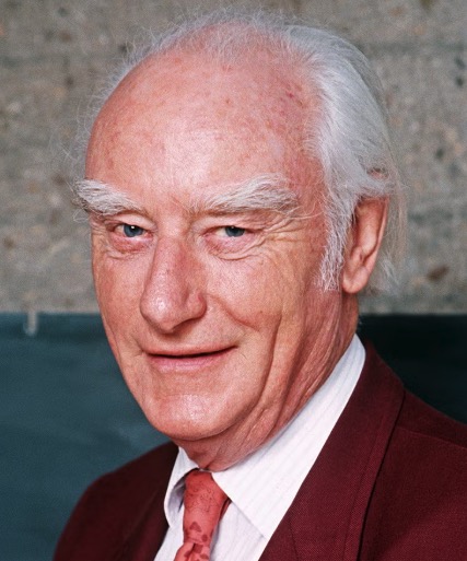 Francis Crick合作论文数Salk Institute for Biological Studies2
 David Wright合作论文数Chesapeake Biological Laboratory, Center for Environmental Science, University of Maryland2
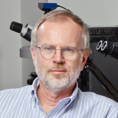 Roeland Nusse合作论文数Department of Developmental Biology, School of Medicine, Stanford University;Institute for Stem Cell Biology and Regenerative Medicine, School of Medicine, Stanford University;Howard Hughes Medical Institute2
 Mark S. Bretscher合作论文数MRC Laboratory of Molecular Biology, Cambridge2
 Ginés Morata合作论文数Autonomous University of Madrid2Gunther Stent合作论文数University of California, Berkeley1HW SAUER合作论文数1Miguel Rovira合作论文数1C V Cabrera合作论文数1Sara Pasalodos-Sanchez合作论文数1MC ALONSO合作论文数1KangBo Ng合作论文数1Jc Gerhart合作论文数1Matt Ridley合作论文数1
 Héctor Herranz合作论文数Department of Cellular and Molecular Medicine, Faculty of Health and Medical Sciences, University of Copenhagen1M Calleja合作论文数1J Brockes合作论文数1T Kornberg合作论文数1




