Abstract Background Leptomeningeal disease (LMD) from breast cancer (BC) is a rare and fatal metastasis to the meninges, with a median survival of 2–6 months and no effective cure. Our ongoing first-in-human Phase 1 trial of intrathecal peptide-pulsed dendritic cell (IT cDC1) therapy (NCT05809752) has demonstrated encouraging clinical benefit, with median overall survival exceeding 19 months as of March 2026. Transcriptomic analysis of cerebrospinal fluid (CSF) from the first six patients revealed robust adaptive immune activation, including increased effector and central memory T cells and memory B cells, consistent with findings in murine BC LMD models. Clonal expansion of T cell receptors and B cell receptors was also observed in mice, suggesting a potential cancer-preventive immune mechanism. Methods BC LMD–cured and systemic BC–cured mice were generated using cDC1 therapy and subjected to LMD or systemic BC rechallenge. Survival was assessed, and tissue and CSF samples were collected for downstream analyses. Results In the HER2+ LMD rechallenge model, IT cDC1–cured mice showed significantly improved survival compared with untreated LMD controls (P < 0.0005), with 61% rejecting tumor engraftment. No survival difference was observed in systemic BC rechallenge; however, 40% of mice exhibited slowed tumor growth in the mammary fat pad. Notably, 90% of systemic BC–cured mice rejected LMD growth. Functional assays demonstrated IFN-γ responses in lymphocytes from cervical tumor-draining lymph nodes following HER2/HER3 peptide stimulation, indicating adaptive immune memory. Transcriptomic analysis revealed increased T and B cell populations and enhanced NKT cell signaling in LMD rechallenge CSF, suggesting a target for combination therapy. Conclusions IT cDC1 therapy induces adaptive immune memory that may protect against LMD recurrence. Ongoing studies aim to define underlying mechanisms and optimize combination strategies, including HER2/HER3-targeted and α-GalCer–loaded cDC1s. Phase 1 and Phase 2 clinical trials are underway.
TPS1140 Background: Patients with triple negative breast cancer (TNBC) are at a high risk to develop brain metastases. The prognosis and quality of life for triple negative brain-metastatic breast cancer remains very poor. Treatment options are limited and improvement in efficacy is needed. Sacituzumab govitecan (SG) has shown blood brain barrier (BBB) penetration in preclinical and clinical settings with anti-tumor activity in the phase III ASCENT Trial (NCT02574455). Radiotherapy can open the BBB and has been shown to be safe in combination with anti-PD-1 therapy in our previous trial (NCT03807765). Therefore, combining radiation with the anti-PD-1 monoclonal antibody, zimberelimab, and SG may provide a synergistic anti-tumor approach. We hypothesize the use of SG and zimberelimab with stereotactic radiosurgery (SRS) among patients with metastatic TNBC with brain metastases will be safe and improve progression free survival compared to treatment with SG alone. Methods: The study is designed as a single-arm, nonrandomized, open-label, phase I/II trial of SG and zimberelimab with SRS among patients with metastatic TNBC with brain metastases. The primary endpoint will be neurologic toxicity defined by CTCAE v5 criteria (phase I) and 12 month PFS (phase II). TNBC patients ≥ 18 years old with ≤ 15 brain metastases with at least one measurable lesion ≥ 0.5 cm per RANO-BM criteria will be enrolled. Treatment will be initiated with SRS followed 1 week later by SG on days 1 and 8 (10 mg/kg) with zimberelimab on day 1 (360 mg IV) repeated every 3 weeks. Follow-up imaging response assessments will be conducted at q9 week intervals. An interim analysis will be performed after 21 patients are enrolled. An additional 10 patients will be enrolled if interim futility criteria are not met. Clinical trial information: NCT06238921 .
Abstract Leptomeningeal disease (LMD) is one of the most devastating complications of advanced cancer, resulting in severe neurological deterioration and poor survival outcomes. Despite this, very little is known about the mechanisms responsible for the exceedingly poor prognosis and severely impaired neurological function of these patients. Here, we report that toxic branched-chain keto acids (BCKAs) accumulate in the cerebrospinal fluid of LMD patients and drive both immunosuppression and neurodegeneration, presenting a novel therapeutic target. Using single-cell transcriptomics and multi-omics profiling of patient specimens, we created a comprehensive LMD atlas that revealed two hallmarks of LMD: an immunosuppressive cellular microenvironment and an environment promoting neurodegeneration. Mechanistically, there is a profound BCKA-mediated disruption of both T cell function and neuronal integrity. BCKA exposure inhibits T cell viability, proliferation, energy metabolism, and effector cytokine release. Strikingly, BCKA-reducing therapy using sodium phenylbutyrate improves survival and neurological outcomes in preclinical models while enhancing the efficacy of CAR-T cell therapy in lymphoma LMD and immunecheckpoint inhibitors in melanoma LMD. Thus, BCKA accumulation is a central mediator of LMD progression and treatment resistance and provides an actionable therapeutic opportunity through drug repurposing. Importantly, our pan-cancer atlas of human LMD delivers a framework for understanding disease mechanisms and developing novel therapeutic strategies for this urgent unmet clinical need.
Background The CARING intervention is designed to help neuro-oncology caregivers recognize existing social support and improve coping. Caregivers received 8 weekly phone-based individual manualized sessions with a navigator. Our objective was to understand the impact of CARING navigation sessions on ratings of stress, frustration, in control, and hopefulness for each session and across the sessions. Methods In this secondary analysis of data from a randomized controlled trial of CARING, caregiver self-report emotional ratings before and after each of 8 navigator sessions were analyzed. Navigators asked caregivers to rate their level of stress, frustration, feeling in control, and feeling hopeful. Responses were documented in the navigation log. Paired t-tests were conducted on each emotional construct for each session. Generalized estimating equations (GEEs) analyzed within- and between-session change for each emotional construct. Results Seventy-three caregivers provided at least 1 emotional rating in a navigation session. There were significant improvements from pre- to post- for all sessions for both stress and frustration ratings (ts < -3.06, Ps < .004). The majority of sessions also had significant improvements for feeling hopeful and in control (5 of 8 sessions; all ts > 2.66, Ps <.02). Generalized estimating equation results mirror t-test findings for within-session ratings and show a significant decrease for feeling in control across sessions (b = -0.08, P = .035). Conclusions Our findings suggest emotional improvement for each navigation session, with more consistent change in negative than in positive ratings. Navigation may offer emotional regulation and coping tools for neuro-oncology caregivers.
Leptomeningeal disease (LMD), a devastating complication of metastatic cancers, has a median survival of weeks to a few months. LMD incidence is highest in melanoma (5-25%), carrying the worst prognosis. Adoptive cell therapy (ACT) using tumor-infiltrating lymphocytes (TILs) has demonstrated complete and durable responses in patients with advanced metastatic melanoma, including regression of brain metastases, and other solid tumors i.e., lung cancer. We are optimizing rapid expansion of CSF-TRT cells as a source of T cells for ACT. We aim to evaluate CSF-TRT cells and determine their tumor reactivity ex-vivo/in-vivo to develop a novel therapeutic approach. Cells from the CSF from melanoma (M-LMD) patients (n=26; via lumbar puncture/Ommaya) were plated following established TIL culture protocols (high-dose IL-2): 1) OKT3, 2) anti-4-1BB-agonist, 3) IL-7+IL-15+IL-21, 4) anti-CD3/CD28-T-activator, 5) anti-CD3/CD28/CD137-T-activator, 6) anti-CD3/CD28-T-expander. After 4-6 weeks, reactivity was assessed with HLA-matched or autologous tumor cells. We are investigating tumor-based genomic correlates with whole exome sequencing (WES) by interrogating tumor samples initially collected from extracranial disease, and response to therapy in vivo in our LMD mouse model. Similar studies are underway for other solid tumors including lung cancer. After initial culture in IL-2, 53.4% of samples showed increased cell yield with average 165.29-fold expansion. PreREP resulted in 37.6% CD8+ T cells. REP was completed with a 96.8% success rate (mean 589.87-fold expansion). Post-REP flow cytometry revealed expansion of CD4+ T cells. Additional cultures produced similar yields with reduced T cell input requirement and demonstrated the potential to enrich for CD8+ T cells. Out of five samples tested for reactivity to HLA-matched melanoma cell lines, three produced varying levels of IFN-y. Results from WES, in vivo experiments, and testing other solid tumors are ongoing with pending results. Results demonstrate successful expansion of T cells ex vivo from CSF in M-LMD, raising the potential to use autologous CSF-derived T-cells as therapeutic strategy for patients with LMD.
Leptomeningeal disease (LMD), a devastating complication of metastatic cancers, has a median survival of weeks to a few months. LMD incidence is highest in melanoma (5-25%), carrying the worst prognosis. Adoptive cell therapy (ACT) using tumor-infiltrating lymphocytes (TILs) has demonstrated complete and durable responses in patients with advanced metastatic melanoma, including regression of brain metastases. We are optimizing the rapid expansion of CSF-TRT cells as a potential source of T cells for ACT. We aim to evaluate CSF-TRT cells and determine their tumor reactivity ex vivo/in vivo to develop a novel therapeutic approach. Cells from the CSF from M-LMD patients (n=26; via lumbar puncture/Ommaya) were plated following established TIL culture protocols (high-dose IL-2): 1) OKT3, 2) anti-4-1BB-agonist, 3) IL-7+IL-15+IL-21, 4) anti-CD3/CD28-T-activator, 5) anti-CD3/CD28/CD137-T-activator, 6) anti-CD3/CD28-T-expander. After 4-6 weeks, reactivity was assessed with HLA-matched or autologous tumor cells. We are investigating tumor-based genomic correlates with whole exome sequencing (WES) by interrogating tumor samples initially collected from extracranial disease, and response to therapy in vivo in our LMD mouse model. After initial culture in IL-2, 53.4% of samples showed increased cell yield with an average 165.29-fold expansion. PreREP resulted in 37.6% CD8+ T cells. REP was completed with a 96.8% success rate (mean 589.87-fold expansion). Post-REP flow cytometry revealed expansion of CD4+ T cells. Additional cultures produced similar yields with reduced T cell input requirement and demonstrated the potential to enrich for CD8+ T cells. Out of five M-LMD samples tested for reactivity to HLA-matched melanoma cell lines, three produced varying levels of IFN-y. Results from WES and our in vivo experiments are ongoing with pending results. Results demonstrate successful expansion of T cells ex vivo from CSF in M-LMD, raising the potential to use autologous CSF-derived T cells as therapeutic strategy for patients with LMD.
Leptomeningeal disease (LMD) arises as a metastatic complication from tumor cells invading the leptomeninges and cerebrospinal fluid. Advances in targeted and immune therapies in the last decade have not translated to this disease and the prognosis for patients is dismal, with most surviving only weeks to months after the diagnosis is made. Melanoma is one of the most common etiologies of LMD, however specific developments for this complication still represent a huge gap in research. Recent clinical trials have been covering different delivery methods (systemic, intrathecal), different types of drugs (immune checkpoint inhibitors, small molecule inhibitors) and different radiation therapy modalities (IFRT, WBRT, pCSI) with varied results. With this review we aimed to summarize recent advances in LMD therapies, current efforts and active clinical trials, focusing on melanoma LMD. We found challenges that need to be overcome to improve research including the limited amount of melanoma LMD patients accrued in trials and limited access to specialized therapies such as pCSI. We also found research gaps that could be further explored, such as tumor characterization with scRNAseq. This could potentially open additional research avenues in the future as we better understand the intricacies of LMD ultimately providing patients with better odds of survival.
The Marie Skłodowska-Curie Symposia on Cancer Research and Care (MSCS-CRC) promote collaborations between cancer researchers and care providers in the United States, Canada and Central and Eastern European Countries (CEEC) to accelerate the development of new cancer therapies, new strategies for early detection and prevention, and improve cancer care and the quality of life for patients and their families. The 4th MSCS-CRC (September 25-27, 2024, Buffalo, New York) brought together 147 participants from the US, Canada, Croatia, Czechia, Lithuania, Poland, Romania and Ukraine, and involved representatives of the US Centers for Disease Control and Prevention (CDC), National Cancer Institute (NCI) and their counterparts from Poland, Ukraine Lithuania and other CEECs. They were accompanied by New York State (NYS) and local representatives of the NYS Empire State Development, and of the Translational Research Consortium of Cancer Centers (TRCCC), involving 13 cancer centers from the Northeastern US and Canada, as well as several Pharma and Biotech companies. The 4th Meeting focused on prevention and early detection of smoking- and HPV-related cancers, reducing disparities in cancer detection-, care and outcomes, and increasing the feasibility and reducing costs of high-end treatments, such as cell therapies for patients with advanced cancers. The second focus area were the available sources of funding of regional and international collaborations in these areas. The relevance of the successful model TRCC to promoting the oncology training and research collaborations in the CEE Countries was discussed. The 5th MSCR-CRC meeting will take place September 3-5, 2025, in Warsaw, Poland.
PURPOSE In CheckMate 204, nivolumab + ipilimumab showed high intracranial (IC) objective response rates (icORRs) in patients with melanoma brain metastases (MBMs). Using icORR as a surrogate for overall survival (OS) has prompted use of alternate response criteria. To set the stage for harmonized MBM trials, the aim of this exploratory analysis was to determine icORR using several response criteria and examine correlations of response with survival. METHODS Patients (N = 119) with ≥one unirradiated MBMs received nivolumab + ipilimumab every 3 weeks (four doses), followed by nivolumab every 2 weeks for ≤24 months. Blinded review icORR was assessed with modified RECIST (mRECIST), Response Assessment in Neuro-Oncology Brain Metastases (RANO-BM; 5 mm and 10 mm cutoffs), and volumetric criteria (5 mm and 10 mm). Using a 6-week response landmark, IC progression-free survival (icPFS) and OS were compared for responders versus nonresponders. RESULTS icORR was higher with mRECIST and volumetric criteria than with RANO-BM or RECIST. mRECIST and volumetric response also showed stronger correlations with icPFS and OS. mRECIST responders who were RANO-BM 5 mm nonresponders (n = 14) had similar OS to RANO-BM 5 mm responders (n = 41). Clinical deterioration affected RANO-BM icORR; however, when assessed only radiographically without deterioration, RANO-BM 5 mm performed similarly to mRECIST. Among 41 patients with target lesions all <10 mm, responder icPFS and OS were similar to those of responders in the total population, indicating that response could be accurately determined in these patients. CONCLUSION This analysis supports mRECIST or radiographic-only RANO-BM 5 mm as reliable assessment scales in MBM trials. Volumetric response correlated with survival, supporting its application in future trials. Response could be accurately determined in patients with MBMs all <10 mm, supporting the inclusion of patients with MBMs ≥5 mm in future trials.
BACKGROUND:Screening of asymptomatic stage IV breast cancer with brain MRIs is currently not recommended by National Comprehensive Cancer Network Guidelines. The incidence of asymptomatic brain metastasis is not well documented. METHODS:The study is designed as a single-arm, phase II trial, with the goal of investigating surveillance brain MRIs in neurologically asymptomatic patients with metastatic breast cancer. Breast cancer patients were classified into triple-negative (TN), HER2+, and hormone receptor (HR)+/HER2-. Patients underwent a surveillance brain MRI and a second brain MRI at 6 months if the baseline MRI was negative. Asymptomatic, stage IV breast cancer patients, ECOG ≤ 2, and life expectancy ≥ 6 months were eligible. The primary objective was to determine the frequency of asymptomatic brain metastasis in metastatic breast cancer. Clinical trial information: NCT05115474. RESULTS:A total of 101 patients completed the surveillance brain MRI including 40 HR+/HER2-, 33 HER2+, and 28 TN patients. The overall frequency of brain metastasis on initial surveillance brain MRI was 14% (n = 14) with rates of 18%, 15%, and 10% in TN, HER2+, and HR+/HER2- patients, respectively. Following the 6-month MRI, the cumulative rates of brain metastasis increased to 25% in TN, 24% in HER2+, and 23% in HR+/HER2- patients. CONCLUSIONS:The highest frequency of brain metastases at baseline was in TN and HER2+ breast cancer. Following the 6-month MRI, the cumulative frequency was approximately a quarter across all subtypes. These results warrant confirmatory trials to refine brain MRI surveillance recommendations for neurologically asymptomatic stage IV breast cancer.
Abstract:BackgroundGIntratumoral and intertumoral heterogeneity combined with immunosuppressive tumor microenvironments (TME) contribute to the poor outcomes associated with glioblastoma (GBM). Well-characterized immunocompetent models that recapitulate human GBM features are urgently needed to identify targets in the TME and develop novel therapeutics. Here, we used multiomic approaches to characterize syngeneic mouse brain tumor stem cell lines in vitro and in orthotopically engrafted tumors. Methods:Whole-genome sequencing, transcriptomics, ATAC-sequencing, and imaging mass cytometry were used to characterize syngeneic brain tumor stem cell lines derived from Trp53+/-/Nf1+/- C57Bl6 mice. Mouse and human bulk, single-cell, and spatial sequencing datasets were analyzed for validation. CRISPR/Cas9 and shRNA were used for gene knockdowns. Tumor growth was investigated using orthotopic engraftment in syngeneic C57Bl6 mice. Results:One of the syngeneic lines, mBT0309, generated tumors with histopathological characteristics of GBM. mBT0309 displayed amplification and high expression of Igf2. Copy number gains at the IGF2 locus were observed in human GBM tumors and stem cell lines. Furthermore, we determined that high IGF2 RNA expression is associated with poor survival in GBM patients. Imaging mass cytometry on mBT0309 tumors showed early infiltration of monocyte-derived macrophages, vascularization, and cell states characteristic of human GBM. Genetic targeting of Igf2 decreased in vitro cell growth, improved survival of engrafted mice, and decreased the percentage of Arginase-1+ macrophages in mBT0309 tumors. Conclusions:mBT0309 is a valuable syngeneic model for studying immunosuppression and therapeutic resistance in GBM. IGF2 offers promise as a valuable therapeutic target to combat tumor growth and immunosuppression in GBM patients.
TPS2088 Background: Brain metastases develop in up to 50% of patients (pts) with metastatic breast cancer. Overexpression of HER3 in brain metastatic breast cancer (BMBC) is a resistance factor to HER2-targeted therapies and a driver of brain metastasis. Disease progression is associated with loss of anti-HER2 and anti-HER3 immunity. Previously, we have demonstrated that glioma-specific peptide-loaded αDC1 which produces CXCL9, CXCL10, CXCL11, and CCL5, the chemokines that attract CXCR3- and CCR5- expressing cytotoxic T-lymphocytes (CTLs) and T-helper 1 (Th1) cells, induce clinical responses and long-term disease stabilization in pts with aggressive recurrent primary brain tumors (Okada et al. JCO 2011. PMID: 21149657). We hypothesized that anti-HER2/3-loaded αDC1 combination with PD1 blockade will result in a strong Th1/CTL response against HER2/3 epitopes (Basu A et al. Cancer Immunol Res. 2022 PMID: 34785506) that will translate into anti-cancer benefit in the central nervous system (CNS) and systemically. Methods: This is a phase II single-arm, non-randomized multicenter study (NCT04348747). Eligibility includes pts with BMBC ≥18 years, ECOG PS ≤1, normal marrow and organ function with asymptomatic untreated brain metastases ≥ 5 mm. The study subjects receive αDC1 q3 weeks x 3 along with pembrolizumab every 3 weeks. Thereafter, αDC1 booster doses can be administered every 3 months until disease progression, intolerable side effects, or withdrawal from study, up to 24 months. Baseline and 9-week post-αDC1 peripheral biopsies (non-CNS) are required for six pts. The primary endpoint is CNS response rate (RR) by RANO-BM criteria. If no CNS response is observed after 12 pts, the study will be terminated. If ≥ 1 response is observed, then 9 more pts will be enrolled, for a total of 21 pts. If ≥ 3 CR are observed, the proposed therapy will be considered promising for further evaluation. Secondary endpoints include non-CNS RR per RECIST v1.1, median CNS, non-CNS and overall progression-free survival, overall survival, and safety. Exploratory endpoints include changes in intratumoral biomarkers (CTLs, PDL1, chemokines) in pre- and post-treatment peripheral tumor biopsies and immune changes in the blood. So far, 7 of the planned 21 pts have been enrolled. Clinical trial information: NCT04348747 .
Background:We developed CARING to support neuro-oncology caregivers. CARING includes eSNAP, a web-based tool to identify social support resources, and 8 weeks of individual phone-based non-clinical navigation focused on support identification and coping skills. This study aimed to evaluate the efficacy of CARING on caregiver and patient psychosocial outcomes. Methods:We compared CARING to a waitlist control (WLC) in an RCT. Neuro-oncology patients and their caregivers were recruited from a National Cancer Institute (NCI)-designated comprehensive cancer center in Florida from February 2020 to June 2024. Eighty-one caregivers were assigned to CARING and 35 to WLC. All participants completed baseline measures, including demographics and health data. Primary outcomes collected at 8 weeks were caregiver support and self-efficacy. Secondary outcomes were caregiver burden, personal gain, and caregiver and patient anxiety and depressive symptoms. General Linear Mixed Models examined changes in outcomes from baseline to 8 weeks by condition. Results:No significant demographic differences existed between conditions. CARING caregivers reported significantly lower ratings of stress/frustration compared to feeling in control/hope (F(1,92) = 6.19, P = .015) at 8 weeks compared to WLC. There was a significant increase in personal gain (M = 0.7, SD = 2.5, t(63) = 2.13, P = .037) and a marginally-significant increase in self-efficacy (M = 1.6, SD = 6.6, t(61) = 1.88, P = .065) for CARING caregivers at 8 weeks. Although the intervention is targeted only at caregivers, patients of CARING caregivers reported marginally significantly fewer depressive symptoms (F(1,51) = 2.89, P = .096) at 8 weeks compared to WLC. Conclusion:CARING decreases the ratio of negative to positive emotions and perceptions of personal gain in caregivers at 8 weeks, compared to WLC.
Leptomeningeal disease (LMD) is a lethal complication of breast cancer (BC), occurring in ~5% of patients and characterized by poor prognosis. The cerebrospinal fluid (CSF) immune environment in LMD is predominantly innate, lacking adaptive immune responses. In murine LMD models, intrathecal (IT) administration of cDC1s was safe, elicited a CD4+ Th1-dominant response, eliminated HER2+ LMD, and prevented recurrence. These findings led us to hypothesize that in BC LMD patients, IT cDC1s could induce an adaptive Th1 immune shift in the CSF, as detected by transcriptomic analysis. We conducted a phase I, single-arm, dose-escalation trial (NCT05809752) in patients with triple-negative or HER2+ BC LMD. Participants received weekly IT cDC1s for up to 12 weeks across four dose levels (1 × 10⁶–5 × 10⁷ cells), using a BOIN design to determine the maximum tolerated dose (MTD). Primary endpoints were safety and MTD; secondary endpoints included clinical response (RANO-LM) and CSF immune profiling, including single-cell RNA sequencing (scRNA-seq). Six patients received doses ranging from 1–2 × 10⁷ cells with no dose-limiting toxicities. Headaches were the most common adverse event (grade 3 in two patients). Five patients (83%) showed stable or improved performance status; median overall survival was 48 weeks. Cytokine analysis revealed elevated Th1 markers (IFN-γ, IL-6, IL-12, TNF-α) without increased IL-4. Transcriptomic analysis of CSF showed a remodeled immune landscape with expanded CD4+, CD8+, and B cell populations and reduced tumor cell burden. Anti-HER2/HER3 antibodies were detected in 80% of patients. IT cDC1s are safe and induce robust Th1-polarized adaptive immune responses in BC LMD, as confirmed by transcriptomic and cytokine analyses. This approach offers a novel strategy to convert the immunologically cold CSF environment into one capable of mounting effective anti-tumor immunity.
Leptomeningeal disease (LMD) is a dreadful complication from cancer and its incidence is uprising with improved patient survivals from expanding therapies and diagnostic studies. There are no approved treatments for patients with LMD and the patient’s survival is dismal, with modest improved survivals achieved in a handful of clinical trials using immunocellular therapies. Renewed optimism has recently re-surfaced amongst a multidisciplinary group of scientists and clinical experts in LMD, in the hopes to expand venues to improve diagnostic tools, research inclusion, and therapies for patients with LMD. At H. Lee Moffitt Cancer Center and Research Institute, we have a great multidisciplinary team with join forces to Advance our LMD Program. We will describe our LMD Program infrastructure involving both clinics and research laboratories, and propose approaches to overcome roadblocks inherent to LMD, building upon the different areas that need to be improved, and most importantly following a patient-centered approach with a multidisciplinary team dedicated to coordinate care for these patients.
The CSF of patients with LMD have an innate, but not adaptive, immune cellular profile. Murine LMD models showed IT cDC1s were safe, induced a Th1 response (CD4+ > CD8+ T cell and B cell dependent), cured most HER2+ LMD, and prevented LMD recurrence. We conducted a Phase I study of IT DCs and hypothesized the CSF would be remodeled to have a Th1 adaptive immunological profile. This is a Phase I single-arm, dose escalation study to establish 1) the safety and 2) associations between clinical outcomes & translational CSF studies. Eligibility includes TNBC or HER+ LMD pts, prior pCSpRT/WBRT, ECOG PS ≤2, and Ommaya reservoir. IT cDC1 IT were administered weekly x 12 at one of 4 dose levels (1 x 10⁶ – 5 x 10⁷) until PD/DLT. Endpoints were 1) safety, MTD RP2D, 2) association between clinical and immune profiles in the CSF. DLTs were defined as ≥ gr. 3 not due to LMD & didn’t respond to intervention. As of 03/15/25, the first 6 patients received 1 x 10⁶ – 2 x 10⁷ DCs with no DLTs. Headaches were common and grade 3 in two (33%) patients. Two (33%) had PD of LMD within 13 weeks. One patient died from leukoencephalopathy. The median OS was 48.0 weeks and five (83%) are alive. CSF cytokines Th1 cytokines included IFN-g, IL-6, IL-12, TNF-a; Th2-related IL-4 was not elevated. Transcriptomics showed a marked increase in CD4+ T, CD8+ T and B cells with a reduction of tumor cells. Antibodies to HER2 or HER3 developed in four of five (80%) patients. Our results suggest this approach activates CD4+ Th1 adaptive immune responses that drives adaptive antitumor activity which is otherwise lacking in LMD (NCT05809752).
Leptomeningeal disease (LMD) occurs when malignant cells seed into the leptomeningeal space and cerebrospinal fluid (CSF), leading to severe neurological symptoms and very short survivals. LMD occurs in 5to15% of cancer patients and is most common in breast, melanoma, lung, and lymphomas. LMD tumors are generally resistant to all standard of care therapies. To better understand LMD biology we used comprehensive multiomic analyses of CSF specimens from LMD patients with different primary tumors to understand its immune and metabolic tumor microenvironment. Single cell RNA sequencing on patient CSF showed a distinct immunosuppressive landscape regardless of the primary histology. We identified significant enrichment of CD4Tregs and exhausted T cells in LMD patients. Lack of active, proliferating T cells and protumorigenic macrophage infiltration were associated with LMD poor survival (<10months, p<0.01). Additionally, proteomic and lipidomic analysis of patients’ CSF showed downregulation in proteins and lipids vital for neuronal development and myelin sheath integrity. Strikingly, the metabolomic analysis demonstrated an accumulation of branched-chain keto acids (BCKA), well known neurotoxins, in CSF of LMD versus controls (p<0.05). Next, we performed correlation analysis between single cell RNA seq data and levels of BCKA in LMD samples (n=8). Results revealed positive correlations between high BCKA concentrations and CD4Tregs (p<0.01), and two exhausted T cell populations (p<0.05), suggesting an immunosuppressive impact of BCKA in LMD patients. Our in-vitro functional data demonstrated that BCKA drastically disrupts the T-lymphocyte viability, proliferation, and effector cytokine secretion. Moreover, BCKA reduced the viability of chimeric antigen receptor CART cells, neurons, and meningeal cells but not tumor cells. These results were validated in immunocompetent LMD animal models. Consistent with BCKA-induced neurotoxicity, we found that LMD mice had a rapid neurological decline (p<0.0001). Furthermore, we found the leptomeningeal layer in LMD mice was compromised and had significantly high levels of BCKA (p<0.01). Highlighting the translatability of this work, we found that phenylbutyrate, a BCKA lowering agent, improved neurological function (p<0.01), survival outcomes (p<0.01), and efficacy of CART therapy in lymphoma LMD model (p<0.05). Phenylbutyrate also improved responses to chemotherapy in breast cancer LMD model. This is the first report describing BCKA accumulation in LMD as a cause of neurotoxicity and immunosuppression and provides a unique strategy to treat these deadly tumors. Repurposing of FDA approved phenylbutyrate improves the quality of life, survival, and efficacy of immune therapies in LMD. Mariam Lotfy Khaled, Ethan Vallebuona, Min Liu, M.Baraa Boozo, Zhihua Chen, Gerald C. Wallace, Yuan Ren, Ronak Kundalia, Hasan Alhaddad, Oscar Ospina, Brittany Evernden, Victoria Izumi, Lancia NF Darville, Ann chen, MacLean Hall, Michael Jain, Shari Pilon-Thomas, Paul Stewart, Fredrick L. Locke, Timothy J. Robinson, John M. Koomen, Peter A. Forsyth, Inna Smalley. Accumulation of branched chain keto acids promotes immunosuppression and neurodegeneration in leptomeningeal disease [abstract]. In: Proceedings of the American Association for Cancer Research Annual Meeting 2025; Part 1 (Regular Abstracts); 2025 Apr 25-30; Chicago, IL. Philadelphia (PA): AACR; Cancer Res 2025;85(8_Suppl_1):Abstract nr 3804.
Background To address the unmet support needs of neuro-oncology family caregivers, our team developed an 8-week intervention, CARING. CARING consists of electronic Social Network Assessment Program (eSNAP), a web-based tool to identify and organize support resources, and manualized navigation sessions for caregivers. Our objective is to evaluate the methodological feasibility of our randomized controlled trial (RCT) study design, and caregiver engagement and acceptability of CARING.Methods The RCT compared CARING to a waitlist control. RCT feasibility was evaluated based on caregiver and patient recruitment and retention. CARING intervention engagement was evaluated via caregiver participation data over 8 weeks. CARING intervention acceptability was evaluated via quantitative satisfaction scales, as well as qualitative debrief interviews of caregiver participants at 8 weeks.Results Of 502 potentially eligible patient-caregiver dyads approached, 148 consented (29%). Although effective, randomization was ended due to mid-study coronavirus disease (COVID)-related administrative issues that impacted recruitment rates. In the last year, all participants were assigned to the intervention condition to prioritize the evaluation of CARING. Of those enrolled, 87% of caregivers and 77% of patients completed the Week-8 assessment. Of the 81 caregivers assigned to CARING, 88% used eSNAP at least once, and 73% engaged with 6+ navigation topics. At 8 weeks, 77% of caregivers reported that they liked CARING and 81% found it helpful. Qualitative data provided additional insights into intervention acceptability.Conclusions Results suggest that the RCT methodology used is feasible, and neuro-oncology caregivers engaged with CARING components and found the intervention acceptable. Future studies can apply key lessons in developing or implementing other caregiver interventions.
Leptomeningeal disease in melanoma (M-LMD) is a rare but fatal metastasis with limited treatment options and a poor prognosis. A major challenge in developing effective therapies is the lack of patient-derived models, such as cerebrospinal fluid-circulating tumor cells (PD-CSF-CTCs). Additionally, collecting and analyzing M-LMD specimens is difficult. To overcome these barriers, we established CSF and tissue collection methods, successfully propagated ex vivo CSF-CTCs, and generated patient-derived xenograft models. These resources enabled proteomic and transcriptomic analyses, revealing unique LMD-specific biological pathways. This study explores PD-CSF-CTCs to identify clinical compounds targeting these pathways with in vitro and in vivo efficacy. PD-CSF-CTCs were derived and propagated from M-LMD patients. A 384-well high-throughput assay screened over 1,400 FDA-approved small-molecule compounds to identify those inhibiting cell proliferation. The most sensitive compound was validated in vivo using intrathecal (IT) delivery in M-LMD xenografts. Of 1,436 FDA-approved compounds, 20 (~1.4%) exhibited complete cytotoxicity in PD-CSF-CTCs and murine melanoma cell lines. The most potent included ponatinib (EC50: 1.85–4.06e-06), sorafenib (EC50: 9.57–9.77e-06), ceritinib (EC50: 1.84–2.05e-06), and homoharringtonine (HHT) (3.63–4.11e-08). In a randomized murine M-LMD study, we selected HHT, a plant-derived, semisynthetic cephalotaxine ester compound that can penetrate the blood-brain and blood-CSF barriers and has cytotoxic effects against multiple cancers, including melanoma. Our results showed that IT HHT (24.0 μg daily) was well-tolerated, significantly prolonged survival (P < 0.001; Mantel-Cox test), and induced a complete response in 27% of mice, maintaining body weight and motor function. This study presents a novel approach for rational therapeutic development in M-LMD. Our data suggest the potential of FDA-approved HHT for M-LMD treatment. Future studies will explore HHT's molecular mechanisms and its efficacy in other LMD types, including breast and lung cancers.
 Nam D Tran合作论文数Medical College of Virginia, Virginia Commonwealth University62
 Etame Arnold B合作论文数60Keiran S. M. Smalley合作论文数Moffitt Cancer Center53Inna Smalley合作论文数Dept Canc Physiol, H Lee Moffitt Canc Ctr & Res Inst49Yolanda Pina合作论文数49Brittany Evernden合作论文数48
 Vincent Law合作论文数Department of Physiology and Biophysics, University of Alabama at Birmingham47Sahebjam Solmaz合作论文数Shriners Hospital for Children, McGill University42
 Brian Czerniecki合作论文数Moffitt Cancer Center39Robert J. B. Macaulay合作论文数36Xueqing Lun合作论文数Department of Clinical Neurosciences, University of Calgary35Kamran A Ahmed合作论文数33John A. Arrington合作论文数31Yu Hsiang-Hsuan Michael合作论文数29
 Y. Ann Chen合作论文数Department of Internal Medicine, School of Medicine, University of Utah;Department of Population Health Sciences, School of Medicine, University of Utah;Huntsman Cancer Institute28
 Oliver Daniel E合作论文数28
 Rajappa Kenchappa合作论文数Division of Molecular and Cellular Neuroscience, National Brain Research Centre26Hyo S Han合作论文数H Lee Moffitt Canc Ctr & Res Inst, Dept Breast Oncol, Tampa, FL 33612 USA25
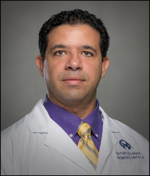 Hatem Soliman合作论文数Center for Women’s Oncology, Moffitt Cancer Center25
 Michael Vogelbaum合作论文数Moffitt Cancer Center;Research Institute, Inc25
 James K.C. Liu合作论文数23
 Matthew N. Mills合作论文数Dept Radiat Oncol, H Lee Moffitt Canc Ctr & Res Inst22
 J. Gregory Cairncross合作论文数Department of Clinical Neurosciences, Cumming School of Medicine, University of Calgary;Arnie Charbonneau Cancer Institute, University of Calgary22
 Grant Mcfadden合作论文数Center for Immunotherapy, Vaccines and Virotherapy , Biodesign InstituteArizona State University 727 E Tyler Street, Room A330E 85281 Tempe AZ USA21
 Nikhil Khushalani合作论文数Department of Cutaneous Oncology, Moffitt Cancer Center20
 Zemp Franz J合作论文数Southern Alberta Canc Res Inst, Univ Calgary20
 Louis Nabors合作论文数Department of Neurology, School of Medicine, The University of Alabama at Birmingham;Department of Cell, Developmental and Integrative Biology, The University of Alabama at Birmingham18Donna L. Senger合作论文数Cancer Biology Research Group, University of Calgary18
 Priya U Kumthekar合作论文数Hematology and Oncologys, Department of Medicine, Feinberg School of Medicine, Northwestern University17Li Jiannong合作论文数17
 Patrick Y. Wen合作论文数Department of Neurology, Harvard Medical School;Center For Neuro-Oncology, Dana-Farber Cancer Institute17Sepideh Mokhtari合作论文数16
 Michael Yu合作论文数16
 Young Chul Kim合作论文数Departmant of Pulmonology and Critical Care Medicine, Chonnam National University Hwasun Hospital16
 Ian F. Parney合作论文数Cancer Center, Mayo Clinic;Department of Neurosurgery, Mayo Clinic16
 Timothy P. Robinson合作论文数Livestock Information, Sector Analysis and Policy Branch, Food and Agriculture Organization of the United Nations16Paulo C. Rodriguez合作论文数Health Sciences Center, Louisiana State University16
 Aixa E. Soyano合作论文数15
 Nicholas Figura合作论文数15
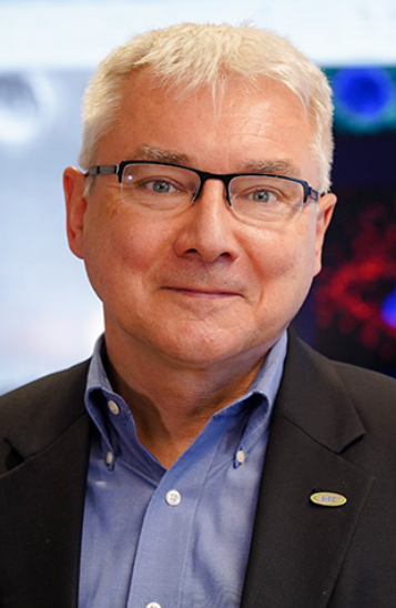 Pawel Kalinski合作论文数Department of Immunology, Roswell Park Comprehensive Cancer Center15
 Prakash Chinnaiyan合作论文数Beaumont School of Medicine, Oakland University15
 Kamran Ahmed合作论文数Department of Radiation Oncology, Moffitt Cancer Center;Department of Immunology, Moffitt Cancer Center;Department of Oncologic Sciences, University of South Florida14
 Zeynep Eroglu合作论文数14
 Hussein A. Tawbi合作论文数Department of Melanoma Medical Oncology, Division of Cancer Medicine, The University of Texas MD Anderson Cancer Center13Clayton Wyatt合作论文数12Manali Phadke合作论文数12Xiaotao Qu合作论文数12Sondak Vernon K合作论文数12




