Purpose: Timely initiation of radiation therapy (RT) following craniotomy for malignant brain tumors is associated with better clinical outcomes. Despite guideline recommendations and best practices, the timely initiation of postoperative RT remains suboptimal. Our objective was to evaluate how structural, social, and payer-related factors independently and interactively influence RT initiation following craniotomy. Methods and Materials: We conducted a retrospective, patient-level cohort study using a large, linked, multisource data set that incorporated all-payer claims from 2023, geospatial hospital data, and indicators of community vulnerability. RT initiation was assessed at 42-, 60-, and 90-day postoperative intervals. Primary predictors included hospital referral region-level RT center density, hospital proximity, payer type, and Centers for Disease Control and Prevention/Agency for Toxic Substance and Disease Registry (CDC/ATSDR) Social Vulnerability Index (SVI) domains. Secondary analyses included region and hospital type. Results: Among 18,885 patients across 548 hospitals, 29.2% initiated RT ≤ 42 days, 45.6% ≤ 60 days, and 48.9% ≤ 90 days. Hospital-level RT initiation ≤42 days ranged from 0% to 87.5%. Hospital referral region per capita RT center density was the strongest structural predictor of RT initiation ≤42 days (adjusted odds ratio [aOR] per IQR, 1.18; 95% CI, 1.11-1.25) and lower odds of noninitiation ≤90 days (aOR, 0.84; 95% CI, 0.78-0.90). Hospital proximity predicted RT initiation ≤42 days (aOR, 1.12; 95% CI, 1.08-1.17) but not ≤90 days. Medicaid coverage predicted lower odds of RT initiation ≤42 days (aOR, 0.58) and higher odds of noninitiation ≤90 days (aOR, 2.29); Medicare coverage predicted lower odds of timely initiation but not of noninitiation (aOR, 0.76). Higher community social vulnerability (Social Vulnerability Index), particularly the share of elderly residents, limited transportation, and housing cost burden, predicted noninitiation, with interaction models showing that hospital density mitigated, but did not eliminate, these disadvantages. Conclusions: Structural access, socioeconomic disadvantage, and insurance coverage independently and interactively predict the timely initiation of postoperative RT, with greater regional RT capacity improving initiation but not fully mitigating social and payer-related barriers.
PURPOSE:Drug-eluting stents fail in up to 20% of patients. In failed cases, intravascular brachytherapy (IVBT) is administered with β-emitting 90Sr90Y through a guidewire. Current clinical dosimetry is water-based, neglecting attenuation from patient-specific materials such as plaques, stents, and the off-centered guidewire, leading to a discrepancy between prescribed and delivered dose. This study retrospectively performed patient-specific IVBT dose calculations using Optical Coherence Tomography (OCT) to quantify uncertainties in clinical dosimetry. METHODS AND MATERIALS:Dose calculations on OCT images from ten patients were performed using RapidBrachyIVBT, a Monte Carlo-based dose calculation software. Heterogeneities, including guidewire(s), stents, and fibrotic and calcified plaques, were contoured and assigned material properties; surrounding tissue was modeled as smooth muscle. Absorbed dose to water and medium were calculated. The prescribed dose to water was 18.4 or 23 Gy at 2 mm from the source, depending on lumen diameter. The dose homogeneity index was defined as the ratio of the maximum to the minimum dose in the target volume. RESULTS:When heterogeneities were included, median maximum dose attenuation was 76.7% (75.0-77.1) in the artery segment and 56.2% (52.2-65.1) in the target volume. The median dose homogeneity index increased from 1.29 in water to 2.93 (2.44-3.33) with patient-specific materials. The guidewire produced asymmetric dose distributions in all patients, with the greatest attenuation where it opposed thick calcified plaques. CONCLUSIONS:Standard water-based IVBT dosimetry is inaccurate due to dose-attenuating materials present during treatment. Personalized, image-guided IVBT planning that accounts for patient-specific heterogeneities may improve treatment accuracy and clinical outcomes.
BACKGROUND:The clinical standard practice of surface brachytherapy (SB) planning has long been to use computed tomography (CT) imaging to visualize applicators for catheter reconstruction in the treatment planning process. Recent work in SB has suggested that magnetic resonance (MR)-guidance can be used in place of CT-guidance in SB planning to utilize the increased soft tissue contrast for visualization of diseased tissue. This soft tissue visualization can be used to verify the target depth for enhanced coverage of the clinical target volume. Two optimized MR sequences (pointwise encoded time reduction with radial acquisition (PETRA) and volumetric interpolated breath-hold examination (VIBE) obtaining Dixon in-phase (DIP) and Dixon opposed-phase (DOP)) have been shown to detect sufficient signal from the silicone-based applicators to perform accurate catheter reconstruction and produce SB treatment plans. PURPOSE:This study compares three in-house MR series optimized for applicator visualization to determine which is best-suited for SB planning based on tissue contrast and applicator visibility. This study then applies this series to produce MR-only SB treatment plans geometrically and dosimetrically comparable to those produced by CT-only for a phantom and eight patients. METHODS:An anthropomorphic phantom (True Phantom Solutions, Canada) with applicators (Elekta, Netherlands) on the foot and hand and eight patients undergoing SB for Dupuytren's Contracture/Palmar fascial fibromatosis were imaged by two optimized MR sequences: 1) PETRA and 2) VIBE obtaining DIP and DOP images. CT scans were acquired for verification. SB planning was performed in Oncentra Brachy (Elekta, Netherlands) treatment planning software using three MR series and CT. MR-based and CT-based plans were compared for geometric and dosimetric accuracy. Geometric accuracy was determined by registering CT-based to MR-based catheter digitizations and calculating distances between corresponding dwell positions. Patient MR images were compared using signal-to-noise ratios (SNR's) and contrast-to-noise ratios (CNR's) for various regions of interest (ROIs) including bone, fat, muscle, and applicator. The series with the greatest tissue contrast and applicator visualization was used to produce treatment plans. MR-based plans were compared to CT-based plans by point-based dose differences (DD's). The MR-based plan was rigidly registered to the CT-based plans, and the isodose volumes were segmented to V150, V125, V100, V95, V90, V80, and V65 and compared using the Dice similarity coefficient (DSC) and volumetric similarity (VS) metric. RESULTS:The distances between the CT-based and MR-based dwell positions were on average 1 mm. The DOP series displayed superior SNR's for all ROIs compared to PETRA and DIP. CNR's for DOP were equivalent to DIP and superior to PETRA. DD's were all below 5% between MR-based and CT-based plans. DSC's were above 0.9 for all segmentations associated with the phantoms and 0.8 for those associated with the patients. VS was above 0.98 for all segmentations across all subjects. CONCLUSIONS:The geometric accuracy of each MR sequence suggests that each can produce accurate treatment plans. The higher SNR's for DOP suggest DOP's suitability for SB, and DOP was utilized to create plans comparable to CT. This novel approach can result in more robust target coverage and potentially improve patient outcomes.
Purpose:Recent advances in surface high-dose-rate (HDR) brachytherapy imaging indicate that flap applicators, human skin, and fibromatosis can be visualized using MRI. Complete MR-only surface brachytherapy workflows would require skin marker identification to define clinical target edges. However, CT markers are not detected on MR images, and common MR markers are unsuitable for continuous surface target tracing. In this paper, we proposed an alternative skin marker that was evaluated for MRI and CT detectability and contourability using a brachytherapy treatment planning system (TPS). Material and methods:Commercially obtained silicone rubber tubes of 2 or 3 mm diameter were taped on the hand of an anthropomorphic phantom, a healthy volunteer, and three palmar fascial fibromatosis patients. Subjects were imaged with an optimized 3D pointwise encoding time reduction with radial acquisition (PETRA) sequence, and a volumetric interpolated breath-hold examination (VIBE) sequence with Dixon reconstruction. Additionally, patients underwent standard CT imaging. Obtained images were reviewed for tube conspicuity, and tubes were tracked on axial views using Oncentra Brachy TPS. Independent tube and muscle reference contours were drawn in MIM for quantitative analysis, considering the three orthogonal imaging planes. Results and Conclusions:Silicone rubber tubes were detected with positive signal on PETRA, VIBE, and CT images. Among the MR series, Dixon VIBE fat-only showed the highest contrast against muscle tissue and the best separation from human skin, followed by DIXON opposed-phase. 3 mm diameter tubes were tracked better by TPS than 2 mm diameter ones. Considering MR images in the three orthogonal planes in MIM was more helpful for localizing the entire tube than using axial images only in TPS. All obtained contour shapes generally agreed with the known tube positions. Overall, solid silicone rubber tubes of 3 mm diameter represent a suitable skin marker alternative to CT markers for MR-only surface HDR brachytherapy.
Purpose Drug-eluting stents are the first-line therapy for in-stent restenosis. However, intravascular brachytherapy (IVBT) is used to treat patients whose drug-eluting stents fail. Current clinical dosimetry for IVBT is water-based, i.e., the absorbed dose in the target volume is calculated by assuming that the patient's artery, calcified plaques, metallic stents, and off-centred guidewire from the IVBT delivery system are all water with unit mass density. We have previously developed a Monte Carlo-based dosimetry software, RapidBrachyIVBT, to account for these heterogeneities and allow for dose calculations on optical coherence tomography images (OCT). This study examines the impact of off-centred guidewires on dose inhomogeneity during irradiation, considering scenarios with multiple guidewires, often overlooked in previous studies. Multiple guidewires are used in cases where the source train passes through a bifurcation of blood vessels. Materials and Methods RapidBrachyIVBT, a Monte Carlo dosimetry software based on the Geant4 toolkit, including the Novoste Beta-Cath 3.5F IVBT device with a 90Sr90Y source train, was used. OCT images from a patient undergoing coronary IVBT for recurrent in-stent restenosis treated at Brigham and Women's Hospital (Boston, Massachusetts) were used to calculate the absorbed dose considering all heterogeneities compared to the dose calculated in water. The patient artery was segmented as water (lumen), fibrotic plaque (around the lumen), calcified plaque (behind the fibrotic plaque), smooth muscle (tunica media) and cobalt-alloy (stents). The source position was assumed to be at the origin of the image, where the OCT imaging device was placed. The guidewire positions were assumed to be at the exact locations used during imaging. Simulations were performed on the Digital Research Alliance of Canada Cedar computing cluster with 200 million decay events to yield less than 1% uncertainty on absorbed dose in the target volume, 2 mm from the source center. The absorbed dose was scored in rectangular voxels of 0.1 × 0.1 × 1.0 mm3 along a 42 mm length, which includes the stents, source train and an additional 2 mm margin. The prescribed dose was 23 Gy to the target volume. The dose homogeneity index, the maximum to minimum dose ratio in the target volume, was calculated in both water and patient cases. Results The dose difference between the water and patient-specific cases was up to 56.2%, 55.8%, and 64.6% in the target volume with one, two, and three guidewires, respectively. The mean dose at the target volume was reduced by 3.4% and an additional 4.1% when adding the second and third guidewire, respectively. The dose homogeneity index was 1.29 in water and 2.96, 2.94, and 3.33 for the respective patient-specific cases. Each guidewire added a cold spot around the IVBT source at the target volume. Conclusions The dose at the target volume in IVBT is significantly reduced when three guidewires are present. Limiting the number of guidewires present during irradiation would reduce cold spots and dose inhomogeneity at the target volume.
Transcriptional profiling demonstrated markedly reduced type I IFN gene expression in untreated mycosis fungoides (MF) skin lesions compared with that in healthy skin. Type I IFN expression in MF correlated with antigen -presenting cell -associated IRF5 before psoralen plus UVA therapy and epithelial ULBP2 after therapy, suggesting an enhancement of epithelial type I IFN. Immunostains confirmed reduced baseline type I IFN production in MF and increased levels after psoralen plus UVA treatment in responding patients. Effective tumor clearance was associated with increased type I IFN expression, enhanced recruitment of CD8 thorn T cells into skin lesions, and expression of genes associated with antigen -specific T -cell activation. IFNk, a keratinocyte-derived inducer of type I IFNs, was increased by psoralen plus UVA therapy and expression correlated with upregulation of other type I IFNs. In vitro, deletion of keratinocyte IFNk decreased baseline and UVA-induced expression of type I IFN and IFN response genes. In summary, we find a baseline deficit in type I IFN production in MF that is restored by psoralen plus UVA therapy and correlates with enhanced antitumor responses. This may explain why MF generally develops in sun -protected skin and suggests that drugs that increase epithelial type I IFNs, including topical MEK and EGFR inhibitors, may be effective therapies for MF.
BackgroundCoronary artery disease is the most common form of cardiovascular disease. It is caused by excess plaque along the arterial wall, blocking blood flow to the heart (stenosis). A percutaneous coronary intervention widens the arterial wall with the inflation of a balloon inside the lesion area and leaves behind a metal stent to prevent re-narrowing of the artery (restenosis). However, in-stent restenosis may occur due to damage to the arterial wall tissue, triggering neointimal hyperplasia, producing fibrotic and calcified plaques and narrowing the artery again. Drug-eluting stents, which slowly release medication to inhibit neointimal hyperplasia, are used to prevent in-stent restenosis but fail up to 20% of cases. Coronary intravascular brachytherapy (IVBT), which uses beta$\beta$-emitting radionuclides to prevent in-stent restenosis, is used in these failed cases to prevent in-stent restenosis. However, current clinical dosimetry for IVBT is water-based, and heterogeneities such as the guidewire of the IVBT device, fibrotic and calcified plaques and stents are not considered.PurposeThis study aimed to develop a Monte Carlo-based dose calculation software, accounting for patient-specific geometry from Optical Coherence Tomography (OCT) images.MethodsRapidBrachyIVBT, a Monte Carlo dose calculation software based on the Geant4 toolkit v. 10.02.p02, was developed and integrated into RapidBrachyMCTPS, a treatment planning system for brachytherapy applications. The only commercially available IVBT delivery system, the Novoste Beta-Cath 3.5F, with a 90Sr90Y$<^>{90}{\rm Sr}<^>{90}{\rm Y}$ source train, was modeled with 30, 40, and 60 mm source train lengths. The software was validated with published TG-149 parameters compared to Monte Carlo simulations in water. The dose calculation engine was tested with OCT images from a patient undergoing coronary IVBT for recurrent in-stent restenosis at Brigham and Women's Hospital in Boston, Massachusetts. Considering the heterogeneities, the images were segmented and used to calculate the absorbed dose to water and the absorbed dose to medium. The prescribed dose was normalized to 23 Gy at 2.0 mm from the source center, which is the target volume in IVBT.ResultsThe dose rate values in water obtained using RapidBrachyIVBT aligned with TG-149 consensus values, showing agreement within a range of 0.03% to 1.7%. Considering the heterogeneities present in the patient's OCT images, the absorbed dose in the entire artery segment was up to 77.5% lower, while within the target volume, it was up to 56.6% lower, compared to the dose calculated in a homogeneous water phantom.ConclusionRapidBrachyIVBT, a Monte Carlo dose calculation software for IVBT, was developed and successfully integrated into RapidBrachyMCTPS, a treatment planning system for brachytherapy applications, where accurate attenuation of the absorbed dose by heterogeneities is considered.
Purpose: Best practices for high-dose-rate surface applicator brachytherapy treatment (SABT) have long relied on computed tomography (CT)-based imaging to visualize diseased sites for treatment planning. Compared with magnetic resonance (MR)-based imaging, CT provides insufficient soft tissue contrast. This work described the feasibility of clinical implementation of MR-based imaging in SABT planning to provide individualized treatment optimization. Material and methods: A 3D-printed phantom was used to fit Freiberg flap-style (Elekta, The Netherlands) ap- plicator. Images were taken using an optimized pointwise encoding time reduction with radial acquisition (PETRA) MR sequence for catheter visualization, and a helical CT scan to generate parallel treatment plans. This clinical study included three patients undergoing SABT for Dupuytren's contracture/palmar fascial fibromatosis imaged with the same modalities. SABT planning was performed in Oncentra Brachy (Elekta Brachytherapy, The Netherlands) treatment planning software. A geometric analysis was conducted by comparing CT-based digitization with MR-based digitization. CT and MR dwell positions underwent a rigid registration, and average Euclidean distances between dwell positions were calculated. A dosimetric comparison was performed, including point-based dose difference calculations and volumetric segmentations with Dice similarity coefficient (DSC) calculations. Results: Euclidean distances between dwell positions from CT-based and MR-based plans were on average 0.68 +/- 0.05 mm and 1.35 +/- 0.17 mm for the phantom and patients, respectively. The point dose difference calculations were on average 0.92% for the phantom and 1.98% for the patients. The D-95 and D-90 DSC calculations were both 97.9% for the phantom, and on average 93.6% and 94.2%, respectively, for the patients. Conclusions: The sub-millimeter accuracy of dwell positions and high DSC's (> 0.95) of the phantom demonstrat- ed that digitization was clinically acceptable, and accurate treatment plans were produced using MR-only imaging. This novel approach, MRI-guided SABT, will lead to individualized prescriptions for potentially improved patient outcomes.
Purpose Despite advances in surgical techniques and multimodality therapy for anal canal and rectal cancers, local recurrence and unresectable disease remain a significant challenge, often associated with poor quality of life. It is not clear how best to address this entity in the absence of large prospective randomized trials. The aim of this study was to retrospectively evaluate the outcomes and toxicities of pelvic low dose rate brachytherapy (LDR) combined with surgical resection in patients with unresectable and locally recurrent anorectal cancers. Materials and Methods Following IRB approval, patients with biopsy-proven anorectal cancers who underwent LDR during surgery for unresectable or locally recurrent anorectal cancers from 2004 to 2022 were included. Patients who had LDR for recurrent gynecological or genitourinary cancers were excluded. For all patients, the intent of surgery was complete resection of all visible disease. This was followed by LDR, either in the surgical bed for recurrent cancers or the site of microscopically positive margins for the unresectable patients. Following LDR mesh fixation with sutures, an omental flap was draped over the site of LDR to prevent seed migration and to minimize dose to organs at risk. Toxicity grading was done using the Common Terminology Criteria for Adverse Events (CTCAE) Version 5. Results Out of 29 eligible patients, 20 underwent Iodine-125 LDR, and 9 received Cesium-131 LDR with an average of 73.8 sources used per patient. The primary site was colorectal in 79.3% and anal canal in 20.7%. 27 (93.1%) of the patients had pelvic only disease at the time of LDR and surgery. 21 (72.4%) patients had at least 1 surgery prior to the implant prior to the implant, whereas 8 (27.6%) patients had no surgery prior to the implant. Brachytherapy was offered at recurrence in 23 patients (79.3%) and in 6 (20.7%) patients who were considered unresectable initially. In those with recurrences, 20 (87.0%) had LDR offered during their first recurrence and the rest were offered LDR at subsequent recurrences. Chemotherapy was offered at first recurrence in 21 (91.3%) patients. No Grade 4 or 5 toxicities were reported. The most common adverse event seen was neuralgia in 12 (41.4%) patients with 2 of those developing Grade 3 neuralgia. The rate of Grade 2 gastrointestinal fistula was 5 (17.2%) and Grade 2 urinary fistula was 3 (10.3%). All of the patients who developed fistulas were diverted at the time of surgery and LDR or before, and did not require additional invasive interventions. The 12 month and 24 month local progression free survival were 55.4% (95% CI: 34.9-71.8) and 41.7% (95% CI: 22.3 - 60.1), respectively. The 12 month and 24 month progression free survival were 38.4% (95% CI: 20.7-55.9) and 25.6% (95% CI: 10.9-43.3), respectively. The 12 month and 24 month overall survival rates were 88.7% (95% CI: 69.0-96.2) and 70.6% (95% CI: 47.7-84.9), respectively. Conclusions LDR in combination with resection of all macroscopic disease in unresectable or locally recurrent anorectal cancers is a viable treatment option. Toxicities are acceptable, with no Grade 4 or 5 adverse events in this cohort. Fistulas were managed expectantly as the patients who developed fistulas were already diverted. Furthermore, it is not clear if the fistulas developed as an adverse event of LDR or were due to local tumor progression. Progression free survival and overall survival at 12 and 24 months align with prior retrospective reports. Our study is subject to the limitations of being a retrospective study with a small sample size. However, the findings are encouraging and warrant further prospective studies to guide the management of these heterogenous and often challenging cases.
Scalp melanomas are associated with poor disease-related and survival outcomes compared with melanomas affecting other anatomical sites, partially due to high rates of in-transit metastases (ITM), defined as localized >2 cm from the primary tumor but not beyond draining lymph nodes.1,2 The standard treatment for ITMs is surgical excision3; however, surgery is often impractical for multiple or diffuse ITMs. Currently, the only Food and Drug Administration-approved therapy, specifically directed at ITMs is talimogene laherparepvec, an injectable modified oncolytic herpes simplex virus, which has shown modest response rates in phase III studies in patients with locally advanced melanoma.
Options for meningiomas that have progressed to a higher grade (World Health Organization grade II/III) after repeated surgical resections, exhausting external beam radiation, and failing medical trials are limited. Although palliative additional resection can be offered, tumor control is short lived. The outcomes of transformed meningiomas to a higher grade are particularly ominous.1,2 Although chemotherapy and immunotherapy clinical trials are being actively investigated, no effective medical treatment exists for patients with recurrent meningioma, which leaves a population of patients with recurrent meningiomas that have maximized external radiation treatment and failed trials of medical therapy.3 Brachytherapy carries several theoretical advantages for the treatment of recalcitrant meningioma, including the delivery of high doses of radiation to the surgical cavity over an extended period, limited exposure of previously radiated brain parenchyma to additional radiation, and in a tumor that is grossly resected, to treat the microscopic disease that remains and is the source of recurrence. In these cases, radioactive seeds can be applied at the highest-risk areas after resection, such as the convexity dural margin, the falx, and parasagittal regions. This represents an attractive and effective treatment option for these patients.4 In this article, we report a case of a multiple recurrent falcotentorial meningioma that has demonstrated malignant progression after 2 prior surgeries, radiosurgery, and stereotactic radiation treatment. The patient underwent gross total resection of the recurrence with placement of Cesium-131 brachytherapy to the resection cavity and margins, with no evidence of tumor recurrence on follow-up. The patient consented for surgery. {"href":"Single Video Player","role":"media-player-id","content-type":"play-in-place","position":"float","orientation":"portrait","label":"VIDEO.","caption":"","object-id":[{"pub-id-type":"doi","id":""},{"pub-id-type":"other","content-type":"media-stream-id","id":"1_z8rmc5c3"},{"pub-id-type":"other","content-type":"media-source","id":"Kaltura"}]}
Background: Limited data exists regarding the efficacy of curative hypofractionated radiotherapy (hypoRT) regimens compared to conventionally-fractionated radiotherapy (conv-RT) for Merkel cell carcinoma (MCC).Methods: A retrospective analysis of 241 patients diagnosed with non-metastatic MCC from 2005-2021 and who received RT at Dana-Farber/Brigham & Women's Cancer Center. The primary outcome was cumulative incidence of in-field locoregional relapse using Gray's test with competing risks of death and isolated out-of-field recurrence. Secondary outcomes included overall survival (OS) and MCC specific survival using log-rank tests, and risk factors of recurrence using Cox-proportional hazards regression.Results: There were 50 (20.6 %) and 193 (79.4 %) courses of hypo-RT and conv-RT, respectively. The hypoRT cohort was older (>73 years at diagnosis: 78.0 % vs 41.5 %, p < 0.01), and received a lower equivalent total RT dose in 2 Gy per fraction (<50 Gy: 58.0 % vs 5.2 %, p < 0.01). Median follow-up was 65.1 months (range: 1.2-194.5) for conv-RT and 25.0 months (range: 1.6-131.3) for hypo-RT cohorts. Two-year cumulative incidence of in-field locoregional relapse was low in both groups (1.1 % conv-RT vs 4.1 % hypo-RT, p = 0.114). While two-year OS was lower for the hypo-RT group (62.6 % vs 84.4 %, p = 0.0008), two-year MCC-specific survival was similar (84.7 % vs 86.6 %, p = 0.743). On multivariable analysis, immunosuppression, clinical stage III disease, and lymphovascular invasion were associated with any-recurrence when controlling for sex, age, and hypo-RT.Conclusions and Relevance: There was no difference in cumulative incidence of in-field locoregional relapse or MCC-specific survival between hypo-RT and conv-RT. Prospective studies are needed to confirm hypo-RT as an efficacious treatment option for MCC.(c) 2022 The Author(s). Published by Elsevier B.V. Radiotherapy and Oncology 173 (2022) 32-40 This is an open access article under the CC BY-NC-ND license (http://creativecommons.org/licenses/by-nc-nd/4.0/).
 Ivan Buzurovic合作论文数Thomas Jefferson University64
 R. Cormack合作论文数61Desmond A. O'Farrell合作论文数47Mandar S. Bhagwat合作论文数29
 Jorgen L Hansen合作论文数25
 Paul L. Nguyen合作论文数Brigham and Women's Hospital;75 Francis Street;Boston, MA 02115/Dana-Farber Cancer Institute;450 Brookline Avenue;Boston, MA 0221522Scott Friesen合作论文数22
 Peter F. Orio III合作论文数Department of Radiation Oncology, Dana-Farber Cancer Institute, Brigham and Women’s Hospital18Damato Antonio L合作论文数Department of Radiation Oncology, Dana-Farber Cancer Institute/Brigham and Women's Hospital17
 Harris C Thomas合作论文数Center for Integrated Nanotechnologies, Sandia National Laboratories16
 Alexandra J Stewart合作论文数16Larissa Lee合作论文数15
 Akila N. Viswanathan合作论文数14King Martin T合作论文数13Elizabeth H. Baldini合作论文数12
 Subhakar Mutyala合作论文数Radiation Oncology, Montefiore Medical Center11
 Thomas S. Kupper合作论文数Dana-Farber Cancer Institute, Inc.;Brigham and Women's Hospital;Harvard Medical School10
 Chandrajit Premanand Raut合作论文数Division of Surgical Oncology, Brigham and Women's Hospital;Harvard Medical School10Nicole R. Leboeuf合作论文数Dana Farber Cancer Institute/Brigham and Women's Hospital, Harvard Medical School10
 Brian A. Bergmark合作论文数Division of Cardiovascular Medicine, Brigham and Women's Hospital;Heart and Vascular Center, Brigham and Women's Hospital;Harvard Medical School9
 Brandon A. Mahal合作论文数Sylvester Comprehensive Cancer Center, Miller School of Medicine, University of Miami;Sylvester Comprehensive Cancer Center;Department of Radiation Oncology, Miller School of Medicine, University of Miami9Kevin Croce合作论文数Cardiovascular Division, Brigham and Women's Hospital9Evangelia Kaza合作论文数8
 Muralidhar Vinayak合作论文数Harvard Medical School, MIT8
 Christian V. Guthier合作论文数Medical Faculty of Mannheim, Heidelberg University7Emily Neubauer Sugar合作论文数6D. A. O'Farrell合作论文数6Atif J Khan合作论文数Rutgers Canc Inst New Jersey6
 Aldelaijan Saad合作论文数Department of Radiation Oncology, McGill University6
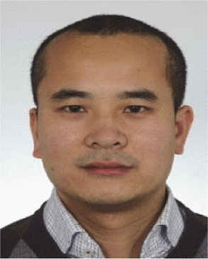 John T O'Malley合作论文数6
 Rachael Ann Clark合作论文数Department Dermatology, Brigham And Women's Hospital;Harvard Medical School6Julia Kazakin合作论文数6
 Caroline Holloway合作论文数6Luciant D. Wolfsberger合作论文数5Niedermayr Thomas合作论文数5Jessica E Teague合作论文数5
 Edward Christopher Dee合作论文数Dept Radiat Oncol, Mem Sloan Kettering Canc Ctr5
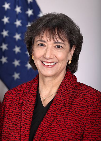 Monica M. Bertagnolli合作论文数National Institutes of Health;Alliance for Clinical Trials in Oncology Foundation;Alliance for Clinical Trials in Oncology5Harris Thomas C合作论文数5
 Suzanne George合作论文数5
 Roy B. Tishler合作论文数Dana-Farber Cancer Institute;Department of Radiation Oncology, Harvard Medical School;Brigham and Women's Hospital5
 Clair Beard合作论文数Division of Genitourinary Radiation Oncology, Department of Radiation Oncology, Harvard Medical School;Dana-Farber/Brigham Cancer Center, Harvard Medical School5Christopher L. Williams合作论文数4Maryam Rahbaran合作论文数4T. C. Harris合作论文数4Jonathan M Cantalino合作论文数4Cecilia Larocca合作论文数4D. O'Farrell合作论文数4Miranda B Lam合作论文数4




