False positive T or B cell flow cytometry crossmatching (FCXM) results present a significant challenge in transplantation decisions. In this multicentre study, we investigated the efficacy of ethylenediaminetetraacetic acid (EDTA) treatment of serum samples to enhance the quality of FCXM results by reducing false positivity at two independent laboratories. Our findings indicate that EDTA treatment of serum effectively eliminated false positivity in B cell non-pronased (NP) FCXM compared with untreated samples, possibly by lowering aggregated IgG formation from in vitro storage of patient sera. In B cell NP FCXM, EDTA-treated serum from both sensitised and non-sensitised patients exhibited comparable efficacy to B cell pronase treated (PT) FCXM, with a sensitivity of 94% and specificity of 96%. Moreover, EDTA-treated serum samples demonstrated superior performance to T cell PT FCXM, effectively reducing false positivity. In addition, EDTA-treated sera performed in par with sera pre-treated with 2-mercaptoethanol, and superior to sera treated with Dithiothreitol or with heat inactivation, in removing false B cell positivity in NP FCXM. Importantly, our analysis of 124 FCXM underscores the simplicity and effectiveness of EDTA treatment, which can be seamlessly integrated into both NP and PT FCXM. Consequently, the adoption of EDTA-treated patient serum in B cell NP FCXM reduces the need for B cell PT FCXM.
OBJECTIVES:A T-cell-positive and B-cell-negative flow cytometry crossmatch result remains a conundrum since HLA class I antigens are expressed on both T and B cells. We investigated the frequency, donor HLA specificity of the antibodies, and mechanisms for these crossmatch results.MATERIALS AND METHODS:We analyzed 3073 clinical flow cytometry crossmatch tests performed in an American Society of Histocompatibility and Immunogeneticsaccredited histocompatibility laboratory. The sera associated with the T-cell positive and B-cell negative flow cytometry crossmatches were also tested for donor HLA immunoglobulin G antibodies using LABScreen single antigen assays.RESULTS:Among the 3073 test results, 1963 were T-cell negative and B-cell negative, 811 were T-cell negative and B-cell positive, 274 were T-cell positive and B-cell positive, and 25 were T-cell positive and B-cell negative. The LABScreen single antigen assay detected HLA class I immunoglobulin G donor-specific antibodies in 23 of 25 sera associated with a T-cell positive and B-cell negative flow cytometry crossmatch result, and donorspecific antibodies directed at not only HLA-Cw but also at HLA-A or HLA-B were observed. In addition, we identified that the B-cell channel shift threshold used to classify a B-cell flow cytometry crossmatch was a potential contributor to a T-cell-positive and B-cellnegative flow cytometry crossmatch result.CONCLUSIONS:Our analysis of 3073 flow cytometry crossmatches, in addition to demonstrating that HLA antibodies directed at the HLA-A, -B, or -Cw locus were associated with a T-cell-positive and B-cell-negative result, identified mechanisms for the surprising T-cell-positive and B-cell-negative flow cytometry crossmatch result.
Background: The aim of this study is to determine the incidence of early acute rejection and graft outcomes in living donor renal transplants with an isolated positive B-Cell Flow-Cytometry Crossmatch (B-FCXM). Methods: We performed a retrospective analysis of graft outcomes in 152 adults who received living donor renal transplants between January 2019 and March 2021 at Mount Sinai Hospital in New York CIty. Pre-transplant histocompatibility was assessed with flow cytometric crossmatch (FCXM) and Luminex single-antigen beads. Positive B- FCXM is was defined as median channel shift of ≥ 40. A mean fluorescence intensity (MFI) ≥ 1400 defined positive donor-specific HLA Class I and/or II antibody (DSA). Most patients received induction immunosuppression consisting of rabbit anti-thymocyte globulin, steroids and intravenous immunoglobulin. Estimated glomerular filtration rate (eGFR) and biopsy-proven rejection at six months’ post-transplant were compared between patients with and without a positive B-FCXM, and positive and negative DSA. Results: Of 152 patients, 89 (59%) were males and 63 (41%) females; the mean age was 44 years (SD ± 13.8). 45 donor B-FCXMs (30%) were positive and 107 (70%) were negative. 42 (28%) positive donor B-FCXM had no DSA and 3 (2%) had DSA. 91 (85%) negative donor B-FCXMs did not have DSA, and 16 (15%) had DSA. Thirty-six (80%) out of 45 patients with positive donor B-FCXM, including the 3 patients with DSA, received intravenous-immunoglobulin (IVIG) as part of induction immunosuppression. None of the patients with both positive donor B-FCXM and DSA had rejection. eGFR and rejection rate at 6 months were similar between patients with positive and negative donor B-FCXM [median 58.9 (IQR= 50.2-70.8) vs 57.6 (48.8-76.6), p=0.88; 6.8% vs 5.6%, p=0.7, respectively]. Comparison by combination of donor B-FCXM and DSA status (B-FCXM- DSA-, B-FCXM- DSA+, B-FCXM+ DSA-, B-FCXM+ DSA+) did not show statistically significant differences in eGFR or rejection rate at 6 months [median 57.4 (IQR= 48.5-74.3) vs 59.3 (52.6-83.3) vs 58.2 (50.1-70.8) vs 76.6 (58.3-102.5) p=0.4; 3.3% vs 18.7% vs 7.3% vs 0% p=0.09, respectively]. Conclusion: In our cohort, there was no statistical difference in early acute rejection and graft function at 6 months between living donor transplant recipients with positive and negative donor B-FCXM even after accounting for DSA status. Peri-transplant treatment with IVIG for a positive donor B-FCXM might be a helpful adjunct in this setting.
Complement dependent cytotoxicity crossmatch (CDC-XM) has been the original standard crossmatch test, whereas, flow cytometry crossmatch (FCXM) is an enhanced and highly sensitive crossmatch assay performed to detect donor specific anti-HLA antibodies (DSA). We analyzed American Society for Histocompatibility and Immunogenetics (ASHI) proficiency testing data (2011-2020) and examined the number of laboratories performing CDC-XM vs. FCXM, the overall efficiency of laboratories in reporting ≥80% consensus CDC-XM vs. FCXM result, and reasons for non-consensus results in the two assays. Of 600 crossmatches in each crossmatch category, the percentage of laboratories reporting T cell CDC-XMs reduced from 40% in 2011 to 13% in 2020, T cell anti-human globulin (AHG) CDC-XM reduced from 56% in 2011 to 21% in 2020, and B cell CDC-XM reduced from 51% in 2011 to 20% in 2020. The percentage of laboratories performing T cell and B cell FCXM remained at approximately 80% throughout. CDC-XM performed on par with FCXM in providing a consensus negative result using negative DSA serum, but under-performed in comparison to FCXM in providing a consensus positive result using positive DSA serum. In addition, only minority of CDC-XMs was reported positive in presence of complement fixing DSA. This study shows that non-consensus CDC-XM was always in presence of HLA IgG DSA and that laboratories may be struggling to interpret the low sensitive CDC-XM results, where highly sensitive solid phase multi-antigen or single antigen assay shows the presence of HLA IgG DSA in serum.
Interfering factors in sera may confound crossmatch (XM) results. Heat inactivation (HI), DTT and dialysis treatment of sera have been utilized to minimize these confounders. We investigated the impact of HI on donor T cell complement dependent cytotoxicity (CDC) XM (T Cell AHG XM) and donor B cell CDC XM (B Cell NIH XM). We queried our electronic database for all donor CDC T cell and B cell XM performed during 2011–2017 in which potential kidney transplant recipient’s serum was tested in parallel with or without HI and using potential donor’s T cells and B cells as target cells. A total of 6295 living donor XMs and 15880 deceased donor XMs performed in our laboratory were reviewed. Table 1 shows the impact of HI on the 22175 consecutive XM results. 394 of the 6295 living donor T Cell AHG XMs were positive using neat sera and 303 were positive following HI at 63C for 6 min, a 23% conversion from positive to negative results. 698 of the 15880 deceased donor T Cell AHG XMs were positive using neat sera and 540 XMs were positive following HI, a 23% reduction. 1372 of the 6295 living donor B Cell NIH XMs were positive using neat sera and 539 were positive following HI, a 61% reduction. 3015 of the 15880 deceased donor B Cell NIH XMs were positive using neat sera and 842 XMs were positive following HI, a 72% reduction from a positive result to negative result. A substantial percentage of XMs are converted from a positive CDC result to negative result and such conversion is more frequent with B cell XM compared to T cell XM. Transplant centers seldom transplant across a positive T cell CDC XM even when HI converts a positive result to a negative result. On the other hand, it is not uncommon to proceed with a kidney transplant when HI converts a positive B cell CDC to a negative CDC provided there are no other immune contraindications. There is an urgent need to establish the clinical significance of XMs converted from a positive to a negative result by heat inactivation.
Aim Emerging data suggest that DSA development is a frequent complication post BKV replication, and most DSA are directed against donor HLA-DQ. In the current investigation, we quantified DQ epitope mismatches between the recipient and donor in individuals with BKVN diagnosis and investigated the association between DQ epitope load and development of DSA. Methods In this pilot study, using the most probable 4-digit HLA typings, we investigated epitope mismatches in individuals who developed DSA post BKVN diagnosis compared to those who did not and correlated them to DSA development and graft dysfunction defined as an increase in serum creatinine by u003e 0.5 mg/dL at 12 months post-BKVN diagnosis. We had SSOP based intermediate HLA typing available for recipients and donors in 13 recipients who developed BKVN confirmed by renal allograft biopsy. Of the 13 recipients, 2 were excluded: 1 for early graft loss and 1 who had zero DQ mismatches. None of the 11 recipients with BKVN diagnosis had a positive XM or DSA at time of transplantation. Results Time from kidney transplantation to BKVN diagnosis was 8.5 ± 5.1 months. Serum creatinine at time of diagnosis was 1.78 ± 0.56. Of the 11 with BKVN, 36% developed de novo DSA. All DSAs were directed against DQ mismatches. Time from BKVN diagnosis to DSA development was 5.5 ± 2.3 months. Table below lists the epitope mismatch number at each locus. Of the 4 patients with de novo DSA, 75% experienced graft dysfunction during the 12 months post BKVN and of the 7 patients without de novo DSA, 29% experienced graft dysfunction by 12 months. Our data demonstrated that the most frequent DQ epitope mismatches in those with de novo DSA were 52PL3, 45EV, 52PQ2, and 52PR. Conclusions In our pilot study, the epitope load was not different between BKVN patients who developed or did not develop de novo DSA. The most frequent target of de novo antibodies were DQ locus mismatched epitopes. Knowledge of epitope targets of de novo DSA in patients with BKVN may facilitate targeted therapies and improve kidney allograft outcomes. Download high-res image (148KB) Download full-size image
Luminex Single Antigen bead assay (LSAB) derived mean fluorescence intensity (MFI) values of anti-HLA antibodies directed at the potential donor’s HLA are the primary parameter used in the virtual crossmatch to predict physical crossmatch (XM) outcome. We aimed to develop statistically validated LSAB MFI cutpoints for predicting CDC XM and FCXM results. We leveraged the ASHI proficiency testing (PT) 80% consensus results of 7156 T FCXM, 6758 B FCXM, 2917 T CDC XM and 2233 B CDC XM as the reference results to investigate whether LSAB MFI of IgG anti-HLA antibodies predict validated physical XM results. LSAB MFI was determined in our laboratory using One Lambda Single Antigen HLA Class I and Class II beads. The 80% consensus results are from 8 consecutive challenges during 2013 to 2016, and 107 laboratories across USA tested sera and HLA typed cells distributed by ASHI, and reported the results to ASHI for assessment of the laboratory’s proficiency. Data analysis included: summing of MFI of IgG antibodies directed at HLA-A, B and C for investigating their association with T FCXM and T CDC XM results; summing of MFI of antibodies directed at HLA-A, B, C, DR, DQ and DP for investigating their association with B FCXM and B CDC XM results; assigning alternate ASHI challenges to the Discovery set and the Validation set; investigating the association between LSAB MFI and physical XM results by logistic regression analysis corrected for overdispersion; and identification of LSAB MFI cutpoint for maximizing the sum of sensitivity and specificity. LSAB MFI cutpoints derived from the Discovery set to predict FCXM and CDC XM results were investigated in an independent validation set. Table 1 demonstrates that LSAB MFI cutpoints from the Discovery set predicts XM outcomes in the Validation set (ROC AUC range from 0.974 to 0.999). We have developed and validated LSAB MFI cutpoints for use in the virtual crossmatch to accurately predict physical T FCXM, B FCXM, T CDC XM and B CDC XM results.
LSAB mean fluorescence intensity (MFI) is often used to predict physical FCXM results, and LSAB MFI cutpoints are used to list unacceptable antigens. We therefore determined the accuracy of LSAB assay MFI cutpoint used to score the LSAB assay as positive or negative in foretelling FCXM results. One hundred and forty-six pre-transplant sera from 146 patients with end stage renal disease were tested against potential kidney donor’s T cells and B cells using a 3-color flow cytometry clinical protocol; the same 146 sera were tested using LSAB assay and the cumulative MFI directed at donor HLA class I and/or II antigens were determined. A T cell FCXM with a median channel shift (MCS) ≥ 40 with the patients serum vs. negative control serum was classified as a positive test; a B cell FCXM with MCS ≥ 50 with the patients serum vs. negative control serum was classified as a positive test. In the LSAB assay, MFI > 2000 directed at donor HLA antigens was scored as positive DSA. These cutpoints are our laboratory’s cutpoints for both ASHI proficiency testing and for clinical use. Statistical analysis demonstrated a significant association between LSAB results and FXCM results (Table 1). LSAB assay results predicted T Cell FCXM results with a sensitivity of 70% and a specificity of 100% (Fischers Exact Test, P < 0.0001, Positive Predictive Value (PPV = 100%, Negative Predictive Value (NPV) = 91%). LSAB assay results also predicted B Cell FCXM results with a sensitivity of 86% and a specificity of 72% (Fischers Exact Test, P < 0.0001, PPV = 62%, NPV = 91%) (Table 1). In a subset of 31 patients, both donor FCXM and auto FCXM results were available and 17 of 31 donor positive FCXM were also auto B cell FCXM positive (17/17) and auto T cell FCXM positive (1/17). LSAB assay results are significantly associated with FCXM results. Auto B cell FCXM and high resolution HLA typing should further refine the relationship between these two high sensitive assays.Download : Download high-res image (138KB)Download : Download full-size image
Luminex Single Antigen bead assays (LSAB) are robust for detecting and identifying IgG anti-HLA antibodies, and the LSAB mean fluorescence intensity (MFI) is currently used to list unacceptable antigens and to predict physical crossmatch (XM) results. Concerns exist regarding the use of MFI as the criterion because MFI thresholds for accurately predicting physical XM results are far from established. We aimed to address this unmet need. We leveraged the XM results from ASHI Proficiency Testing (PTs) to investigate whether LSAB MFI cutpoints are predictive of physical XM results. ASHI graded (⩾80% consensus) XM results from 8 consecutive ASHI PTs in which up to 30 labs tested 40 sera and 16 cells distributed by ASHI were used to examine whether LSAB MFI data we generated in the same PTs predict physical XM results. Cumulative MFIs of DSA directed at HLA-A, B, and C were tested for their ability to predict T cell Flow XM and T cell AHG XM results; cumulative MFIs of DSA directed at HLA-A, B, C, DRB1, DRB3/4/5, DQB and DP were tested for their ability to predict B cell Flow XM and B cell CDC XM results. Our data analysis identified that cumulative MFI of DSA less than 6000 directed at HLA-class I antigens predicted T cell Flow XM results with a 100% negative predictive value (NPV) and MFI > 7000 predicted T cell Flow XM results with a 100% positive predictive value (PPV) (Chi-Square for trend, X2 = 70, df = 1, P < 0.0001) (Table 1). Cumulative MFI of DSA less than 5000 directed at HLA-class I and II antigens predicted B cell Flow XM result with a 100% NPV and MFI > 8000 predicted B Flow XM result with a 100% PPV (X2; =; 67, df = 1, P < 0.0001) (Table 1). Cumulative MFIs predicted T cell AHG XM results and B cell CDC XM results with a slightly lesser accuracy (Table 1).Download : Download high-res image (343KB)Download : Download full-size image Our data demonstrating the feasibility of developing LSAB MFI cutpoints for the accurate prediction of T cell Flow XM, B cell Flow XM, T cell AHG XM, and B cell CDC XM results advance a strategy for the interpretation of virtual crossmatches and the prediction of physical XM results.
Early and late post transplant complications have been associated with antibodies (Abs) directed against donor HLA; however, the impact of pre-transplant Abs directed against donor HLA-C remains unresolved. In view of diminished display of HLA-C antigens on the cell surface compared to HLA-A or-B antigens, we hypothesized that the impact of pre-transplant Abs to donor HLA-C will be lesser compared to Abs directed at donor HLA-A and B. In the current investigation we studied the impact of circulating pre-transplant IgG antibodies to donor HLA-C locus antigens alone as compared to Abs to HLA- or B locus antigens. We conducted a retrospective review of records of 1252 kidney allograft recipients transplanted at our center between January 2010 and January 2016 to identify patients with circulating pre-transplant IgG Abs directed at kidney donor HLA-A, B or C locus antigens. Abs were detected and reported using the Luminex Single Antigen Bead assay for HLA Class I. Pre and post-transplant data were collected and graft outcomes of 16 kidney graft recipients with Abs to HLA-C locus antigens alone were compared to the outcomes of 56 recipients with Abs to HLA-A or B locus antigens. The mean pre-transplant MFI value of DSA in those with Abs to HLA- A or B antigens was 3880 ± 3023 and 4331 ± 5540 in those with Abs to HLA- C antigens (P = 0.4). The one-year acute rejection rate was 6% in those with DSA to HLA-C antigens and 20% in those with DSA to HLA-A or B antigens (Fig. 1A). Incidence of AMR was increased in those with DSA-MFI value greater than 6000 (Fig. 1B). Kidney allograft survival rate was 100% in those with DSA to HLA-C antigens and 95% in those with DSA to HLA-A or B locus antigens (P > 0.05). Our observations support the concept that circulating pre-transplant IgG antibodies directed at kidney donor HLA-C locus alone do not negatively impact kidney allograft outcomes and suggests that listing unacceptable HLA C locus antigens based on MFI of Single Antigen Bead Assay requires additional scrutiny and due caution.Download : Download high-res image (125KB)Download : Download full-size image
CD4+ T helper (Th) cell subset generation in vivo requires T cell receptor activation and surface CD28 co-stimulation in the presence of one or more cytokines. Similarly, Th cells can be generated in vitro by activating naïve CD4+CD25- T cells with plate bound-anti-CD3 monoclonal antibody (mAb) (pbCD3) and soluble-anti-CD28 mAb (sCD28) in the presence of polarizing recombinant (r) cytokines and anti-cytokine mAbs. In comparison to in vitro CD4+CD25- T cells, memory CD4+CD25-CD45RO+ T cells have been shown to convert to Th9 cells more efficiently. Here, protocol for in vitro generation of human Th9 cells by activating CD4+CD25-CD45RO+ memory T cells with pbCD3 and sCD28 in the presence of polarizing recombinant interleukin-4 (rIL-4) and transforming growth factor (rTGF-β) is described.
The relationship between circulating pre-transplant immunoglobulin G (IgG) antibodies to donor human leukocyte antigen (HLA) -C locus determined antigens alone and acute rejection, kidney allograft function, and graft survival is not fully defined. Also, the impact of circulating pre-transplant IgG antibodies to donor HLA-C locus antigens alone on these outcomes has not been compared with the impact of circulating pre-transplant IgG antibodies to donor HLA-A or -B locus antigens. We conducted a retrospective review of records of 1252 kidney allograft recipients transplanted at our center between January 2010 and January 2016 to identify patients with circulating pre-transplant IgG antibodies directed at kidney donor HLA-A, -B, or -C locus determined antigens. Antibodies were detected and reported using the LABScreen Single Antigen Bead assay with microbeads coated with single HLA class I antigens. Pre-transplant and post-transplant data were collected and the graft outcomes of 16 kidney graft recipients with antibodies to HLA-C locus antigens were compared to the outcomes in 56 recipients with antibodies to HLA-A or -B locus determined antigens. The one-year acute rejection rate was 6% in those with donor-specific antibodies (DSA) to HLA-C locus antigens and 20% in those with DSA to HLA-A or -B locus antigens. The graft survival rate was 100% in those with DSA to HLA-C locus antigens and 95% in those with DSA to HLA-A or -B locus antigens. None of the numerical differences were statistically significant (p>0.05). The presence of circulating pre-transplant IgG antibodies directed at kidney donor HLA-C locus antigens alone may not be associated with an increased risk of acute rejection or a decreased graft survival rate. Our observations support the concept that circulating pre-transplant IgG antibodies directed at kidney donor HLA-C locus antigens alone do not negatively impact kidney allograft outcomes and that the mean fluorescence intensities of the antibodies directed at HLA-C locus alone should not be used to list unacceptable HLA-C locus antigens for kidney allocation. A study with a larger cohort is needed to investigate our hypothesis.
Identification of biomarkers that assess posttransplant risk is needed to improve long‐term outcomes following heart transplantation. The Clinical Trials in Organ Transplantation (CTOT)‐05 protocol was an observational, multicenter, cohort study of 200 heart transplant recipients followed for the first posttransplant year. The primary endpoint was a composite of death, graft loss/retransplantation, biopsy‐proven acute rejection (BPAR), and cardiac allograft vasculopathy (CAV) as defined by intravascular ultrasound (IVUS). We serially measured anti‐HLA‐ and auto‐antibodies, angiogenic proteins, peripheral blood allo‐reactivity, and peripheral blood gene expression patterns. We correlated assay results and clinical characteristics with the composite endpoint and its components. The composite endpoint was associated with older donor allografts (p < 0.03) and with recipient anti‐HLA antibody (p < 0.04). Recipient CMV‐negativity (regardless of donor status) was associated with BPAR (p < 0.001), and increases in plasma vascular endothelial growth factor‐C (OR 20; 95%CI:1.9–218) combined with decreases in endothelin‐1 (OR 0.14; 95%CI:0.02–0.97) associated with CAV. The remaining biomarkers showed no relationships with the study endpoints. While suboptimal endpoint definitions and lower than anticipated event rates were identified as potential study limitations, the results of this multicenter study do not yet support routine use of the selected assays as noninvasive approaches to detect BPAR and/or CAV following heart transplantation.
Flow cytometry crossmatch (FCXM) is a high sensitive tool to identify donor cell specific antibodies in recipient's serum. However, the procedure is time consuming and fraught with inter-assay and inter-operator variabilities. To optimize instrumentation for automated tagging (AT) in FCXM that will both quicken the procedure and exclude inter-operator variability. Serum, spleen, or peripheral blood mononuclear cells were isolated from human subjects. AT was performed using a combination of Precision-XS and EL406 instruments (BioTek Instruments, Inc.) and compared to manual tagging (MT) (both in 96 well plates). FITC-goat α-human IgG channel shift (CS) of >40 for T cells and >50 for B cells was considered positive. AT of two cell specimens took 90 min vs. 100 min by MT. AT did not cause cell loss. Automated Tagging vs. Manual Tagging: AT and MT of specimens (N = 6) provided similar CS results (T cell-(P = 0.8) and B cell-FCXM (P = 0.9); (Fig 1A)) and median channel intensity results for T cell FCXM (%CV: negative control (NC) = 8 vs. 11; positive control (PC) = 10 vs. 14) and B cell FCXM (%CV: NC = 6 vs. 17; PC = 10 vs. 9). Intra-assay variability: When AT was performed in duplicates in a plate (N = 3), no intra-assay variability was noted in CS results (T cell FCXM: P = 0.1; B cell FCXM: P = 0.1 (Fig 1B)) or in median channel intensity results [T cell FCXM (%CV: NC = 1 vs. 5; PC = 4 vs. 2) and B cell FCXM (%CV: NC = 2 vs. 1; PC = 5 vs. 4).] Inter-assay variability: When AT was performed in two different plates (N = 3), no inter-assay variability was noted in CS results (T cell FCXM: P = 0.8; B cell FCXM: P = 0.9 (Fig 1C)) or in median channel intensity results [T cell FCXM (%CV: NC = 8; and PC = 6) and B cell FCXM (%CV: NC = 11; PC = 5)]. Inter-operator variability: Inter-operator variability was not noted when FCXM were performed by AT (Fig 1D). Combination of Precision-XS- and EL406-instruments helps making a FCXM faster and automatous, and provides reliable results with inter-assay and inter-operator consistency.Download : Download full-size image
Effective means to identify anti-donor immune activity before the transplant organ is damaged and rejected has been an important goal in transplantation research. Development of sensitive and non-invasive diagnostic methods that probe the immune status of the recipient as well as the resilience of the donor organ should enable personalized application of immunosuppressive drugs. With a non-invasive biomarker for rejection, it should be possible to selectively treat the patients that are rejecting the graft and wean the tolerant patients from immunosuppression. Although A20 is also expressed by activated CD4+ T cells and CD8+ T cells, its expression by mouse tubular cells has been shown to play an important role in protecting allografts from ischemia/reperfusion (I/R) injury and rejection. Using quantitative (real-time) reverse transcriptase polymerase chain reaction (qt-RT-PCR), we showed that expression levels of A20, heme oxygenase (HO)-1, other anti-apoptotic molecules, granzyme-B (GZMB), perforin (PRF1), CD3 and other immune molecules in renal transplant biopsies, urinary cells and peripheral blood cells are predictive of transplantation outcomes. Measuring A20 at mRNA and protein levels has the potentiality to be diagnostic and prognostic of transplantation outcomes and thereby help in timely therapeutic interventions to prolong graft life.
pact of hepatitis C infection on outcomes after heart transplantation. Transplantation 2009; 88: 1137. 3. Lee I, Localio R, Brensinger CM, et al. Decreased post-transplant survival among heart transplant recipients with pre-transplant hepatitis C virus positivity. J Heart Lung Transplant 2011; 30: 1266. 4. Fried MW, Shiffman ML, Reddy KR, et al. Peginterferon alfa-2a plus ribavirin for chronic hepatitis C virus infection. N Engl J Med 2002; 347: 975. 5. Fagiuoli S, Pevere S, Minniti F, et al. Natural leukocyte interferon alfa for the treatment of chronic viral hepatitis in heart transplant recipients. Transplantation 2003; 75: 982. 6. Kim EY, Ko HH, Yoshida EM. A concise review of hepatitis C in heart and lung transplantation. Can J Gastroenterol 2011; 25: 445. 7. Slaughter MS, Pagani FD, Rogers JG, et al. Clinical management of continuousflow left ventricular assist devices in advanced heart failure. J Heart Lung Transplant 2010; 20: S1.
 Terry B. Strom合作论文数19
 Sharma Vijay Kumar合作论文数Yong Loo Lin School of Medicine, National University of Singapore12
 Darshana M Dadhania合作论文数Joan and Sanford I. Weill Department of Medicine, Weill Cornell Medicine, Cornell University12
 Rex Friedlander合作论文数12
 Manikkam Suthanthiran合作论文数Weill Cornell Medicine, Cornell University;Department of Transplantation Medicine, The New York-Presbyterian-Weill Cornell Medical Center;Division of Nephrology and Hypertension, The New York-Presbyterian-Weill Cornell Medical Center12Arvind Menon合作论文数10Wenda Gao (高闻达)合作论文数Antagen (Beijing) Biotechnologies Co., Ltd.10
 Manoj Bhasin合作论文数Institute of Microbial Technology5
 Thangamani Muthukumar合作论文数Joan and Sanford I. Weill Department of Medicine, Weill Cornell Medicine, Cornell University5Qiang Zhou合作论文数4
 Towia Aron Libermann合作论文数Divisions of Immunology and Nephrology, Beth Israel Deaconess Medical Center;Harvard Medical School, Harvard University4
 Simon Robson合作论文数Division of Gastroenterology, Department of Medicine, Beth Israel Deaconess Medical Center;Department of Anesthesia, Beth Israel Deaconess Medical Center;Division of Medical Sciences, Harvard Griffin Graduate School of Arts and Sciences, Harvard University;Liver Center, University of Cape Town;Harvard Medical School, Harvard University;Inner Mongolian Medical College;Department of Medicine, New England Deaconess Hospital4
 Zhigang Fan合作论文数SKR Labs, LLC4
 Maria Koulmanda合作论文数4Sue Tiongko合作论文数3
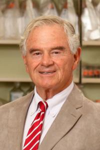 David H Sachs合作论文数Vagelos College of Physicians and Surgeons, Columbia University Irving Medical Center3Andi Qipo合作论文数3Dusan Hanidziar合作论文数3Hang Shi合作论文数3
 Hans Link合作论文数Department of Neurology, Karolinska Institutet3
 Tatsuo Kawai (河合達男)合作论文数Cardiovascular Research Center, Lewis Katz School of Medicine, Temple University3
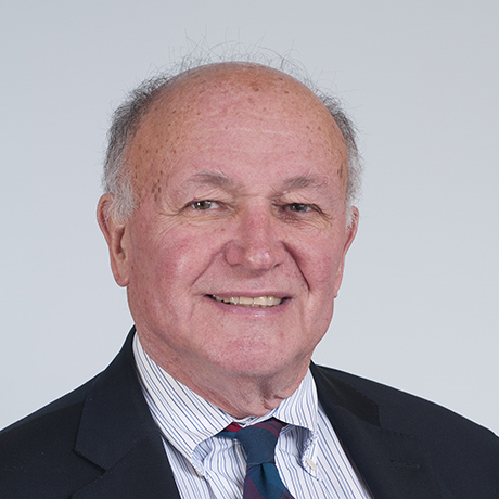 Benedict Cosimi合作论文数Mass General Research Institute, Massachusetts General Hospital, Mass General Brigham;Mass General Cancer Center, Massachusetts General Hospital, Mass General Brigham;Harvard Medical School, Harvard University3Svjetlan Boskovic合作论文数3Vijay Sharma合作论文数2Cindy H. Park合作论文数2
 Essa Abuhelaiqa合作论文数2Lorraine Thompson合作论文数2Nasim Kassam合作论文数2Gabor Bodonyi-Kovacs合作论文数2Hugh Auchincloss合作论文数2YM Huang合作论文数2chaquetta grace合作论文数2jeannette knight合作论文数2
 Su Metcalfe合作论文数2
 Elzbieta Kaczmarek合作论文数2
 P Hjelmström合作论文数Camurus AB, Ideon Sci Pk2
 Tarek Fahmy合作论文数Biomedical Engineering, School of Engineering & Applied Science, Yale University2
 Scott M. Damrauer合作论文数Surgery Faculty, Perelman School of Medicine, University of Pennsylvania2
 Susan Bonner-Weir合作论文数Joslin Diabetes Center, Harvard Medical School;Harvard Stem Cell Institute, Harvard University2Maccariello Elizabeth R合作论文数2
 Cleide Goncalves da Silva合作论文数2
 Jan Hillert合作论文数Department of Clinical Neuroscience, Karolinska Institutet2Ferran Christiane合作论文数2
 Rex Neal Smith合作论文数Harvard Medical School, Harvard University;Department of Pathology, Massachusetts General Hospital2
 Robert Colvin合作论文数Harvard Medical School;The Immunopathology Research Laboratory, Center for Transplantation Sciences, Massachusetts General Hospital;Transplantation Biology Research Center, Massachusetts General Hospital2
 Jeffrey S. Flier合作论文数Blavatnik Institute, Harvard Medical School2Vijay K. Sharma合作论文数2Yan Wu合作论文数2
 Joyce Popoola合作论文数Department of Nephrology;Leicester General Hospital;Clinical Microbiology and Public Health Laboratory2




