BackgroundUltrasonography is an important imaging method for clinical breast cancer screening. As the original echo signals of ultrasonography, ultrasound radiofrequency (RF) signals provide abundant tissue macroscopic and microscopic information and have important development and utilization value in breast cancer detection.MethodsIn this study, we proposed a deep learning method based on bispectrum analysis feature maps to process RF signals and realize breast cancer detection. The bispectrum analysis energy feature maps with frequency subdivision were first proposed and applied to breast cancer detection in this study. Our deep learning network was based on a weight sharing network framework for the input of multiple feature maps. A feature map attention module was designed for multiple feature maps input of the network to adaptively learn both feature maps and features that were conducive to classification. We also designed a similarity constraint factor, learning the similarity and difference between feature maps by cosine distance.ResultsThe experiment results showed that the areas under the receiver operating characteristic curves of our proposed method in the validation set and two independent test sets for benign and malignant breast tumor classification were 0.913, 0.900, and 0.885, respectively. The performance of the model combining four ultrasound bispectrum analysis energy feature maps in breast cancer detection was superior to that of the model using an ultrasound grayscale image and the model using a single bispectrum analysis energy feature map in this study.ConclusionThe combination of deep learning technology and our proposed ultrasound bispectrum analysis energy feature maps effectively realized breast cancer detection and was an efficient method of feature extraction and utilization of ultrasound RF signals.
BACKGROUND:This study explored the feasibility of radiofrequency (RF)-based radiomics analysis techniques for the preoperative prediction of programmed cell death protein 1 (PD-1) in patients with hepatocellular carcinoma (HCC).METHODS:The RF-based radiomics analysis method used ultrasound multifeature maps calculated from the RF signals of HCC patients, including direct energy attenuation (DEA) feature map, skewness of spectrum difference (SSD) feature map, and noncentrality parameter S of the Rician distribution (NRD) feature map. From each of the above ultrasound maps, 345 high-throughput radiomics features were extracted. Then, the useful radiomics features were selected by the sparse representation method and input into support vector machine (SVM) classifier for PD-1 prediction.RESULTS AND CONCLUSION:Among all the RF-based prediction models and the ultrasound grayscale comparative model, the RF-based model using all of the three ultrasound feature maps had the highest prediction accuracy (ACC) and area under the curve (AUC), which were 92.5% and 94.23%, respectively. The method proposed in this paper is effective for the meaningful feature extraction of RF signals and can effectively predict PD-1 in patients with HCC.
Breast cancer is a high incidence of malignancy in women, with a higher mortality rate. Accurate screening is helpful to early detection and improve the treatment success rate and patient survival rate. This study is based on low-cost ultrasound, using ultrasound multifeature maps based on the original radiofrequency (RF) signals and radiomics analysis method to evaluate the benign and malignant of breast tumors. The three ultrasound multifeature maps of breast tumor are composed of direct energy attenuation coefficient (AC), standard deviation of image intensity (SD) and Rician distribution parameters (RD). From the above multifeature maps, high-throughput radiomics features were extracted, then sparse representation method was used for feature selection, and then support vector machine was used to predict the benign and malignant of breast tumors. Eight groups of comparative experiments were established by using ultrasound gray-scale image, single ultrasound feature map and two ultrasound feature maps. The results from 164 patients with breast tumor showed that the AUC, accuracy and sensitivity of the radiomics classification model with feature maps of AC, SD and RD can reach 93.61%, 93.94% and 100%, respectively. The use of RF based ultrasound multifeature maps combined with radiomics could effectively predict the benign and malignant of breast tumors in this study.
Objectives To propose a transfer learning (TL) radiomics model that efficiently combines the information from gray scale and elastogram ultrasound images for accurate liver fibrosis grading. Methods Totally 466 patients undergoing partial hepatectomy were enrolled, including 401 with chronic hepatitis B and 65 without fibrosis pathologically. All patients received elastography and got liver stiffness measurement (LSM) 2–3 days before surgery. We proposed a deep convolutional neural network by TL to analyze images of gray scale modality (GM) and elastogram modality (EM). The TL process was used for liver fibrosis classification by Inception-V3 network which pretrained on ImageNet. The diagnostic performance of TL and non-TL was compared. The value of single modalities, including GM and EM alone, and multimodalities, including GM + LSM and GM + EM, was evaluated and compared with that of LSM and serological indexes. Receiver operating characteristic curve analysis was performed to calculate the optimal area under the curve (AUC) for classifying fibrosis of S4, ≥ S3, and ≥ S2. Results TL in GM and EM demonstrated higher diagnostic accuracy than non-TL, with significantly higher AUCs (all p < .01). Single-modal GM and EM both performed better than LSM and serum indexes (all p < .001). Multimodal GM + EM was the most accurate prediction model (AUCs are 0.950, 0.932, and 0.930 for classifying S4, ≥ S3, and ≥ S2, respectively) compared with GM + LSM, GM and EM alone, LSM, and biomarkers (all p < .05). Conclusions Liver fibrosis can be staged by a transfer learning modal based on the combination of gray scale and elastogram ultrasound images, with excellent performance. Key Points • Transfer learning consists in applying to a specific deep learning algorithm that pretrained on another relevant problem, expected to reduce the risk of overfitting due to insufficient medical images. • Liver fibrosis can be staged by transfer learning radiomics with excellent performance. • The most accurate prediction model of transfer learning by Inception-V3 network is the combination of gray scale and elastogram ultrasound images.
Background: To evaluate the accuracy of radiomics algorithm based on original radio frequency (ORF) signals for prospective prediction of microvascular invasion (MVI) in hepatocellular carcinoma (HCC) lesions. Methods: In this prospective study, we enrolled 42 inpatients diagnosed with HCC from January 2018 to December 2018. All HCC lesions were proved by surgical resection and histopathology results, including 21 lesions with MVI. Ultrasound ORF data and grayscale ultrasound images of HCC lesions were collected before operation for further radiomics analysis. Three ultrasound feature maps were calculated using signal analysis and processing (SAP) technology in first feature extraction. The diagnostic accuracy of model based on ORF signals was compared with the model based on grayscale ultrasound images. Results: A total of 1,050 radiomics features were extracted from ORF signals of each HCC lesion. The performance of MVI prediction model based on ORF was better than those based on grayscale ultrasound images. The best area under curve, accuracy, sensitivity, and specificity of ultrasound radiomics in prediction of MVI were 95.01, 92.86, 85.71, and 100%, respectively. Conclusions: Radiomics algorithm based on ultrasound ORF data combined with SAP technology can effectively predict MVI, which has potential clinical application value for non-invasively preoperative prediction of MVI in HCC patients.
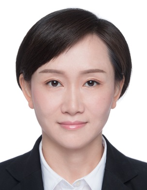 Jinhua Yu (余锦华)合作论文数School of Information Science and Technology, Fudan University5
 Wenping Wang (王文平)合作论文数Zhongshan Hospital, Fudan University3
 Meng Dai合作论文数2Tianlei Xiao合作论文数2Leyin Li合作论文数Fudan Univ, Dept Elect Engn, Shanghai, Peoples R China2Xiaohong Jia合作论文数Shanghai Jiao Tong Univ, Shanghai Ruijin Hosp, Med Sch, Dept Ultrasound, Shanghai, Peoples R China2
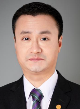 Yao Zhao (赵曜)合作论文数Department of Neurosurgery, Huashan Hospital, Fudan University;Institute of Neurosurgery, Fudan University2Yi Dong (董怡)合作论文数Huashan Hospital, Fudan University2
 Qi Zhang (张奇)合作论文数Shanghai Key Laboratory of Data Science, School of Computer Science, Fudan University;Natural Language Processing Group, Fudan University2Shanghai Institute of Medical Imaging合作论文数1Shiquan Zhang合作论文数1Zhuoyun Jiang合作论文数1
 Ting Luo合作论文数1Shujun Xia合作论文数1Yu-Li Zhu合作论文数1
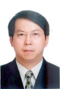 Yuanyuan Wang (汪源源)合作论文数School of Information Science and Technology, Fudan University1
 Qian Li合作论文数1
 Hong Ding (丁红)合作论文数Huashan Hospital, Fudan University1
 Jianqiao Zhou (周建桥)合作论文数Ruijin Hospital, School of Medicine, Shanghai Jiao Tong University1
 Xiao Yang (杨晓)合作论文数Academy of Military Sciences1Li-Yun Xue (薛立云)合作论文数1




