Background:Sustained exposures to high atmospheric levels of PM2.5 at population scale are associated with increased risks for pulmonary inflammatory diseases. These are marked by activation of the TRPC6 (Transient Receptor Potential Canonical type 6) calcium channel, increased reactive oxygen species (ROS) and oxidative stress. Long term exposures are associated with reduced life span, and increased incidences of cardiovascular diseases, dementia, Parkinson's and Alzheimer disease, and increased risk of autism and autism spectrum disorders. It has been proposed that the PM2.5 toxin is benzo[a]pyrene (B[a]P) that is adsorbed to the surface of the PM2.5 particle.. But the mechanism by which B[a]P might drive pulmonary inflammatory diseases, or any other of the indications above, are not known. Hypothesis:B[a]P was recently reported to bind irreversibly and destructively to the β2 Adrenergic Receptor (β2AR) in the lung. We have therefore hypothesized that B[a]P is the adsorbed PM2.5 toxin, and that β2AR is the B[a]P receptor responsible for TRPC6 activation in lung epithelial cells. Results:To test this hypothesis, we exposed a polarized organoid model of normal human lung epithelia, polarized lung epithelial 16HBE14o- cells, and tracheobronchial slice cultures from ferret lung to either PM2.5 or B[a]P. We found that both PM2.5 and B[a]P: (i) irreversibly activated of β2AR signaling via Gi to PI3K/AKT; (ii) increased NFκB-activated release of proinflammatory cytokines through IKKαβ activation by PI3K/AKT, which was suppressed by the PI3K inhibitor LY 294002 (iii) desensitized and destroyed the activated β2AR receptor by endocytic recycling; (iv) also destroyed β2AR's signalplex partner CFTR by the same process; (v) activated the CFTR-bound calcium channel protein TRPC6 due to loss of inhibitory CFTR; leading to (vi) increased cytosolic [Ca2+] concentration; (vii) increased ROS due to mitochondrial uncoupling; and (viii) increased expression of oxidative stress. Treatment with the TRPC6 inhibitor BI 749327 blocked steps (vi-viii), and preserved CFTR from endocytic loss. Treatment of tracheobronchial slice cultures of ferret lung with either PM2.5 or B[a]P resulted in increased secretion of IL-6, increased expression of TRPC6, and reduced expression of β2AR and CFTR. Finally, we found that exposure of lung organoids to B[a]P significantly reduced expression of the same five microRNAs (miR-126a-3p, miR-30b-5p, miR-103a-3p, miR-26a-5p, and miR-766-3p) previously identified in sera from service members exposed to PM2.5 from burn pit emissions during deployment to Iraq and Afghanistan. Conclusion:PM2.5 and the PM2.5 toxin benzo[a]pyrene (B[a]P) induce inflammation and oxidative stress in the airway by increased expression of TRPC6 and inactivation of β2AR/CFTR signaling. These discoveries mark the first identification of a mechanism by which exposure to PM2.5 or the PM2.5 toxin B[a]P itself can induce inflammation and TRPC6-dependent oxidative stress in lung epithelia.
Abstract SARS-CoV-2-contributes to sickness and death in COVID-19 patients partly by inducing a hyper-proinflammatory immune response in the host airway. This hyper-proinflammatory state involves activation of signaling by NFκB, and unexpectedly, ENaC, the epithelial sodium channel. Post-infection inflammation may also contribute to "Long COVID"/PASC. Enhanced signaling by NFκB and ENaC also marks the airway of patients suffering from cystic fibrosis, a life-limiting proinflammatory genetic disease due to inactivating mutations in the CFTR gene. We therefore hypothesized that inflammation in the COVID-19 airway might similarly be due to inhibition of CFTR signaling by SARS-CoV-2 spike protein, and therefore activation of both NFκB and ENaC signaling. We used western blot and electrophysiological techniques, and an organoid model of normal airway epithelia, differentiated on an air–liquid-interface (ALI). We found that CFTR protein expression and CFTR cAMP-activated chloride channel activity were lost when the model epithelium was exposed to SARS-CoV-2 spike proteins. As hypothesized, the absence of CFTR led to activation of both TNFα/NFκB signaling and α and γ ENaC. We had previously shown that the cardiac glycoside drugs digoxin, digitoxin and ouabain blocked interaction of spike protein and ACE2. Consistently, addition of 30 nM concentrations of the cardiac glycoside drugs, prevented loss of both CFTR protein and CFTR channel activity. ACE2 and CFTR were found to co-immunoprecipitate in both basal cells and differentiated epithelia. Thus spike-dependent CFTR loss might involve ACE2 as a bridge between Spike and CFTR. In addition, spike exposure to the epithelia resulted in failure of endosomal recycling to return CFTR to the plasma membrane. Thus, failure of CFTR recovery from endosomal recycling might be a mechanism for spike-dependent loss of CFTR. Finally, we found that authentic SARS-CoV-2 virus infection induced loss of CFTR protein, which was rescued by the cardiac glycoside drugs digitoxin and ouabain. Based on experiments with this organoid model of small airway epithelia, and comparisons with 16HBE14o- and other cell types expressing normal CFTR, we predict that inflammation in the COVID-19 airway may be mediated by inhibition of CFTR signaling by the SARS-CoV-2 spike protein, thus inducing a cystic fibrosis-like clinical phenotype. To our knowledge this is the first time COVID-19 airway inflammation has been experimentally traced in normal subjects to a contribution from SARS-CoV-2 spike-dependent inhibition of CFTR signaling.
To initiate SARS-CoV-2 infection, the Receptor Binding Domain (RBD) on the viral spike protein must first bind to the host receptor ACE2 protein on pulmonary and other ACE2-expressing cells. We hypothesized that cardiac glycoside drugs might block the binding reaction between ACE2 and the Spike (S) protein, and thus block viral penetration into target cells. To test this hypothesis we developed a biochemical assay for ACE2:Spike binding, and tested cardiac glycosides as inhibitors of binding. Here we report that ouabain, digitoxin, and digoxin, as well as sugar-free derivatives digitoxigenin and digoxigenin, are high-affinity competitive inhibitors of ACE2 binding to the Original [D614] S1 and the α/β/γ [D614G] S1 proteins. These drugs also inhibit ACE2 binding to the Original RBD, as well as to RBD proteins containing the β [E484K], Mink [Y453F] and α/β/γ [N501Y] mutations. As hypothesized, we also found that ouabain, digitoxin and digoxin blocked penetration by SARS-CoV-2 Spike-pseudotyped virus into human lung cells, and infectivity by native SARS-CoV-2. These data indicate that cardiac glycosides may block viral penetration into the target cell by first inhibiting ACE2:RBD binding. Clinical concentrations of ouabain and digitoxin are relatively safe for short term use for subjects with normal hearts. It has therefore not escaped our attention that these common cardiac medications could be deployed worldwide as inexpensive repurposed drugs for anti-COVID-19 therapy.
Castration Resistant Prostate Cancer (CRPC) is thought to be driven by a collaborative mechanism between TNFα/NFκB and TGFβ signaling, leading to inflammation, Epithelial-to-Mesenchymal-Transition (EMT), and metastasis. Initially, TGFβ is a tumor suppressor, but in advanced metastatic disease it switches to being a tumor promoter. TGFBR2 may play a critical role in this collaboration, as its expression is driven by NFκB and it is the primary receptor for TGFβ. We have previously reported that the cardenolide drug digitoxin blocks TNFα/NFκB-driven proinflammatory signaling. We therefore hypothesized that digitoxin might break the collaborative process between NFκB and TGFβ by also inhibiting expression of TGFBR2. We therefore tested whether TGFβ-driven EMT and resulting metastases would be suppressed. Here we show, in vitro, that digitoxin inhibits NFκB-driven TGFBR2 expression, as well as Vimentin, while elevating E-cadherin expression. Digitoxin also significantly reduces HSPB1 mRNA and the HSPB1/RBFOX2 mRNA ratio in PC3 cells. In vivo, in a syngeneic, immune competent rat model of metastatic CRPC, we show that digitoxin also suppresses Tgfbr2 expression, as well as expression of other genes classically driven by NFκB, and of multiple EMT genes associated with metastasis. Concurrently, digitoxin suppresses tumor growth and metastasis in these animals, and prolongs survival. Gross tumor recurrence following tumor resection also appears prevented in ca 30% of cases. While the existence of a collaboration between NFκB and TGFβ to drive EMT and metastasis has previously been appreciated, we show here, for the first time, that chronic, low concentrations of digitoxin are able to block CRPC tumor progression, EMT and the ensuing metastatic disease.
BACKGROUND/AIMS:Chronic lung infection in cystic fibrosis leads to an inflammatory response that persists because of the chronic presence of bacteria and ultimately leads to a catastrophic failure of lung function.METHODS:We use a combination of biochemistry, cell and molecular biology to study the interaction of TRADD, a key adaptor molecule in TNFα signaling, with CFTR in the regulation of NFκB.RESULTS:We show that Wt CFTR binds to and colocalizes with TRADD. TRADD is a key signaling intermediate connecting TNFα with activation of NFκB. By contrast, ΔF508 CFTR does not bind to TRADD. NF-κB activation is higher in CFBE expressing ΔF508 CFTR than in cells expressing Wt CFTR. However, this differential effect is abolished when TRADD levels are knocked down. Transfecting Wt CFTR into CFBE cells reduces NF-κB activity. However the reduction is abolished by the CFTR chloride transport inhibitor-172. Consistently, transfecting in the correctly trafficked CFTR conduction mutants G551D or S341A also fail to reduce NFκB activity. Thus CFTR must be functional if it is to regulate NF-κB activity. We also found that TNFα produced a greater increase in NF-κB activity in CFBE cells than in the same cell when Wt CFTR-corrected. Consistently, the effect is also abolished when TRADD is knocked down by shRNA. Thus, Wt CFTR control of TRADD modulates the physiological activation of NF-κB by TNFα. Based on studies with proteosomal and lysosomal inhibitors, the mechanism by which Wt CFTR, but not ΔF508 CFTR, suppresses TRADD is by lysosomal degradation.CONCLUSION:We have uncovered a novel mechanism whereby Wt CFTR regulates TNFα signaling by enhancing TRADD degradation. Thus by reducing the levels of TRADD, Wt CFTR suppresses downstream proinflammatory NFκB signaling. By contrast, suppression of NF-κB activation fails in CF cells expressing ΔF508 CFTR.
Background: The discovery of small molecules to correct the trafficking defect of the mutant ΔF508-CFTR protein has been challenging. Results: C4-ceramide rescues and stabilizes ΔF508-CFTR. High basal secretion of interleukin-8 is also suppressed. Conclusion: Results identify a novel mechanism by which C4-ceramide activates the PDK1/SGK1 pathway, thereby rescuing ΔF508-CFTR. Significance: C4-ceramide may be a novel candidate therapeutic for CF patients. Cystic fibrosis (CF) is due to a folding defect in the CF transmembrane conductance regulator (CFTR) protein. The most common mutation, ΔF508, prevents CFTR from trafficking to the apical plasma membrane. Here we show that activation of the PDK1/SGK1 signaling pathway with C4-ceramide (C4-CER), a non-toxic small molecule, functionally corrects the trafficking defect in both cultured CF cells and primary epithelial cell explants from CF patients. The mechanism of C4-CER action involves a series of mutual autophosphorylation and phosphorylation events between PDK1 and SGK1. Detailed mechanistic studies indicate that C4-CER initially induces autophosphorylation of SGK1 at Ser422. SGK1[Ser(P)422] and C4-CER coincidently bind PDK1 and permit PDK1 to autophosphorylate at Ser241. Then PDK1[Ser(P)241] phosphorylates SGK1[Ser(P)422] at Thr256 to generate fully activated SGK1[Ser422, Thr(P)256]. SGK1[Ser(P)422,Thr(P)256] phosphorylates and inactivates the E3 ubiquitin ligase Nedd4-2. ΔF508-CFTR is thus free to traffic to the plasma membrane. Importantly, C4-CER-mediated activation of both PDK1 and SGK1 is independent of the PI3K/Akt/mammalian target of rapamycin signaling pathway. Physiologically, C4-CER significantly increases maturation and stability of ΔF508-CFTR (t½ ∼10 h), enhances cAMP-activated chloride secretion, and suppresses hypersecretion of interleukin-8 (IL-8). We suggest that candidate drugs for CF directed against the PDK1/SGK1 signaling pathway, such as C4-CER, provide a novel therapeutic strategy for a life-limiting disorder that affects one child, on average, each day.
Background : Cardiac glycosides such as digitoxin have been shown to directly cause apoptotic death of cancer cells both in vitro, and in vivo. However, the mechanism connecting cardiac glycoside action to apoptosis is not known. It has been reported that compounds resembling digitoxin are able to reduce c-MYC expression. Furthermore, it has been previously shown that the transcription of c-MYC depends on nuclear factor of activated T-cells (NFAT) binding sites in the c-MYC promoter. We have therefore hypothesized that NFAT might mediate digitoxin effects on c-MYC mRNA message. Materials and Methods : We have chosen to study this process in HeLa cells where structurally intact c-MYC genes in 8q24 co-localize with human papilloma virus 18 at all integration sites. Results : Here we show that within the 1 st h following treatment with digitoxin, a significant reduction in c-MYC mRNA occurs. This is followed by a precipitous loss of c-MYC protein, activation of caspase 3, and subsequent apoptotic cell death. To test the NFAT-dependence mechanism, we analyzed the effects of digitoxin on NFAT isoform-dependent auto-activation of a NFAT-luciferase expression system. Drug dependent effects on expression varied according to each of the four canonical NFAT isoforms (1, 2, 3 or 4). The most digitoxin-sensitive NFAT isoform was NFAT1. Using c-MYC chromatin immune precipitation, we find that digitoxin inhibits interaction of NFAT1 with the proximal c-MYC promoter. Conclusions : These results suggest that the carcinotoxic activity of digitoxin includes suppression of NFAT-driven c-MYC expression.
Tumour necrosis factor receptor (TNFR)1 is the main receptor responsible for TNF‐induced diverse cellular events. In this study, we report that TNFR1 has a crucial role in endoplasmic reticulum (ER) stress‐induced Jun amino‐terminal kinase (JNK) activation. Although ER stress leads to JNK activation in wild‐type mouse embryo fibroblasts, we failed to detect any JNK activation in TNFR1−/− cells. ER stress‐induced JNK activation is restored in TNFR1−/− cells when TNFR1 expression is reconstituted. We also found that TNFR1 functions downstream of IRE1 and that IRE1 is present in the same complex with TNFR1 under ER stress condition. Therefore, our study shows a novel role of TNFR1 in mediating ER stress‐induced JNK activation.
Insulin-like growth factor I (IGF-I) plays an important role in cell survival, proliferation, and differentiation. Diverse kinases, including AKT/ protein kinase B, extracellular signal-regulated kinase (ERK), and c-Jun N-terminal kinase (JNK), can be activated by IGF-I. Here, we show that the receptor-interacting protein (RIP), a key mediator of tumor necrosis factor-induced NF-kappa B and JNK activation, plays a key role in IGF-I receptor signaling. IGF-I induced a robust JNK activation in wild type but not RIP null (RIP-/-) mouse embryonic fibroblast cells. Reconstitution of RIP expression in the RIP-/- cells restored the induction of JNK by IGF-I, suggesting that RIP is essential in IGF-I-induced JNK activation. Reconstitution experiments with different RIP mutants further revealed that the death domain and the kinase activity of RIP are not required for IGF-I-induced JNK activation. Interestingly, the AKT and ERK activation by IGF-I was normal in RIP-/- cells. The phosphatidylinositol 3-kinase inhibitor, wortmannin, did not affect IGF-I-induced JNK activation. These results agree with previous studies showing that the IGF-I-induced JNK activation pathway is distinct from that of ERK and AKT activation. Additionally, physical interaction of ectopically expressed RIP and IGF-IR beta was detected by co-immunoprecipitation assays. More importantly, RIP was recruited to the IGF-I receptor complex during IGF-I-induced signaling. Furthermore, we found that IGF-I-induced cell proliferation was impaired in RIP-/- cells. Taken together, our results indicate that RIP, a key factor in tumor necrosis factor signaling, also plays a pivotal role in IGF-I-induced JNK activation and cell proliferation.
Digitoxin and structurally related cardiac glycoside drugs potently block activation of the TNF-alpha/NF-kappaB signaling pathway. We have hypothesized that the mechanism might be discovered by searching systematically for selective inhibitory action through the entire pathway. We report that the common action of these drugs is to block the TNF-alpha-dependent binding of TNF receptor 1 to TNF receptor-associated death domain. This drug action can be observed with native cells, such as HeLa, and reconstituted systems prepared in HEK293 cells. All other antiinflammatory effects of digitoxin on NF-kappaB and c-Jun N-terminal kinase pathways appear to follow from the blockade of this initial upstream signaling event.
Cystic fibrosis (CF) is a common, lethal genetic disease, which is due to mutations in the CFTR gene. The CF lung expresses a profoundly proinflammatory phenotype, due to constitutive hypersecretion of IL-8 from epithelial cells lining the airways. In a systematic search for candidate drugs that might be used therapeutically to suppress IL-8 secretion from these cells, we have identified a potent and efficacious series of amphiphilic pyridinium salts. The most potent of these salts is MRS2481, an (R)-1-phenylpropionic acid ester, with an IC50 of ca. 1μM. We have synthesized 21 analogues of MRS2481, which have proven sufficient to develop a preliminary structure–activity relationship (SAR). For optimal activity, we have found that the ester must be connected to the pyridinium derivative by an eight-carbon chain. An optical isomer of the lead compound, containing an (S)-1-phenylpropionic acid ester, has been found to be a much less active. The mechanism of action of MRS2481 appears to involve inhibition of signaling of the NFκB and AP-1 transcription factors to the IL-8 promoter. MRS2481 is a potent inhibitor of TNFα-induced phosphorylation and proteosomal destruction of IκBα. Inasmuch as IκBα is the principal inhibitor of the NFκB signaling pathway, preservation of intact IκBα would serve to keep the IL-8 promoter silent. We also find that MRS2481 blocks TNFα-activated phosphorylation of JNK, the c-JUN kinase. The IL-8 promoter is also activated by an AP-1 site, which requires a phospho-c-JUN/c-FOS dimer for activity. We therefore interpret these data to suggest that the mechanism of MRS2481 action is to inhibit both NFκB and AP-1 signaling on the IL-8 promoter. Given the medicinally promising properties of water-solubility, potency in the low μM concentration range, and high efficacy, we anticipate that MRS2481, or a further optimized derivative, may find an important place in the armamentarium of pharmaceutical strategies yet to be arrayed against the inflammatory phenotype of the CF lung.
Cystic fibrosis (CF) is a fatal, autosomal, recessive genetic disease that is characterized by profound lung inflammation. The inflammatory process is believed to be caused by massive overproduction of the proinflammatory protein IL-8, and the high levels of IL-8 in the CF lung are therefore believed to be the central mechanism behind CF lung pathophysiology. We show here that digitoxin, at sub nM concentrations, can suppress hypersecretion of IL-8 from cultured CF lung epithelial cells. Certain other cardiac glycosides are also active but with much less potency. The specific mechanism of digitoxin action is to block phosphorylation of the inhibitor of NF-kappa B (I kappa B alpha). I kappa B alpha phosphorylation is a required step in the activation of the NF-kappa B signaling pathway and the subsequent expression of IL-8. Digitoxin also has effects on global gene expression in CF cells. Of the informative genes expressed by the CF epithelial cell line IB-3, 58 are significantly (P < 0.05) affected by gene therapy with wild-type (CFTR CF transmembrane conductance regulator). Of these 58 genes, 36 (62%) are similarly affected by digitoxin and related active analogues. We interpret this result to suggest that digitoxin can also partially mimic the genomic consequences of gene therapy with CF transmembrane conductance regulator. We therefore suggest that digitoxin, with its lengthy history of human use, deserves consideration as a candidate drug for suppressing IL-8-dependent lung inflammation in CF.
The mechanism of tumor necrosis factor (TNF)-induced nonapoptotic cell death is largely unknown, although the mechanism of TNF-induced apoptosis has been studied extensively. In wild-type mouse embryonic fibroblast cells under a caspase-inhibited condition, TNF effectively induced cell death that morphologically resembled necrosis. In this study, we utilized gene knockout mouse embryonic fibroblasts cells and found that tumor necrosis factor receptor (TNFR) I mediates TNF-induced necrotic cell death, and that RIP, FADD, and TRAF2 are critical components of the signaling cascade of this TNF-induced necrotic cell death. Inhibitors of NF-kappaB facilitated TNF-induced necrotic cell death, suggesting that NF-kappaB suppresses the necrotic cell death pathway. JNK, p38, and ERK activation seem not to be required for this type of cell death because mitogen-activated protein kinase inhibitors did not significantly affect TNF-induced necrotic cell death. In agreement with the previous reports that the reactive oxygen species (ROS) may play an important role in this type of cell death, the ROS scavenger butylated hydroxyanisole efficiently blocked TNF-induced necrotic cell death. Interestingly, during TNF-induced necrotic cell death, the cellular ROS level was significantly elevated in wild type, but not in RIP(-/-), TRAF2(-/-), and FADD(-/-) cells. These results suggest that RIP, TRAF2, and FADD are crucial in mediating ROS accumulation in TNF-induced necrotic cell death.
The transcription factor NF-kappaB is activated when cells are exposed to genotoxic stress. It has been suggested that DNA damage will trigger a cytoplasmic signaling that leads to the activation of IKK and NF-kappaB, but the signaling components upstream of IKK have not yet been identified. Here we report that the receptor interacting protein, RIP, is the IKK upstream component, essential for the activation of NF-kappaB by DNA damage. Also, our findings suggest that this NF-kappaB activation by DNA damage is not mediated by autocrine or TNF-R1 signaling pathway. In wild-type fibroblasts, DNA damage induced by agents such as adriamycin, campthothecin, and ionizing radiation induces NF-kappaB activation. We found, however, that DNA damage failed to activate NF-kappaB in RIP-/- fibroblasts. The induction of IkappaBalpha degradation by DNA damage was normal in TNF-R1-/-, TRAF2-/-, TRAF5-/- and FADD-/- fibroblasts or when de novo protein synthesis was blocked. More importantly, the reconstitution of RIP expression in RIP-/- cells restores DNA damage-induced NF-kappaB activation. We also found that RIP forms a complex with IKK in response to DNA damage. Therefore, our study provides a possible mechanism for the initiation of the cytoplasmic signaling to activate NF-kappaB in response to DNA damage.
INTRODUCTION. NFκB is a transcription factor implicated in the regulation of inflammation and in cellular processes associated with the development of cancer. These processes include tumor promotion, angiogenesis, metastasis, anti-apoptosis/survival, and resistance to chemotherapeutic drugs (1). NFκB has therefore become one of the major targets for the development of anti-cancer drugs (1,2). We have recently reported that cardiac glycoside drugs such as digitoxin and oleandrin are able to suppress constitutive hypersecretion of IL-8 from lung epithelial IB3-1 cells by inhibition of the NKκB signaling (3). However, where the active cardiac glycosides act in the NFκB signaling pathway is not yet known. We hypothesized that the mechanism for cardiac glycoside activity may be found through a systematic analysis of the entire TNFα/NFκB signaling pathway. METHODS. To test this hypothesis we have used Western blot, NFκB luciferase assay, kinase assay and other relevant assays to search for the upstream site(s) of cardiac glycoside action in TNFα-activated HeLa cells. The active cardiac glycosides, digitoxin ("II") and oleandrin("I"), have been used as the positive inhibitors, and the inactive digoxigenin 3,12-diAc ("VIII") has been used as a negative control. RESULTS. We find that pretreatment of HeLa cells with oleandrin or digitoxin for 3 or 6 hours prior to activation with TNFα effectively blocks degradation of IκBα. The same pretreatment strategy was also found to block the upstream kinase activity of IκB kinase (IKK), suggesting an upstream site of cardiac glycoside action (see Fig.1A). We also found that cardiac glycoside pretreatment blocked TNFα induced phosphorylation and kinase activity of c- Jun amino (N)-terminal kinase (JNK)(See Fig. 1B). This result suggested that the site of cardiac glycoside action might be on the upstream pathway
 Harvey B Pollard合作论文数School of Medicine, Uniformed Services University of the Health Sciences12Meera Srivastava合作论文数Laboratory of Cell Biology and Genetics, NIDDK National Institutes of Health5Bette S. Pollard合作论文数5
 Yong Lin合作论文数Lovelace Respiratory Research Institute5
 Catherine Jozwik合作论文数5
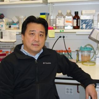 Zhenggang Liu合作论文数Laboratory of Immune Cell Biology, Center for Cancer Research, National Cancer Institute, National Institutes of Health4
 Clifton L Dalgard合作论文数School of Medicine, Uniformed Services University3
 Roopa Biswas合作论文数3Hung Caohuy合作论文数3Gang Min Hur合作论文数3Nathan Walton合作论文数2Lewis Joseph合作论文数2Jian Zhang合作论文数2Ting-Hua Chen合作论文数2Kim You-Sun合作论文数Ajou University School of Medicine, Ajou University2
 Kenneth A. Jacobson合作论文数Molecular Recognition Section, Laboratory of Bioorganic Chemistry, National Institute of Diabetes and Digestive and Kidney Diseases, National Institutes of Health2Mirta Glasman合作论文数2
 Hanming Shen (沈漢明)合作论文数Faculty of Health Sciences, University of Macau2Ofer Eidelman合作论文数2William R. Wolter合作论文数1Andrew S. Herbert合作论文数1S. Nedospasov合作论文数1Tony T. Wang合作论文数1Greg P. Mueller合作论文数1Ognoon Mungunsukh合作论文数1William Gillette合作论文数1W. Huang合作论文数1James Hartley合作论文数1Xijun Zhang合作论文数1Shufeng Liu合作论文数Ctr Biol Evaluat & Res, US FDA1
 Catalina Florez合作论文数Department of Chemistry and Life Science, United States Military Academy at West Point1
 Liudmila Cebotaru合作论文数Department of Physiology, School of Medicine, Johns Hopkins University1
 William Guggino合作论文数Department of Physiology, Johns Hopkins University School of Medicine1Starr Joshua M合作论文数Uniformed Services University, Uniformed Services University of the Health Sciences (USUHS),1
 David M. Jacobowitz合作论文数Department of Anatomy, Physiology, and Genetics, Uniformed Services University School of Medicine, Uniformed Services University of the Health Sciences1
 Raymond A. Frizzell合作论文数Cystic Fibrosis Research Center, Departments of Pediatrics and Cell Biology, University of Pittsburgh1Glover Matthew合作论文数1Xu Andrew E合作论文数1Ha Thien-An合作论文数1Eudy Yvonne合作论文数1Lee Ha Won合作论文数1
 Nakano Hiroyasu (中野裕康)合作论文数Department of Biochemistry, School of Medicine, Toho University1
 Kumar Parameet合作论文数1Wang Hua合作论文数1
 Wei Huang (黄薇)合作论文数Ruijin Hospital, Shanghai Jiaotong University School of Medicine;Shanghai Institute for Biomedical and Pharmaceutical Technologies1
 Swati Choksi合作论文数National Cancer Institute, National Institutes of Health1




