Mount Everest is climbed by an increasing number of mountaineers. Although climbing techniques have changed in recent years, the impact on patterns of mortality is unclear. To compare mortality patterns from 1921 to 2006 with those from 2007 to 2024, we performed a retrospective observational study of mountaineers climbing Everest. We extracted data from the Himalayan Database and examined accounts of deaths. The overall mortality rate above base camp decreased from 1.4% to 0.7% (P < 0.001). From 2007 to 2024, the mean altitude of fatal incidents on the standard routes involving climbers was higher than those involving Sherpas (7970 ± 917 m vs. 6894 ± 1195 m, P = 0.0003). Most climbers (76.5%) died on summit day, whereas most Sherpas (82.2%) died during route preparation. Climber mortality during summit descent on the standard routes during spring 1982-2006 vs. 2007-2024 decreased from 3.0% to 0.8% (P < 0.0001). The spring summit descent mortality for 2007 to 2024 was higher amongst climbers than Sherpas (0.8% vs. 0.1%, P < 0.0001). Mortality rates have decreased on Everest, but further improvements in safety at extreme altitude may be possible. KEY POINTS: Climbing techniques on Mount Everest have changed in recent years, which may impact patterns of mortality. Overall mortality rates from 1921 to 2006 vs. those from 2007 to 20024 decreased from 1.4% to 0.7%. Most climbers still die on summit day, but mortality rates during descent from the summit decreased from 3.0% to 0.8%. Above 8000 m, there are less death involving falls or climbers separated from their group, but profound exhaustion and impaired consciousness are still frequent symptoms of those who died in this hypoxic environment. Awareness of the persistent patterns of mortality may help mountaineers avoid fatal situations.
Chronic exposure to high altitude leads to increases in hemoglobin mass (Hbmass), which may improve exercise performance and decrease acute mountain sickness (AMS) symptoms. We evaluated the influence of intravenous iron or erythropoietin (EPO) treatment on Hbmass, exercise performance, and AMS during a 14-day exposure to 3,094 m. Thirty-nine participants (12 F) completed the study conducted in Eugene, Oregon [sea level (SL), 130 m] and Leadville, Colorado (3,094 m). Participants were dosed with either a placebo (saline; n = 13), iron [Fe(III)-hydroxide sucrose 200 mg, 2 times; n = 14], or EPO (epoetin alfa 50 IU/kg, 3 times/wk; n = 12) at SL for ∼3.5 wk. Hbmass, exercise performance, and AMS symptoms were measured at SL before treatment and on days 1, 2, 7, 13, and 14 at altitude. Absolute Hbmass (g) increased from SL and day 1 to day 13 (P < 0.0001) with no differences between treatment groups (P = 0.3868). Five-kilometer run times were slower at altitude compared with SL (P < 0.0001) and did not improve over the 14-day period at altitude for any group (P > 0.05). Hike times improved with acclimatization from day 2 to day 14 (P = 0.0018) in all groups, independent of treatment. The incidence and severity of AMS remained low across all participants and time points, regardless of treatment. In the current study and dosing, intravenous iron and EPO did not lead to improvements in Hbmass, exercise performance, or AMS with rapid ascent and residence at 3,100 m.NEW & NOTEWORTHY We investigated the influence of iron or erythropoietin treatment on hemoglobin mass, exercise performance, and acute mountain sickness (AMS) during a 14-day exposure to 3,094 m. With the given drug doses of iron [Fe(III)-hydroxide sucrose 200 mg, 2 times over 4 wk] and erythropoietin (epoetin alfa 50 IU/kg, 3 times/wk for 3 wk), we observed no differences in hemoglobin mass, aerobic exercise performance, or symptoms of AMS between groups relative to a placebo.
IntroductionAcute mountain sickness (AMS) is a common altitude illness that occurs when individuals rapidly ascend to altitudes ≥2,500 m without proper acclimatization. Genetic and genomic factors can contribute to the development of AMS or predispose individuals to susceptibility. This study aimed to investigate differential gene regulation and biological pathways to diagnose AMS from high-altitude (HA; 4,300 m) blood samples and predict AMS-susceptible (AMS+) and AMS-resistant (AMS─) individuals from sea-level (SL; 50 m) blood samples.MethodsTwo independent cohorts were used to ensure the robustness of the findings. Blood samples were collected from participants at SL and HA. RNA sequencing was employed to profile gene expression. Differential expression analysis and pathway enrichment were performed to uncover transcriptomic signatures associated with AMS. Biomarker panels were developed for diagnostic and predictive purposes.ResultsAt HA, hemoglobin-related genes (HBA1, HBA2, and HBB) and phosphodiesterase 5A (PDE5A) emerged as key differentiators between AMS+ and AMS− individuals. The cAMP response element-binding protein (CREB) pathway exhibited contrasting regulatory patterns at SL and HA, reflecting potential adaptation mechanisms to hypoxic conditions. Diagnostic and predictive biomarker panels were proposed based on the identified transcriptomic signatures, demonstrating strong potential for distinguishing AMS+ from AMS− individuals.DiscussionThe findings highlight the importance of hemoglobin-related genes and the CREB pathway in AMS susceptibility and adaptation to hypoxia. The differential regulation of these pathways provides novel insights into the biological mechanisms underlying AMS. The proposed biomarker panels offer promising avenues for the early diagnosis and prediction of AMS risk, which could enhance preventive and therapeutic strategies.
Ventilatory acclimatization to high altitude is an important adaption to prolonged time at altitude. Changes in breathing variability over a 16 day acclimatization period to altitude, when awake, were quantified. We tested the hypothesis that the increase in ventilatory loop gain with acclimatization would exacerbate breathing variability at rest and during exercise and that these changes would be correlated with the changes in cerebrovascular CO2, chemoreceptor sensitivity and successful ventilatory acclimatization. Breath-by-breath ventilation, partial pressure of end-tidal O2 and CO2 in 21 healthy lowlanders were recorded at sea level (SL), at day 1 at high altitude (ALT1) and after 16 days of acclimatization (ALT16) to 5260 m at rest and during submaximal and maximal exercise. Power spectrum density (PSD) and the frequency of ventilatory oscillations (fmax) were compared across conditions. At rest, PSD of oscillations were significantly greater at ALT1 and ALT16 compared to SL and fmax of oscillations was significantly lower at ALT16 compared to SL and ALT1. During submaximal exercise PSD of these oscillations was also significantly greater at ALT1 and ALT16 compared to SL. Their fmax were not different between SL, ALT1 and ALT16. The area under the curve (AUC) of ventilation at SL rest and the AUC of ventilation increase from SL to ALT1 was positively associated with the increase in ventilation from SL to ALT16. These data highlight the intrinsic oscillatory nature of the closed loop ventilatory system during and after altitude acclimatization. The AUC metric at SL and acute altitude may be important for predicting successful ventilatory acclimatization with chronic altitude. KEY POINTS: The amplitude of the ventilatory oscillations in acute and chronic hypoxia were greater than at sea level (SL). The frequency of oscillations was slower after acclimatization. These changes may be a result of the increased chemosensitivity and to the closed loop system of the control of ventilation. These changes may also contribute to the adaptation of this closed loop to chronic altitude by limiting the decrease in arterial P O 2 ${{P}_{{{{\mathrm{O}}}_2}}}$ . The area under the curve (AUC) of ventilation at SL rest and the change in AUC from SL to day 1 at high altitude (ALT 1) were both positively associated with the greatest increases in VE from SL to ALT 16, highlighting the potential importance of this metric in predicting successful ventilatory acclimatization to high altitude.
Hypoxia and certain chronic diseases can increase the work of breathing, potentially impacting the muscle hyperemic response during exercise and overall exercise performance. Respiratory muscle training (RMT) has been observed to decrease perceived effort under hypoxic conditions; however, it remains unclear if RMT affects exercise performance in acute hypoxia. We investigated whether 4 weeks of RMT could enhance submaximal and maximal exercise performance in hypoxia (barometric pressure = 425 mmHg, ~4876 m). Seven adult males underwent a 250 kJ submaximal (target RPE 15-17) cycling trial in hypoxia, followed by an incremental test to maximal exertion. After 4 weeks of RMT under normoxic conditions, these tests were repeated in hypoxia. After RMT, RPE showed no significant difference during the submaximal test (Pre-RMT: 16 ± 0.45, Post-RMT: 16 ± 0.45, p > 0.05) despite an increase in HR (Pre-RMT:154 ± 16, Post-RMT: 161 ± 12, p < 0.05). There were no significant differences in submaximal exercise performance. However, RMT did raise maximal oxygen uptake (V̇O2max) peak power (Pre-RMT: 243 ± 35, Post-RMT: 252 ± 38 W, p = 0.04, Cohen's dz. = 0.97). Future investigations should explore the potential effects of RMT on perceived exertion and exercise tolerance at altitude. The results of this pilot study suggest that RMT may improve peak power at V̇O2max, however given the limited sample size of this investigation, further research is required to determine if RMT impacts performance in other domains of exercise intensity. Furthermore, whether RMT impacts performance at altitude in females remains unknown.
Hypoxia and certain chronic diseases can intensify the work of breathing, potentially impacting the muscle hyperemic response during exercise and overall exercise performance. Respiratory muscle training (RMT) has been observed to decrease perceived effort under hypoxic conditions; however, it remains unclear if RMT boosts exercise performance in acute hypoxia. We investigated whether 4 weeks of RMT could enhance maximal and submaximal exercise performance in hypoxia (PB=425 mmHg, ~4876 m). Seven adult males underwent a 250 kJ submaximal (RPE 15-17) cycling trial in hypoxia, followed by an incremental test to maximal exertion. After 4 weeks of RMT under normoxic conditions, these tests were repeated in hypoxia. Although six of the participants displayed improvements in submaximal exercise performance, these numerical differences failed to reach statistical significance. However, RMT did raise VO2max peak power (Pre-RMT: 242.62 ± 35, Post-RMT: 251.85 ± 38 W, p=0.04). Moreover, even with an increased HR during the submaximal exercise trial after RMT (Pre-RMT:154.14 ± 16, Post-RMT: 160.86 ± 12, p<0.05), the RPE showed no significant difference during the test (Pre-RMT: 17.43 ± 0.79, Post-RMT: 17.43 ± 0.53, P>0.05). Despite this, no substantial increase in hypoxic submaximal exercise performance was observed, likely due to the small sample size. These findings suggest RMT might increase hypoxic exercise tolerance by reducing perceived exertion for a given HR. This work was supported by funding from the United States Special Operations Command (USSOCOM) and the National Institutes of Health (2L30HL134123-03). This is the full abstract presented at the American Physiology Summit 2024 meeting and is only available in HTML format. There are no additional versions or additional content available for this abstract. Physiology was not involved in the peer review process.
Acute hypoxia increases pulmonary arterial (PA) pressures, though its effect on right ventricular (RV) function is controversial. The objective of this study was to characterize exertional RV performance during acute hypoxia. Ten healthy participants (34 ± 10 years, 7 males) completed three visits: visits 1 and 2 included non-invasive normoxic (fraction of inspired oxygen ( F i O 2 ${F_{{\mathrm{i}}{{\mathrm{O}}_{\mathrm{2}}}}}$ ) = 0.21) and isobaric hypoxic ( F i O 2 ${F_{{\mathrm{i}}{{\mathrm{O}}_{\mathrm{2}}}}}$ = 0.12) cardiopulmonary exercise testing (CPET) to determine normoxic/hypoxic maximal oxygen uptake ( V ̇ O 2 max ${\dot V_{{{\mathrm{O}}_{\mathrm{2}}}{\mathrm{max}}}}$ ). Visit 3 involved invasive haemodynamic assessments where participants were randomized 1:1 to either Swan-Ganz or conductance catheterization to quantify RV performance via pressure-volume analysis. Arterial oxygen saturation was determined by blood gas analysis from radial arterial catheterization. During visit 3, participants completed invasive submaximal CPET testing at 50% normoxic V ̇ O 2 max ${\dot V_{{{\mathrm{O}}_{\mathrm{2}}}{\mathrm{max}}}}$ and again at 50% hypoxic V ̇ O 2 max ${\dot V_{{{\mathrm{O}}_{\mathrm{2}}}{\mathrm{max}}}}$ ( F i O 2 ${F_{{\mathrm{i}}{{\mathrm{O}}_{\mathrm{2}}}}}$ = 0.12). Median (interquartile range) values for non-invasive V ̇ O 2 max ${\dot V_{{{\mathrm{O}}_{\mathrm{2}}}{\mathrm{max}}}}$ values during normoxic and hypoxic testing were 2.98 (2.43, 3.66) l/min and 1.84 (1.62, 2.25) l/min, respectively (P < 0.0001). Mean PA pressure increased significantly when transitioning from rest to submaximal exercise during normoxic and hypoxic conditions (P = 0.0014). Metrics of RV contractility including preload recruitable stroke work, dP/dtmax, and end-systolic pressure increased significantly during the transition from rest to exercise under normoxic and hypoxic conditions. Ventricular-arterial coupling was maintained during normoxic exercise at 50% V ̇ O 2 max ${\dot V_{{{\mathrm{O}}_{\mathrm{2}}}{\mathrm{max}}}}$ . During submaximal exercise at 50% of hypoxic V ̇ O 2 max ${\dot V_{{{\mathrm{O}}_{\mathrm{2}}}{\mathrm{max}}}}$ , ventricular-arterial coupling declined but remained within normal limits. In conclusion, resting and exertional RV functions are preserved in response to acute exposure to hypoxia at an F i O 2 ${F_{{\mathrm{i}}{{\mathrm{O}}_{\mathrm{2}}}}}$ = 0.12 and the associated increase in PA pressures. KEY POINTS: The healthy right ventricle augments contractility, lusitropy and energetics during periods of increased metabolic demand (e.g. exercise) in acute hypoxic conditions. During submaximal exercise, ventricular-arterial coupling decreases but remains within normal limits, ensuring that cardiac output and systemic perfusion are maintained. These data describe right ventricular physiological responses during submaximal exercise under conditions of acute hypoxia, such as occurs during exposure to high altitude and/or acute hypoxic respiratory failure.
PURPOSE: Oxygen delivery to muscle increases during exercise to match metabolic demand. Hypoxic conditions necessitate adaptations to maintain oxygen delivery but also increase red blood cell (RBC) oxidative stress. The purpose of this study was to examine the response of RBC metabolism to acute hypoxic vs normoxic exercise. METHODS: Nine healthy subjects (35 ± 10 years, 6 males) exercised on upright cycle ergometer under normoxic (FiO2 = 0.21) and hypoxic (FiO2 = 0.12; Patm = 623 mmHg) conditions. Subjects completed three visits including normoxic and hypoxic maximal exercise tests and invasive exercise test. During invasive exercise test, venous blood was collected during normoxic rest and submaximal exercise (50% normoxic VO2max) and hypoxic rest, submaximal (50% hypoxic VO2max), and maximal exercise. RBC metabolomics were analyzed using ultra-high-performance liquid chromatography coupled to mass spectrometry. RESULTS: Maximal oxygen uptake (VO2max) in hypoxic conditions was significantly reduced compared to normoxic conditions (26.2 ± 4.9 vs 41.6 ± 7.4 ml/kg/min; p < 0.05). During hypoxic exercise, medium- (5-12 carbons) and long-chain (≥14 carbons) acylcarnitines (AC; named according to number of carbons, number of double bonds, and presence/absence of hydroxyl group) accumulated in RBCs (p < 0.05 for AC 8:0, AC 8-OH, AC 10:0, AC 10-OH, AC 12-OH, AC 14:1, AC 14:1-OH, AC 14-OH, AC 16:2, AC 16:1-OH). Glycolytic intermediates decreased but lactate increased (4.7 ± 1.9E+06 relative ion count [integrated peak area of extracted ion chromatogram] during normoxic submaximal exercise to 7.9 ± 4.3E+06 during hypoxic submaximal exercise, p < 0.05). Glycolytic intermediate 2-3-bisphosphoglycerate was significantly reduced at maximal hypoxic exercise (8.1 ± 1.3E+06 at hypoxic rest vs 5.9 ± 1.4E+06 at hypoxic maximal exercise, p < 0.05) and varied inversely with lactate (r = -0.54, p < 0.05). CONCLUSIONS: Exercise in hypoxic conditions is associated with RBC metabolic changes including acylcarnitine accumulation and rapid glycolytic flux. Accumulating acylcarnitines may function to repair RBC membranes damaged in the setting of oxidative stress, and rapid glycolytic flux generates ATP during exercise in acute hypoxia. FUNDING: NIH/NCATS UL1TR002535
Acute altitude exposure lowers arterial oxygen content (CaO2) and cardiac output (Qc) at peak exercise, whereas O2 extraction from blood to working muscles remains similar. Acclimatization normalizes CaO2 but not peak Qc nor peak oxygen consumption (Vo2peak). To what extent acclimatization impacts muscle O2 extraction remains unresolved. Twenty-one sea-level residents performed an incremental cycling exercise to exhaustion near sea level (SL), in acute (ALT1) and chronic (ALT16) hypoxia (5,260 m). Arterial blood gases, gas exchange at the mouth and oxy- (O2Hb) and deoxyhemoglobin (HHb) of the vastus lateralis were recorded to assess arterial O2 content (CaO2), Qc, and Vo2. The HHb-Vo2 slope was taken as a surrogate for muscle O2 extraction. During moderate-intensity exercise, HHb-Vo2 slope increased to a comparable extent at ALT1 (2.13 +/- 0.94) and ALT16 (2.03 +/- 0.88) compared with SL (1.27 +/- 0.12), indicating increased O2 extraction. However, the HHb/CaO2 ratio increased from SL to ALT1 and then tended to go back to SL values at ALT16. During high-intensity exercise, HHb-Vo2 slope reached a break point beyond which it decreased at SL and ALT1, but not at ALT16. Increased muscle O2 extraction during submaximal exercise was associated with decreased CaO2 in acute hypoxia. The significantly greater muscle O2 extraction during maximal exercise in chronic hypoxia is suggestive of an O2 reserve. NEW & NOTEWORTHY During incremental exercise muscle deoxyhemoglobin (HHb) and oxygen consumption (Vo2) both increase linearly, and the slope of their relationship is an indirect index of local muscle O2 extraction. The latter was assessed at sea level, in acute and during chronic exposure to 5,260 m. The demonstrated presence of a muscle O2 extraction reserve during chronic exposure is coherent with previous studies indicating both limited muscle oxidative capacity and decrease in motor drive.
relevance in children with ARF but not PARDS. Recent investigations in adults with ARF without ARDS also support the existence of two subphenotypes with characteristics similar to those seen in patients with ARDS, again distinguishable by inflammatory biomarkers and by clinical outcomes (5, 8). Thus, these two subphenotypes share overlap across the adult and pediatric ARF spectra, offering a rationale for innovative trial enrollment strategies across ages. Our findings emphasize the challenge of syndromic definitions such as PARDS in specific, and ARF in general, which struggle to identify and characterize complex pathophysiologic processes that culminate frommultiple inciting diagnoses. Our data suggest that the complex inflammatory pathways and inflammatory-related subphenotypes associated with PARDS (3, 9) are also involved in children with ARF, and recent data suggest that they may also be observed in other critically ill children (10). In conclusion, these data suggest that parsimonious model subphenotype assignment can enable robust prognostic enrichment compared with prior risk stratification schema. Earlier identification of high-risk subphenotypes, particularly in pediatric patients, could result in earlier escalation of care in patients at greater risk for complex course. After validation, these data may impact future clinical trial design, including the expansion of subphenotype identification to the broader pediatric ARF cohort.
New Findings What is the central question to this study? Is there a relationship between a patent foramen ovale and the development of acute mountain sickness and an exaggerated increase in pulmonary pressure in response to 7–10 h of normobaric hypoxia? What is the main finding and its importance? Patent foramen ovale presence did not increase susceptibility to acute mountain sickness or result in an exaggerated increase in pulmonary artery systolic pressure with normobaric hypoxia. This suggests hypobaric hypoxia is integral to the increased susceptibility to acute mountain sickness previously reported in those with patent foramen ovale, and patent foramen ovale presence alone does not contribute to the hypoxic pulmonary pressor response. AbstractAcute mountain sickness (AMS) develops following rapid ascent to altitude, but its exact causes remain unknown. A patent foramen ovale (PFO) is a right‐to‐left intracardiac shunt present in ∼30% of the population that has been shown to increase AMS susceptibility with high altitude hypoxia. Additionally, high altitude pulmonary oedema (HAPE) is a severe type of altitude illness characterized by an exaggerated pulmonary pressure response, and there is a greater prevalence of PFO in those with a history of HAPE. However, whether hypoxia per se is causing the increased incidence of AMS in those with a PFO and whether a PFO is associated with an exaggerated increase in pulmonary pressure in those without a history of HAPE is unknown. Participants (n = 36) matched for biological sex (18 female) and the presence or absence of a PFO (18 PFO+) were exposed to 7–10 h of normobaric hypoxia equivalent to 4755 m. Presence and severity of AMS was determined using the Lake Louise AMS scoring system. Pulmonary artery systolic pressure, cardiac output and total pulmonary resistance were measured using ultrasound. We found no significant association of PFO with incidence or severity of AMS and no association of PFO with arterial oxygen saturation. Additionally, there was no effect of a PFO on pulmonary pressure, cardiac output or total pulmonary resistance. These data suggest that hypobaric hypoxia is necessary for those with a PFO to have increased incidence of AMS and that presence of PFO is not associated with an exaggerated pulmonary pressor response.
Due to lack of nuclei and de novo protein synthesis, post-translational modification (PTM) is imperative for erythrocytes to regulate oxygen (O2) delivery and combat tissue hypoxia. Here, we report that erythrocyte transglutminase-2 (eTG2)-mediated PTM is essential to trigger O2 delivery by promoting bisphosphoglycerate mutase proteostasis and the Rapoport-Luebering glycolytic shunt for adaptation to hypoxia, in healthy humans ascending to high altitude and in two distinct murine models of hypoxia. In a pathological hypoxia model with chronic kidney disease (CKD), eTG2 is critical to combat renal hypoxia-induced reduction of Slc22a5 transcription and OCNT2 protein levels via HIF-1α-PPARα signaling to maintain carnitine homeostasis. Carnitine supplementation is an effective and safe therapeutic approach to counteract hypertension and progression of CKD by enhancing erythrocyte O2 delivery. Altogether, we reveal eTG2 as an erythrocyte protein stabilizer orchestrating O2 delivery and tissue adaptive metabolic reprogramming and identify carnitine-based therapy to mitigate hypoxia and CKD progression.
Ventilatory acclimatization is a hallmark physiological response to high altitude. In ~30% of the population, a shunt located between the right and left atrium called patent foramen ovale (PFO) is present. People with a PFO have a reduced degree of ventilatory acclimatization which may contribute to their increased risk for high altitude illnesses. Recently, the development of a steady‐state chemoreflex drive (SS‐CD) index has provided a novel approach to assessing the overall ventilatory drive at altitude. Using data from a previous high‐altitude expedition (AltitudeOmics), the SS‐CD index (SS‐CD = Minute ventilation (VE), / (arterial partial pressure of carbon dioxide (PaCO2) / arterial oxygen saturation (SaO2)) was utilized to quantify differences in the degree of ventilatory acclimatization between groups with a PFO, (PFO+) and without a PFO, (PFO‐). We aimed to test two hypotheses: 1) PFO‐ subjects would have a higher SS‐CD than PFO+ subjects 2) PFO+ subjects would have no significant change in their SS‐CD from ALT1 to ALT16. Twenty‐one participants (n=11 PFO+, n=10 PFO‐) participated in a high‐altitude study during which they spent 16 days at 5260 meters in Bolivia. VE, PaCO2, and SaO2 data were collected at sea level (SL), day 1 at 5260m (ALT1), and day 16 at 5260m (ALT16). VE was obtained using a pneumotach. PaCO2 and SaO2 were obtained from a radial artery catheter. A mixed‐model 2 way repeat measure ANOVA was performed for statistical analysis with Tukey's multiple comparisons test. There was a main effect of time on the SS‐CD (p<0.0001), but no significant difference between PFO+ and PFO‐ subjects (p=0.2). Within PFO+ and PFO‐ subject groups the SS‐CD significantly increased from SL to ALT16 (PFO+ : 33.5 ± 5.9 vs 80.9 ± 47.8, p=0.0006; PFO‐ : 37.7 ± 10.3 vs 103.1 ± 44.2, p<0.0001) and from ALT1 and ALT16 (PFO+ : 43.16 ± 9.2 vs 80.9 ± 47.8, p=0.0077; PFO‐ : 52.08 ± 14.3 vs 103.1 ± 44.2, p=0.0052). Using the SS‐CD to quantify chemoreceptor drive between groups, our findings suggest the overall chemoreflex drive from ALT1 to ALT16 in both groups reflects significant changes in ventilation. Additionally, PFO+ subjects exhibit no differences in their overall chemoreflex response to altitude from PFO‐ subjects, despite significant differences in a more classical index of drive (i.e., change in VE/change in SaO2). Future research should aim to investigate this inconsistency in chemoreflex responses in PFO+ and PFO‐ subjects at altitude.
 Andrew W Subudhi合作论文数Dept Biol, Univ Colorado77
 Peter H. Hackett合作论文数35Andrew T Lovering合作论文数31Bengt Kayser合作论文数26JA LOEPPKY合作论文数17RB SCHOENE合作论文数16
 Justin Lawley合作论文数16
 Colleen Julian合作论文数Department of Biomedical Informatics, School of Medicine, University of Colorado;Ludeman Family Center for Women's Health Research, School of Medicine, University of Colorado15
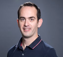 Nicolas Bourdillon合作论文数Institute of Sports Sciences and Department of Physiology, University of Lausanne15Andrew M. Luks合作论文数14
 Philip N. Ainslie合作论文数Centre for Heart, Lung & Vascular Health, School of Health and Exercise Sciences, Faculty of Health and Social Development, University of British Columbia Okanagan14
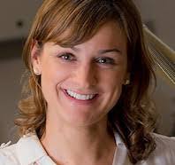 Tatum Simonson合作论文数Department of Medicine, School of Medicine, University of California, San Diego13
 Angelo D'Alessandro合作论文数Department of Biochemistry and Molecular Genetics, School of Medicine, University of Colorado13
 Jonathan Elliott合作论文数12WJ MILLS合作论文数12Song Anren合作论文数12Milton V Icenogle合作论文数12Dimmen Andrew C合作论文数11
 Nemkov Travis合作论文数From the ‡Department of Biochemistry and Molecular Genetics, University of Colorado Denver11
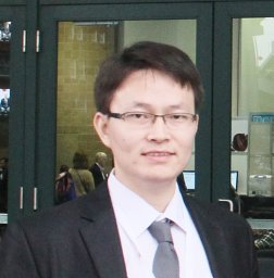 Sun Kaiqi合作论文数University of Texas Health Science Center11Yang Xia合作论文数Department of Biochemistry and Molecular Biology, University of Texas at Houston Medical School11Luciano Bernardi合作论文数11Benjamin Honigman合作论文数10
 Hong Liu (刘洪)合作论文数Big Data Institute, Central South University;Xiangya Hospital, Central South University10Evero Oghenero合作论文数10
 Rodney E. Kellems合作论文数Department of Biochemistry and Molecular Biology, McGovern Medical School, The University of Texas Health Science Center at Houston;Graduate School of Biomedical Sciences, The University of Texas MD Anderson Cancer Center10
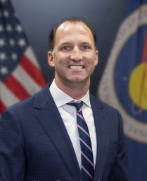 Laurie Steven S合作论文数9
 Kirk Hansen合作论文数Trauma Research Center, Department of Surgery, School of Medicine,University of Colorado Anschutz;University of Colorado Denver;Omix Technologies, Inc.9
 Jui-Lin Fan合作论文数Otago School of Medical Science, University of Otago9Yujin Zhang合作论文数9ER GREENE合作论文数8Goodman Randall D合作论文数8
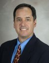 Michael R. Blackburn合作论文数Pulmonary Center of Excellence, McGovern Medical School, University of Texas Health Science Center at Houston8
 Holger Eltzschig合作论文数Department of Anesthesiology, Critical Care and Pain Medicine, McGovern Medical School, The University of Texas Health Science Center;Center for Perioperative Medicine, Department of Anesthesiology, McGovern Medical School, The University of Texas Health Science Center8
 Ronney Panerai合作论文数Department of Cardiovascular Sciences, University of Leicester8D Maes合作论文数7Beasley Kara M合作论文数7
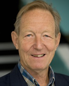 Bengt Saltin合作论文数Copenhagen Muscle Research Centre7Jose A. L. Calbet合作论文数7
 Pietro Scotto合作论文数7Julia Kern合作论文数6Peter Hackett合作论文数6
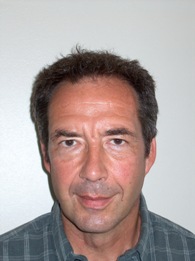 Jean-Marc Vesin合作论文数Electrical Engineering Section, School of Engineering, École Polytechnique Fédérale de Lausanne;ETH Zurich6
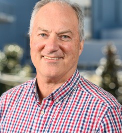 Rob Robergs合作论文数Faculty of Health, School of Exercise and Nutrition Sciences, Queensland University of Technology6
 Harry Karmouty-Quintana合作论文数Meakins-Christie Laboratories, McGill University6K. Riboni合作论文数6D. P. Maes合作论文数6Todd Bull合作论文数6
 Sasan Yazdani合作论文数Swiss Federal Institute of Technology (EPFL)6




