Reconstruction of hard tissue continuity defects caused by ablative tumor surgery has been traditionally reconstructed with autogenous cancellous marrow grafts or microvascular free flaps. Although results have been predictable from both methods of reconstruction, the morbidity associated with bone harvest is quite significant for the patient. With the advances made in tissue engineering, it is our opinion that successful and predictable results can be obtained with using a combination of 100% cadaver bone, Bone Marrow Aspirate Concentrate (BMAC) and rhBMP in immediate reconstruction for benign tumor extirpations.
Statistically, significant numbers of central bone specimens of suppurative osteomyelitis of the jaws (SOJ), bisphosphonate induced osteonecrosis of the jaws (BIONJ), and osteoradionecrosis of the jaws (ORNJ) were compared. All three evidenced the common finding of necrotic bone with empty osteocytic lacunae, Haversian and Volkmann canals, but each showed a distinctive histopathologic pattern indicating a different disease mechanism and treatment options. Suppurative osteomyelitis was characterized by intense marrow inflammation and marrow vessel thrombosis with retention of viable osteoclasts and periosteum. Bisphosphonate induced osteonecrosis was characterized by an empty marrow space with empty Howship's lacunae and an absence of osteoclasts but viable periosteum. Osteoradionecrosis was characterized by a collagenous hypocellular, hypovascular marrow space and nonviable periosteum. Histologic evidence in SOJ indicates a microorganism provoked intense inflammation and marrow vascular thrombosis creating an environment conducive to continual bacterial proliferation. BIONJ is seen as a non-inflammatory drug toxicity to bone by osteoclastic death leading to over suppression of bone renewal, and ORN as another non-inflammatory condition caused by a high linear energy transfer that impairs or kills numerous cell types in the field of radiation including periosteum, bone, and all soft tissue.
Platelet-rich plasma is an autologous source of platelet-derived growth factor and transforming growth factor beta that is obtained by sequestering and concentrating platelets by gradient density centrifugation. This technique produced a concentration of human platelets of 338% and identified platelet-derived growth factor and transforming growth factor beta within them. Monoclonal antibody assessment of cancellous cellular marrow grafts demonstrated cells that were capable of responding to the growth factors by bearing cell membrane receptors. The additional amounts of these growth factors obtained by adding platelet-rich plasma to grafts evidenced a radiographic maturation rate 1.62 to 2.16 times that of grafts without platelet-rich plasma. As assessed by histomorphometry, there was also a greater bone density in grafts in which platelet-rich plasma was added (74.0% +/- 11%) than in grafts in which platelet-rich plasma was not added (55.1% +/- 8%; p = 0.005).
The objective in treating patients with injuries sustained in craniofacial trauma is to reinstate preinjury facial projection and function. The capability of providing spatially related facial reconstruction is predicated on basic craniofacial surgical principles, generally accepted as the standard of care. These principles include early surgical intervention, immediate bone grafting, and the use of internal rigid fixation. The introduction of osseointegrated dental implants has significantly improved the overall reconstruction of patients with cranio-maxillofacial injuries, including soft tissue repair and cosmetic surgery. The purpose of this article is to review the utilization of dental implants in the context of maxillofacial trauma, using three cases to document the clinical procedure.
Over the past two decades, oral and maxillofacial surgeons have gained a greater appreciation for the biology of allogeneic bone healing, resulting in a dramatic increase in its indications and use. Unfortunately, this time period has also ushered in near epidemic proportions of HIV-infected persons, some of whom might be considered as potential donors of allogeneic bone. As this article will discuss, surgeons and tissue bank teams alike must be aware of the clinical and serologic criteria associated with an acceptable donor. Only in this way can contamination-free specimens be obtained and surgically implanted.
Small and medium-sized benign mandibular tumors such as ameloblastoma, odontogenic myxoma, and odontogenic keratocysts are generally resected intraorally when possible. This leaves extraoral soft tissues at the future recipient site untouched and simplifies dissection during delayed mandibular reconstruction. Superior results can be anticipated because of better blood supply and less scar tissue.
The professional literature is the historic depository of data that helps guide the clinician in the treatment of pathologic conditions. Single reports, case series, and retrospective studies all have their influence on the surgical approach to treatment. Individual practitioners and training programs frequently use a literature search in preparation for the management of lesions or in resident teaching. Directed by this assumption, the Section on Research of the American Association of Oral and Maxillofacial Surgeons conducted a panel discussion on the “Management of AmeloblastomaConservative vs Aggressive” (AAOMS Clinical Congress, 1988). The panel found that the absence of standardized terminology for methods of excision and the omission of critical details were major deficiencies in the surgical literature on ameloblastoma. The two teams concluded that the literature was inadequate to allow valid analysis of treatment modalities and outcome. As a result of this debate, a committee was appointed to develop definitions for terms applicable to methods of excision of any lesion in bone. The committee recognized that other parameters of documentation in the surgical literature also were deficient or lacking and should be addressed. Review of the literature shows that accurate,
Surgeons and the lay public have recently expressed concern over the safety of allogeneic dura as it relates to the transmission of Creutzfeldt-Jakob Disease. Indeed, two cases have resulted from use of tissue procured from a commercial agency that did not adhere to criteria accepted by the American Association of Tissue Banks or the Southeast Organ Procurement Foundation. This review discusses the risks and safety of allogeneic dura. The findings should reassure the surgeon of the safety of allogeneic dura when it is properly processed and catalogued by abona fide, reputable tissue bank. To date, there have been no documented cases reported to the Center for Disease Control in which Creutzfeldt-Jakob Disease was transmitted from allogeneic dura obtained from a registered tissue bank.
Today's reconstruction of head and neck cancer related deformities is based on an in-depth knowledge of soft and hard tissue repair coupled with the technical ability to transplant and reorient soft and hard tissues into a functional and cosmetic improvement for each patient. The recent advances of (1) the science of bone regeneration, (2) myocutaneous flaps, (3) hyperbaric oxygen, and (4) allogeneic bone cribs have resulted in reconstructions which are now predictable outcomes with few complications and minimal morbidity. This writing will discuss the contribution and mechanism of each of these four advances and how their application can be used to consistently reconstruct even large deformities with both hard and soft tissue loss, as well as in tissues which have been severely radiated.
Reconstructive oral and maxillofacial surgeons often find the need for additional soft tissue even when a myocutaneous flap has been previously placed. This article introduces the principles, technique, and results of 32 “walk-up” muscle flaps derived from existing myocutaneous flaps. The anastomotic vascular ingrowth at the original myocutaneous flap's distal end permits its proximal detachment and axial vessel ligation to rotate the proximal two thirds to a tissue-deficient site. In our experience at the University of Miami, these flaps have been predictable (97% viable transfer rate), have provided vascular soft tissue in deficient areas, and have been skin grafted at their surface to increase oral lining or skin cover, among several other uses.
The pectoralis major myocutaneous flap is the most commonly employed muscle skin transfer used in soft-tissue reconstruction of defects of the upper neck and jaw region. This article presents conceptual and technical changes in the development of the pectoralis major myocutaneous flap that preserve a greater vascular pedicle and enhance the flap's arc of rotation. Data from 54 consecutive cases using this modified approach show a reduction in complications, a greater range of use, and consistent healing in radiated and nonradiated tissues without requiring sectioning or removing the clavicle or causing significant chest deformities. These modifications have produced a more predictable transfer compared with other reported techniques.
This study was accomplished in an irradiated rabbit model to assess the angiogenic properties of normobaric oxygen and hyperbaric oxygen as compared with air-breathing controls. Results indicated that normobaric oxygen had no angiogenic properties above normal revascularization of irradiated tissue than did air-breathing controls (p = 0.89). Hyperbaric oxygen demonstrated an eight- to ninefold increased vascular density over both normobaric oxygen and air-breathing controls (p = 0.001). Irradiated tissue develops a hypovascular-hypocellular-hypoxic tissue that does not revascularize spontaneously. Results failed to demonstrate an angiogenic effect of normobaric oxygen. It is suggested that oxygen in this sense is a drug requiring hyperbaric pressures to generate therapeutic effects on chronically hypovascular irradiated tissue.
The radiobiology of osteoradionecrosis is a complex of cellular death and cellular functional impairments from radiation energy transfers. Four studies of irradiated patients and a data base from 536 patients with osteoradionecrosis revealed separate pathophysiologic conditions for osteoradionecrosis induced by early trauma, osteoradionecrosis induced by late trauma, and spontaneous osteoradionecrosis. A large body of data suggested useful clinical guidelines for the management of irradiated patients. The guidelines, in part, include a recommendation for deferring radiation treatment for 21 days after tissue wounding, if possible; a relative contraindication to wounding tissue during a radiation course; a recommendation for the use of hyperbaric oxygen before wounding; and a strong recommendation to provide comprehensive dental care to the irradiated patient.
 Pairot Tayapongsak合作论文数4SN KLINE合作论文数3Ramzey Tursun合作论文数3William J. Ehler合作论文数3TI MALININ合作论文数2Marco J. Morales合作论文数2RD HEIMBACH合作论文数1M J Morales合作论文数1michael moody合作论文数1Rb Smith合作论文数1L GOLD合作论文数1
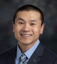 James C Melville合作论文数Department of Oral and Maxillofacial Surgery, University of Texas Health Science Center at Houston1Gottlieb, C.F.合作论文数1Buck B E合作论文数1Johnson R P合作论文数1
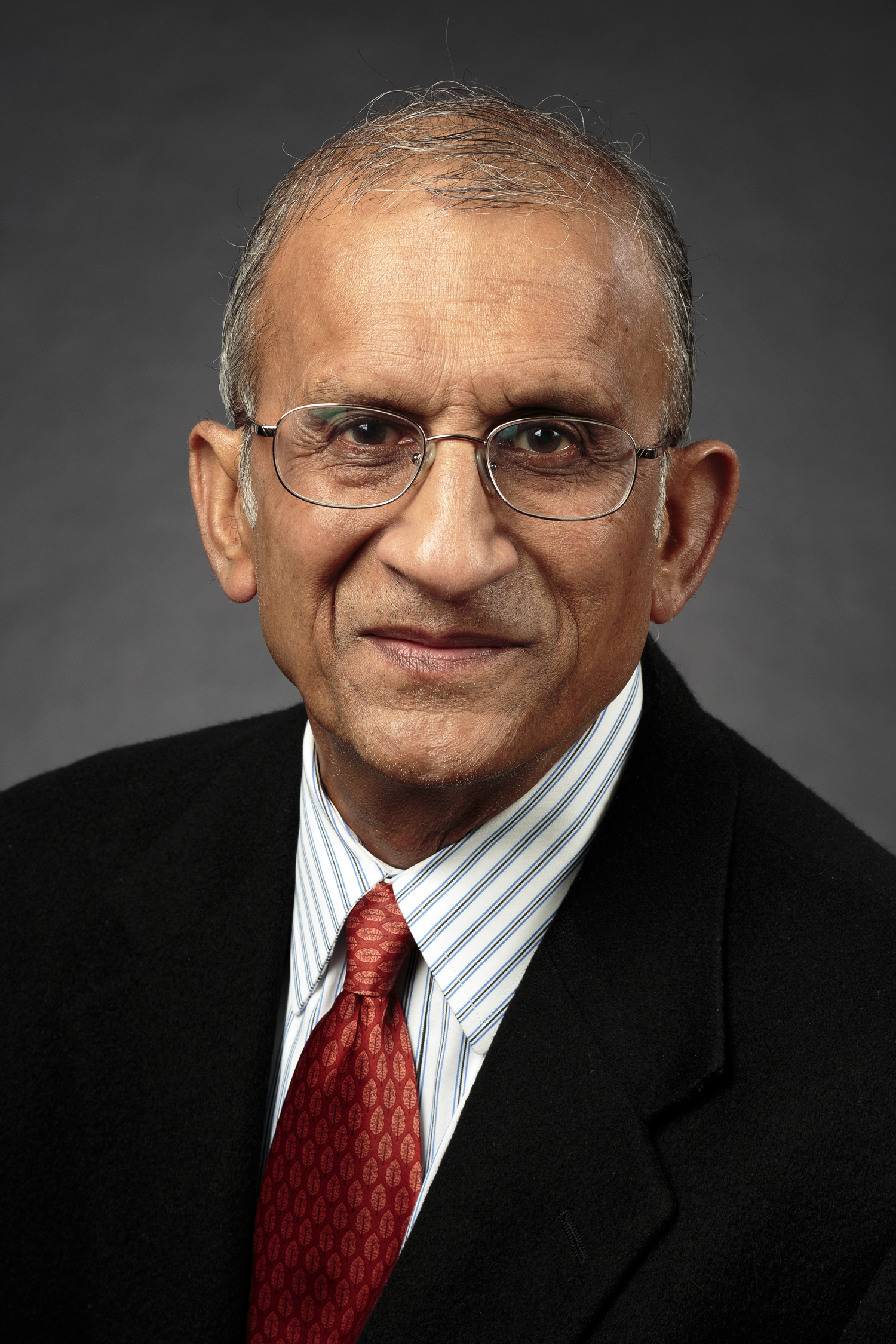 Arun Garg合作论文数Department of Occupational Science & Technology, College of Health Sciences, University of Wisconsin-Milwaukee1Robert P. Johnson合作论文数1James E Strauss合作论文数1
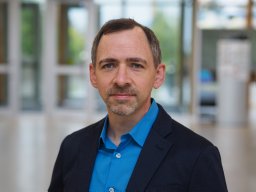 Mark R. Stevens合作论文数Charles River Analytics, Cambridge, MA1Geoffrey M. Jones合作论文数1Leonard W. Pierce合作论文数1
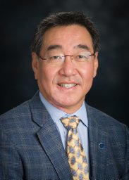 Mark E. Wong合作论文数Department of Oral and Maxillofacial Surgery, University of Texas Health Science Center at Houston1Karen R Georgeff合作论文数1J Ames合作论文数1Steven R Schimmele合作论文数1Gerald W. Upton合作论文数1Ralph M Eichstaedt合作论文数1G HUBBARD合作论文数1




