By definition, amyloids regardless of primary sequence of the subunit, share common structural features. Encouraged by the previous identification of anti–amyloid mAbs that recognize multiple amyloids and data showing that anti–amyloid antibodies can enhance clearance of light chain amyloid in mice, we tested the hypothesis that immunization with heterologous amyloids (hAs) formed from peptides that lack homology to any human or mouse protein could induce a generic anti–amyloid response. We find that vaccination of mice with hAs induces a robust anti–amyloid Ab response and that the anti–amyloid Abs are capable of recognizing multiple amyloids regardless of subunit. Our original rationale for this approach was largely driven by trying to avoid a harmful T–cell response. In most cases, we are immunizing with amyloids composed of subunits lacking homology to human or mouse proteins; thus MHC presentation and activated T–cells would be targeting non–self peptides. Furthermore, we find that amyloids formed from a 6 amino acid peptide induce a generic anti–amyloid response. In this latter case, sequence homology becomes less of an issue; such small peptides theoretically lack the ability to bind to MHC. We had originally speculated that such an approach might avoid self–tolerance issues and induce a stronger anti–amyloid immune response than fAβ42 vaccination. However, this speculation was largely based on the idea that an anti–amyloid response was induced by a thymus dependent (TD) mechanism. Current data suggests that amyloid is not a TD antigen but a thymus independent type 2 (TI2) antigen. The rules or immune recognition of a TI2 antigen are distinct from those for TD responses, and little is known about self–tolerance with respect to TI2 responses. By definition, TI2 antigens do not require help from αβ T–cells or antigen presentation by MHC class II. Therefore, to determine if fAβ42 and hAs are TI2 antigens, we are currently vaccinating TcR αβ knockout mice and MHC class II knockout mice with several hAs and fAβ42. This novel approach enables testing of the hypothesis that immunotherapy targeting amyloid and, potentially, a preamyloid aggregate is beneficial in mouse models of AD.
Passive immunotherapy using fibril-reactive mAbs has been shown experimentally to reduce amyloid formation and also accelerate amyloidolysis. We now report that human sera, as well as various sources of pooled human IgG, including pharmacologic formulations of immune globulin i.v. (IGIV), contain Abs that specifically recognize fibrils formed from light chains and other amyloidogenic precursor proteins, including serum amyloid A, transthyretin, islet amyloid polypeptide, and amyloid β 1–40 peptide, but notably, do not react with these molecules in their native nonfibrillar forms. After isolation of the Abs from IGIV via fibril-conjugated affinity column chromatography, the EC50-binding value for light chains and amyloid β 1–40 peptide fibrils was ∼15 nM–a magnitude ∼200 and 70 times less than that of the unbound fraction and unfractionated product, respectively. Comparable reactivity was found in the case of those formed from serum amyloid A, transthyretin, and islet amyloid polypeptide. The purified Abs immunostained human amyloid tissue deposits and could inhibit fibrillogenesis, as shown in fibril formation and extension assays. Most importantly, in vivo reactivity was evidenced in a murine model when the enriched Abs were used to image amyloid, as well as expedite its removal. These promising experimental results suggest that fibril affinity-purified IGIV has potential as a diagnostic and therapeutic agent for patients with amyloid-associated disease.
Calcifying epithelial odontogenic tumors (CEOTs), also known as Pindborg tumors, are characterized by the presence of squamous-cell proliferation, calcification, and, notably, amyloid deposits. On the basis of immunohistochemical analyses, the amyloidogenic component had heretofore been deemed to consist of cytokeratin-related or other molecules; however, its chemical composition had never been elucidated. We have used our microanalytic techniques to characterize the protein nature of CEOT-associated amyloid isolated from specimens obtained from 3 patients. As evidenced by the results of amino-acid sequencing and mass spectrometry, the fibrils were found to be composed of a polypeptide of approximately 46 mer. This component was identical in sequence to the N-terminal portion of a hypothetical 153-residue protein encoded by the FLJ20513 gene cloned from the human KATO III cell line. That the amyloid protein was derived from this larger molecule was demonstrated by reverse transcription-polymerase chain reaction amplification of tumor-cell RNA where a full-length FLJ20513 transcript was found. Furthermore, immunohistochemical analyses revealed that the amyloid within the CEOTs immunostained with antibodies prepared against a synthetic FLJ20513-related dodecapeptide. Our studies provide unequivocal evidence that CEOT-associated amyloid consists of a unique and previously undescribed protein that we provisionally designate APin.
The human amyloidoses represent a heterogeneous group of disorders characterized by the deposition of fibrillar protein in vital organs. Given the fact that at least 20 different molecules can form fibrils, the unambiguous identification of the type of amyloid deposited is critical to the correct diagnosis and treatment of patients with these disorders. Heretofore, this information has been inferred from particular clinical features of the disease, ancillary laboratory tests, and results of immunohistochemical analyses. However, to establish unequivocally the kind of protein that is deposited as amyloid, it is necessary to determine its chemical composition through amino acid sequencing or mass spectroscopy of material extracted from fibrillar deposits. We have developed a micromethod whereby such studies can be performed readily using sections of formalin-fixed, paraffin-embedded biopsy specimens. The ability to identify precisely the nature of the tissue deposits has diagnostic, therapeutic, and prognostic implications for patients with amyloid-associated disorders.
Primary light-chain-associated (AL) amyloidosis is characterized by the deposition in tissue of monoclonal light chains as fibrils. With rare exception, this process is seemingly irreversible and results in progressive organ dysfunction and eventually death. To determine whether immune factors can effect amyloid removal, we developed an experimental model in which mice were injected with amyloid proteins extracted from the spleens or livers of patients with AL amyloidosis. Notably, the resultant amyloidomas were rapidly resolved, as compared to controls, when animals received injections of an anti-light-chain monoclonal antibody having specificity for an amyloid-related epitope. The reactivity of this monoclonal antibody was not dependent on the V-L or C-L isotype of the fibril, but rather seemed to be directed toward a P-pleated sheet conformational epitope expressed by AL and other amyloid proteins. The amyloidolytic response was associated with a pronounced infiltration of the amyloidoma with neutrophils and putatively involved opsonization of fibrils by the antibody, leading to cellular activation and release of proteolytic factors. The demonstration that AI. amyloid resolution can be induced by passive administration of an amyloid-reactive antibody has potential clinical benefit In the treatment of patients with primary amyloidosis and other acquired or inherited amyloid-associated disorders.
Certain types of human light chains have the propensity to deposit pathologically as amyloid fibrils as evidenced by the preferential association of monoclonal lambda 6 proteins with AL amyloidosis. However, the molecular features that render such proteins amyloidogenic have not been elucidated. Based upon the demonstrated relationship between the thermodynamic stability of light chains and their propensity to aggregate in vitro, we have initiated studies where the thermodynamic properties and fibrillogenic potential of two recombinant (r) V lambda 6 molecules were compared. The first protein was generated from cDNA cloned from marrow-derived plasma cells from a patient (Wil) who had AL amyloidosis and renal amyloid deposits; the second was from a patient (Jto) with multiple myeloma in whom the lambda 6 protein was deposited not as amyloid but in the form of renal tubular casts. The thermodynamic stabilities of rV lambda 6Wil and -Jto were determined from chaotropic and thermal denaturation studies. Based upon the Delta GH2O, Delta H, Delta G25 degrees C, Tm, and Cm values, the rV lambda 6Wil was less stable than its nonamyloidogenic counterpart, rV lambda 6Jto. Measurement of fibril formation using a novel in vitro fibril forming assay demonstrated that although both rV lambda 6 proteins formed fibrils in vitro, Wil had a shorter lag time and exhibited faster kinetics under physiologic conditions. Comparative amino acid sequence analyses of these two components and other lambda 6 amyloid-associated light chains revealed that the Jto protein had certain primary structural features that we posit contributed to its increased stability and thus rendered this protein nonamyloidogenic. Our studies provide the first evidence that stabilizing interactions within the V L domain can influence the kinetics of light chain fibrillogenicity.
This article described micromethods useful for the extraction, purification, and amino acid sequencing of amyloid proteins contained in minute specimens obtained from patients with systemic forms of amyloidosis. We posit that these procedures can also be applied to the biochemical characterization of cerebral amyloid deposits. The selection of the techniques is dependent on the type of sample to be extracted (fresh or formalin fixed) as well as the amount of congophilic material present. Although amyloid proteins are isolated and purified more easily from fresh tissue, it must be noted that formalin-fixed specimens are available more readily for analysis due to the common diagnostic use of fine needle tissue biopsies and are therefore, important for both current and retrospective studies. Remarkably, despite the expected difficulties associated with formalin treatment we were able to extract and sequence amyloid proteins from fixed tissues presumably due to the resistance of amyloid to formalin cross-linking. Through the continued development of techniques for small-scale protein separation and application of highly sensitive microsequencing and mass spectral methods, exact identification of the protein contained in fibrillar amyloid deposits can be determined. Such information has therapeutic and prognostic relevance and can increase our understanding of the pathogenesis of amyloidosis.
AA amyloidosis can be induced in mice experimentally through injection of certain chemical or biological compounds. However, the usefulness of this approach is limited by its dependence on exogenous inflammatory agents that stimulate cytokines to increase the synthesis of precursor serum amyloid A (SAA) protein and the transitory nature of the pathological fibrillar deposits. We now report that transgenic mice carrying the human interleukin 6 gene under the control of the metallothionein-I promoter had markedly increased concentrations of SAA and developed amyloid in the spleen, liver, and kidneys by 3 months of age. At the time of death about 6 months later, organs obtained from these animals had extensive amyloid deposits. This disease process was apparent radiographically using small-animal computer axial tomography and magnetic resonance imaging equipment. The AA nature of the amyloid was evidenced immunohistochemically and was unequivocally established by sequence analysis of protein extracted from the fibrils. The availability of this unique in vivo experimental model of AA amyloidosis provides the means to assess the therapeutic efficacy of agents designed to reduce or prevent the fibrillar deposits found in AA and other types of amyloid-associated disease.
Light chain-associated amyloidosis is characterized by the deposition as fibrils of monoclonal light chain-related components consisting predominately of the variable domain (VL) or the VL plus up to approximately 60 residues of the constant domain (CL). Here, we describe a patient (designated BIF) with light chain-associated amyloidosis and kappa Bence Jones proteinuria in whom, notably, >80% of the amyloid deposits were comprised of CL-related material. The extracted amyloid protein consisted of 99 aa residues identical in sequence to the main portion of the Ckappa region (positions 109-207) of the precursor Bence Jones protein. Remarkably, the CLs from both molecules contained a Ser-->Asn substitution at position 177. This heretofore undescribed Ckappa alteration did not result from somatic mutation but rather was germline encoded. When tested in our in vitro fibrillogenic kinetic assay, Bence Jones protein BIF was highly amyloidogenic. Notably, endopeptidase treatment of amyloid fibrils prepared from the native light chain revealed the VL to be markedly susceptible to enzymatic digestion, whereas the CL was protease-resistant. Our findings provide evidence that the fragmented light chains typically present in this disease result from proteolytic degradation and suggest that, in this case, conformational differences in VL/CL packing within the fibrils may account for the unusual composition of the amyloid deposits. Additionally, we posit that the previously unrecognized Asn177 substitution represents yet another Ckappa allotype, provisionally designated Km4.
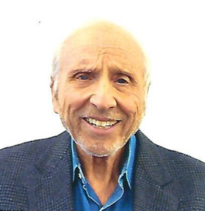 Alan Solomon合作论文数Department of Religious Studies, College of Arts & Sciences, The University of Tennessee, Knoxville17
 Jonathan S Wall合作论文数13
 Deborah T. Weiss合作论文数11Cl Murphy合作论文数8Maria Schell合作论文数8Craig Wooliver合作论文数3S. D. Macy合作论文数2DA Wolfenbarger合作论文数2Fj Stevens合作论文数2D Wolfenbarger合作论文数2S Macy合作论文数2Todd E. Golde合作论文数2
 Knut Sletten合作论文数2Charles L. Murphy合作论文数2
 Pritam Das合作论文数Mayo Clinic2
 Per Westermark合作论文数Department of Immunology, Genetics and Pathology, Uppsala University2K Weaver合作论文数1m eulitz合作论文数1
 Robert Donnell合作论文数1
 Brian O'nuallain合作论文数1McGavin M D合作论文数1Boris Kaplan合作论文数1
 George W. Kabalka合作论文数Department of Chemistry, College of Arts & Sciences, University of Tennessee1
 M.J. Paulus合作论文数1Gunilla T. Westermark合作论文数Dept Med Cell Biol, Uppsala Univ1Manfred Eulitz合作论文数1R. Donnell合作论文数1




