See also: Endoscopic internal drainage and low negative-pressure endoscopic vacuum therapy for anastomotic leaks after oncologic upper gastrointestinal surgeryEndoscopy 2022; 54(01): 71-74DOI: 10.1055/a-1375-8151 See also: Endoscopic internal drainage versus endoscopic vacuum therapy for upper gastrointestinal leaks: what's the real deal?Endoscopy 2023; 55(01): 100-100DOI: 10.1055/a-1888-4116
Sarcopenia, defined as decreased muscle mass and strength, can be evaluated by a computed tomography (CT) examination and might be associated with reduced survival in patients with carcinoma. The prognosis of patients with metastatic pancreatic carcinoma is poor. The FOLFIRINOX (a combination of 5-fluorouracil, irinotecan, and oxaliplatin) chemotherapy regimen is a validated first-line treatment option. We investigated the impact of sarcopenia on overall survival (OS) and progression-free survival (PFS) in patients with metastatic pancreatic carcinoma. Clinical data and CT examinations of patients treated with FOLFIRINOX were retrospectively reviewed. Sarcopenia was estimated using baseline CT examinations. Seventy-five patients were included. Forty-three (57.3%) were classified as sarcopenic. The median OS of non-sarcopenic and sarcopenic patients were 15.6 and 14.1 months, respectively (p = 0.36). The median PFS was 10.3 in non-sarcopenic patients and 9.3 in sarcopenic patients (p = 0.83). No differences in toxicity of FOLFIRINOX were observed. There was a trend towards a higher probability of short-term death (within 4 months of diagnosis) in sarcopenic patients. In this study, the detection of sarcopenia failed to predict a longer OS or PFS in selected patients deemed eligible by a physician for triplet chemotherapy and receiving the FOLFIRINOX regimen in a first-line setting, confirming the major importance of a comprehensive patient assessment by physicians in selecting the best treatment option.
Objectives: Endoscopic mucosal resection (EMR) and endoscopic submucosal dissection (ESD) allow endoscopic resection of early esophageal adenocarcinoma. The choice between the two techniques takes into account the morphology of the lesion, and the experience of the endoscopist. The aim of this study was to compare EMR to ESD for the treatment of early esophageal adenocarcinoma. Methods: Patients who underwent an endoscopic resection for esophageal adenocarcinomas between March 2015 and December 2019 were included. ESD was compared to EMR in terms of clinical, procedural, histologic, and oncologic outcomes.Results: 85 patients were included: 57 ESD and 28 EMR. The median (IQR) diameter of the lesion was 20(15-25) mm in the ESD group, and 15(8-16) mm in the EMR group, p<0.01. ESD allowed en bloc resection in 100% of cases, and EMR in 39% of cases, p<0.001. The R0 and curative resection rate in the ESD group versus the EMR group were 88% and 67%, respectively, versus 21% and 11%, p<0.001. We recorded one severe adverse event, in the EMR group. After a median (IQR) follow-up of 27.5 (14.5-38.7) months, the local recurrence rate was 23% vs. 18% (p = 0.63), and the overall survival 89% vs. 86% (p = 0.72), in the ESD and EMR groups, respectively.Conclusion: ESD was as safe as EMR and allowed higher en bloc, R0 and curative resection rates. Although these results did not translate into long-term outcomes, these data prompt for a broader adoption of ESD for the resection of esophageal lesions suspected of harboring early esophageal adenocarcinoma.(c) 2023 Elsevier Masson SAS. All rights reserved.
A 78-year-old man, with a history of T2N0M0 esophageal adenocarcinoma treated with chemoradiotherapy 2 years before, was referred to our department with recurrent upper gastrointestinal bleeding (hemoglobin level of 8.8 g/dl), attributed to radiation esophagitis. He had been admitted three times before for red blood cell transfusion and endoscopic treatment with argon plasma coagulation. Initial esophagogastroduodenoscopy showed diffuse telangiectasias of the lower esophagus and gastroesophageal junction with signs of recent bleeding (▶Fig. 1 a). Radiofrequency ablation (RFA) of the lower esophagus and gastroesophageal junction was performed using a double 10 J/cm2 ablation with a 360° Express balloon catheter (Medtronic, Minneapolis, Minnesota, USA) (▶Video 1, ▶Fig.1b), followed by double-dose proton pump inhibitors. Despite no recurrent digestive bleeding, persistent iron deficiency anemia prompted repeat esophagogastroduodenoscopies, including two RFA treatment sessions every 6 months, using a triple 12 J/cm2 RFA application with a focal Halo 90 catheter. The 15-month follow-up esophagogastroduodenoscopy showed a significant regression of the esophageal telangiectasias (▶Fig. 1 c). No recurrent bleeding occurred and hemoglobin level remained stable. Radiation esophagitis occurs in up to 42% of patients after radiation therapy for lung, breast, or esophageal carcinomas or mediastinal lymphomas [1]. Whereas acute radiation esophagitis typically presents with self-limiting dysphagia and odynophagia, chronic radiation esophagitis may lead to complications such as bleeding of esophageal telangiectasias, stenosis, impaired motility or esophagorespiratory fistula. Esophageal telangiectasias are generally managed by argon plasma coagulation (APC). As for other angioectasias occurring elsewhere in the gastrointestinal tract (gastric antral vascular ectasia, radiation proctitis), radiofrequency ablation might be offered as a rescue therapy [2]. To our knowledge, this is the second patient with radiation E-Videos
We read with great interest the study by McKay et al highlighting the topic of appendiceal involvement in patients with serrated polyposis syndrome (SPS).1 Considering a 68% prevalence of serrated lesions of the appendix in a series of 34 patients with SPS treated by colectomy,2 and the difficulty of ruling out an invasion of the appendiceal lumen in case of periappendiceal serrated lesions, the authors advocated prophylactic appendectomy in patients with SPS.\n\nSPS is the most frequent polyposis syndrome, with a prevalence of 0.42% in primary screening cohorts.3 While surgery remains needed for a third of the patients at inception due to the presence of an adenocarcinoma or a lesion burden not amenable …
Background and study aims Evidence for the modes of transmission of SARS-CoV-2 remains controversial. Recently, the potential for airborne spread of SARS-CoV-2 has been stressed. Air circulation in gastrointestinal light source boxes and endoscopes could be implicated in airborne transmission of microorganisms. Methods The ENDOBOX SC is a 600 × 600 mm cube designed to contain any type of machine used during gastrointestinal endoscopy. It allows for a 100-mm space between a machine and the walls of the ENDOBOX SC. To use the ENDOBOX SC, it is connected to the medical air system and it provides positive flow from the box to the endoscopy room. The ENDOBOX SC uses medical air to inflate the digestive tract and to decrease the temperature induced by the microprocessors or by the lamp. ENDOBOX SC has been investigated in different environments. Results An endoscopic procedure performed without ventilation was interrupted after 40 minutes to prevent computer damage. During the first 30 minutes, the temperature increased from 18 °C to 31 °C with a LED system. The procedure with fans identified variations in temperature inside the ENDOBOX SC from 21 to 26 °C (± 5 °C) 1 hour after the start of the procedure. The temperature was stable for the next 3 hours. Conclusions ENDOBOX SC prevents the increase in temperature induced by lamps and processors, allows access to all necessary connections into the endoscopic columns, and creates a sterile and positive pressure volume, which prevents potential contamination from microorganisms.
Duodenal polyps are found in 0.1 % to 0.8 % of all upper endoscopies. Duodenal adenomas account for 10 % to 20 % of these lesions. They can be sporadic or occur in the setting of a hereditary predisposition syndrome, mainly familial adenomatous polyposis. Endoscopy is the cornerstone of management of duodenal adenomas, allowing for diagnosis and treatment, primarily by endoscopic mucosal resection. The endoscopic treatment of duodenal adenomas has a high morbidity, reaching 15 % in a prospective study, consisting of bleeding and perforations, and should therefore be performed in expert centers. The local recurrence rate ranges from 9 % to 37 %, and is maximal for piecemeal resections of lesions > 20 mm. Surgical resection of the duodenum is flawed with major morbidity and considered a rescue procedure in cases of endoscopic treatment failures or severe endoscopic complications such as duodenal perforations. In this paper, we review the existing evidence on endoscopic diagnosis and treatment of non-ampullary duodenal adenomas.
Background: Gastric linitis plastica (GLP) is a diffuse infiltrating type of gastric adenocarcinoma. It is associated with a poor prognosis and a five-year survival of 3-10%. The infiltrating profile of this tumor explains the low yield of the superficial mucosal biospies. The objective of this study was to investigate the role of endoscopic ultrasound-fine needle biopsy (EUS-FNB) in the diagnosis of GLP. Methods: We performed a retrospective analysis including all patients who had an EUS-FNB, at a tertiary referral center, over the last 3 years. The primary outcome was the sensitivity of EUSFNB in patients with suspected GLP. Results: Between January 2017 and December 2020, 34 patients had an EUS-FNB for suspected GLP. Ten patients had a diagnostic of GLP. This diagnosis was obtained by EUS-FNB in 90% (9/10) of the cases. Eight patients had at least one previous esophagogastroduodenoscopy (EGD) with negative mucosal biopsies. Gastric EUS-FNB helped diagnose other serious conditions in 47% (16/34) of cases with inconclusive mucosal biopsies. Conclusion: Gastric EUS-FNB in patients with suspected GLP and normal endoscopic mucosal biopsies may lead to a positive diagnosis of GLP in 90% of cases without notable adverse events. This technique should be considered as a second step in the setting of suspicion of GLP after inconclusive mucosal biopsies. (c) 2022 Elsevier Masson SAS. All rights reserved.
Aims Zenker's diverticulum usually presents in the elderly population with symptoms of dysphagia. The endoscopic diverticulotomy can be performed using either cap or diverticuloscope assistance. The abandonment of the diverticuloscope for the treatment of diverticula allows to perform a complete septotomy of the diverticular wall in particular in the case of large diverticula (greater than 5 cm). The aim of this study was to evaluate the safety and feasibility of complete septotomy in the setting of large Zenker's diverticula.
The British Society of Gastroenterology has produced guidance highlighting the importance of manual cleaning of the duodenoscope with specific instructions regarding the removal of the bridge mechanism at the distal tip prior to brushing. This is likely to reduce the risks of bacterial transmission via duodenoscopes, thereby reducing strength of the case for singleuse endoscopes. As members of Green Endoscopy (Twitter, @Green Endoscopy), we champion environmental sustainability in GI endoscopy and are raising awareness of the carbon footprint of endoscopy. We note that Bang et al have not addressed the environmental impact of singleuse duodenoscopes in their paper, and we highlight the significant potential environmental harm from increasing plastic waste through the use of singleuse endoscopes. A significant amount of waste is already generated from an endoscopic procedure (up to 1.5 kg) of which only a fraction is recyclable with the rest going to landfill or being incinerated. The disposal of a singleuse bronchoscope is equivalent to 349 g of household waste. The increasing availability of singleuse plastic disposable endoscopes in Europe and the USA (Exalt duodenoscope, Boston Scientific Corp) will add to this waste. There are approximately 500 000 ERCPs carried out in the USA and over 50 000 procedures in the UK annually. It is unthinkable that each and every one would be conducted with a singleuse scope, notwithstanding the substantial increase in the healthcare costs of these procedures. A comparative study of the environmental impact of reusable and singleuse bronchoscopes has reported on the need to compare the cost of disposing a singleuse plastic bronchoscope to that of sterilising a reusable bronchoscope with the labour, disinfecting equipment and consumable costs. There is also the risk that the development of singleuse duodenoscopy gets translated into other routine endoscopic procedures which have even more negligible contamination risk. We propose that all stakeholders need to consider environmental safety and environmental cost effectiveness in developing and adopting new endoscopes and endoscopic devices. There is a need for research into the assessment of the environmental impact of all new endoscopic technology as well as developing upfront processes for minimising environmental harm so that we as a GI community can do all we can to reduce the impact of healthcare on climate change. The endoscopy community must jointly lead initiatives on climate change being developed by all industries by advocating, educating, amplifying, promoting and organising sustainable endoscopy practices. This needs to be underpinned by research quantifying the carbon cost effectiveness of endoscopic equipment and procedures to reduce our carbon footprint.
Endoscopic mucosal resection (EMR) is the recommended treatment for superficial non-ampullary duodenal epithelial tumors larger than 6 mm. This endoscopic technique carries a high risk of adverse events. Our aim was to identify the risk factors for adverse events following EMR for non-ampullary duodenal adenomatous lesions. We retrospectively analyzed a prospectively collected database of consecutive endoscopic resections for duodenal lesions at a tertiary referral center for therapeutic endoscopy. We analyzed patients with non-ampullary duodenal adenomatous lesions ≥ 10 mm resected by EMR, and searched for factors associated with adverse events after EMR. 167 duodenal adenomatous lesions, with a median size of 25 (25–40) mm, were resected by EMR between January 2015 and December 2020. Adverse events occurred in 37/167 (22.2%) after endoscopic resection, with 29/167 (17.4%) delayed bleeding, 4/167 (2.4%) immediate perforation and 4/167 (2.4%) delayed perforation. In logistic regression, the size of the lesion was the only associated risk factor of adverse events (OR = 2.81, 95% CI [1.27; 6.47], p = 0.012). Adverse events increased mean hospitalization time (7.7 ± 9 vs. 1.9 ± 1 days, p < 0.01). None of the currently recommended preventive methods, particularly clips, affected the adverse event rate. EMR of centimetric and supracentimetric duodenal adenomatous lesions carries a high risk of adverse events, increasing with the size of the lesion and with no benefit from any preventive method. These results suggest that these procedures should be performed in expert centers, and underline the need for novel endoscopic tools to limit the rate of adverse events.
Aims Despite its undeniable carcinologic advantages, endoscopic submucosal dissection (ESD) has not replaced piecemeal endoscopic mucosal resection in Western countries because of the technical difficulty, long procedure duration, high complication rate, and steep learning curve. Differentiation of easy and difficult lesions would promote use of ESD. The objective of this study was to try to create a score that predict outcomes after colorectal ESD for large superficial lesions.
Background Endoscopic internal drainage (EID) with double-pigtail stents or low negative-pressure endoscopic vacuum therapy (EVT) are treatment options for leakage after upper gastrointestinal oncologic surgery. We aimed to compare the effectiveness of these techniques. Methods Between 2016 and 2019, patients treated with EID in five centers in France and with EVT in Gottingen, Germany were included and retrospectively analyzed using univariate analysis. Pigtail stents were changed every 4 weeks; EVT was repeated every 3-4 days until leak closure. Results 35 EID and 27 EVT patients were included, with a median (interquartile range [IQR]) leak size of 0.75cm (0.5-1.5). Overall treatment success was 100% (95% confidence interval [CI] 90%-100%) for EID vs. 85.2% (95%CI 66.3%-95.8%) for EVT ( P =0.03). The median (IQR) number of endoscopic procedures was 2 (2-3) vs. 3 (2-6.5; P =0.003) and the median (IQR) treatment duration was 42 days (28-60) vs. 17 days (7.5-28; P <0.001), for EID vs. EVT, respectively. Conclusion EID and EVT provide high closure rates for upper gastrointestinal anastomotic leaks. EVT provides a shorter treatment duration, at the cost of a higher number of procedures.
Introduction: Proton pump inhibitors (PPIs) are one of the most prescribed classes of drugs worldwide as a first-line treatment of acid-related disorders. Although adverse effects are rare and rapidly reversible after a short exposure, concerns have been recently raised about a greater toxicity on cardiovascular health after a longer exposure, especially when combined with clopidogrel. We aimed to evaluate the safety of long-term PPI use on cardiovascular health in patients with known atheromatous cardiovascular disease. Methods: A literature search was conducted in the PubMed, Embase, and Cochrane Library databases and grey literature in April 2022. Articles published between 2014 and 2022 were considered relevant if they were designed as randomized controlled trials (RCTs) that included post hoc analyses or prospective observational studies and if they investigated clinical cardiovascular outcomes associated with PPI use for 6 months or more in patients suffering from cardiovascular disease requiring antiplatelet agent therapy and/or coronary angioplasty. Statistical analyses were performed using RevMan 5.4 software (Computer program, the Cochrane Collaboration, 2020, London, UK). The risk of bias was assessed using the Cochrane risk-of-bias tool for the RCTs and the Newcastle–Ottawa scale for the observational studies. Results: A total of 10 full-text articles involving 53,302 patients were included. Substantial heterogeneity was found among the 10 included studies. The primary analysis showed no significant differences between the PPI group and the control group for the risks of major adverse cardiovascular events (MACEs), all-cause death (ACD), or target vessel revascularization (TVR) using a random-effects model (OR 1.15, 95% CI 0.98–1.35, p = 0.08, I2 = 73%; OR 1.24, 95% CI 0.94–1.65, p = 0.13, I2 = 63%; and OR 1.19, 95% CI 0.76–1.87, p = 0.45, I2 = 61%, respectively). The primary analysis yielded similar results for the risks of myocardial infarction (MI), stroke, and cardiovascular death (CVD) using a fixed-effects model (OR 0.98, 95% CI 0.88–1.09, p = 0.66, I2 = 0%; OR 1.02, 95% CI 0.90–1.17, p = 0.73, I2 = 0%; and OR 1.04, 95% CI 0.94–1.16, p = 0.44, I2 = 35%, respectively). Likewise, a subgroup analysis based on eight randomized controlled trials failed to identify any association between PPI use and the risks of MACEs, MI, stroke, TVR, ACD, or CVD using a fixed-effects model (overall pooled OR 1.01, 95% CI 0.96–1.06; p = 0.66; I2 = 0%). The pulled data from the two included observational studies (OS) demonstrated a significantly increased risk of MACEs in the PPI group (OR 1.42, 95% CI [1.29–1.57], p <0.001; I2 = 0%). In another subgroup analysis, no evidence of an increased risk of adverse cardiovascular events in the co-therapy PPI/clopidogrel versus clopidogrel alone groups was found with the exception of the risk of ACD (OR 1.50, 95% CI 1.23–1.82, p = 0.001, I2 = 0%). Nevertheless, after performing a sensitivity analysis reaching heterogeneity I2 = 0%, the co-prescription of PPIs and clopidogrel was at increased risk of MACEs (p < 0.001), CVD (p = 0.008), and TVR (p < 0.001) but remained statistically non-significant for the risk of MI (p = 0.11). Conclusions: The overall results of this meta-analysis showed that long-term PPI use was not associated with an increased risk of adverse cardiovascular events. However, inconsistent results were found for combined PPI/clopidogrel therapy. These results should be considered with caution in light of the significant heterogeneity, the limited number of included studies, and the lack of adjustment for potential confounders.
Aims SNADETs are rare lesions, with poorly standardized resection techniques. The complication rates after EMR range from 10.5 to 41.3 %. The aim of our work was to evaluate the efficacy and safety of EMR of non-ampullary duodenal adenomas.
Objectives: Adenocarcinomas of the esophagus and of the gastric cardia are regarded as a same clinical entity in oncology. For endoscopic resection however, endoscopic mucosal resection is recommended for esophageal adenocarcinoma, while endoscopic submucosal dissection (ESD) is advocated for gastric adenocarcinomas. Our aim was to compare the outcomes of ESD in both types of esophagogastric junction adenocarcinomas. Methods: Between March 2015 and December 2019, we included all patients who underwent an ESD for early adenocarcinoma of the esophagogastric junction at a French tertiary referral center. Esophageal and gastric cardia adenocarcinomas were compared in terms of clinical, procedural and histological outcomes. Results: 57 esophageal and 19 gastric cardia adenocarcinomas were included in the analysis, for a total of 76 patients. The median (IQR) size of the resections was 40 (40-57.5) and 50 (35-55) mm, p = 0.96, respectively. En bloc resection was achieved in 100% and 89% for adenocarcinomas of the esophagus and the gastric cardia, p = 0.06. Late adverse events occurred in 14% and 5.3%, respectively, p = 0.44, with no severe adverse event. Curative resection rates were 67% and 63% for adenocarcinomas of the esophagus and the gastric cardia, respectively, p = 0.89. Conclusion: ESD is a safe treatment for T1 adenocarcinomas of the esophagogastric junction, curative in two thirds of the patients, in tumors arising from the esophagus or from the stomach. ESD should be considered for the routine resection of esophageal adenocarcinomas. (C) 2021 Elsevier Masson SAS. All rights reserved.
Background and study aims Endoscopic management of esophagorespiratory fistulas (ERF) is challenging and currently available options (stents, double pigtail, endoscopic vacuum therapy) are not very effective. We report the feasibility and efficacy of endoscopic placement of Amplatzer cardiovascular occluders for this indication. Patients and methods This was a single- center, prospective study (June 2019 to September 2020) of all patients with non-malignant ERF persistent after conventional management with esophageal and/or tracheal stents. The primary outcome was the technical feasibility of Amplatzer placement. Secondary outcomes were clinical success defined by effective ERF occlusion and resolution of respiratory symptoms allowing oral food intake. Results Endoscopic placement of Amplatzer occluders was feasible in 83% of patients (5/6), with a 50% (3/6) clinical success rate at 9 months. The mortality rate was 33% (2/6). Conclusions An Amplatzer cardiac or vascular occluder is a feasible and safe treatment option for refractory ERF, with a 50% short-term clinical success.
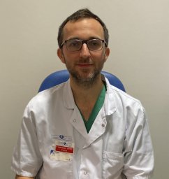 Maximilien Barret合作论文数Faculté Paris Descartes, Hôpital Européen Georges Pompidou26
 Chaussade Stanislas合作论文数24
 Romain Coriat合作论文数20
 Arthur Belle合作论文数17Einas Abou Ali合作论文数14Anna Pellat合作论文数Université Paris Cité8Dhooge Marion合作论文数8Benoît Terris合作论文数7
 Anthony Dohan合作论文数6Felix Corre合作论文数6Antoine Assaf合作论文数6
 Jacques Jérémie合作论文数6Romain Legros合作论文数5Frédéric Prat合作论文数4Goéré Diane合作论文数Inst Gustave Roussy4
 Elie Rassy合作论文数Faculty of Medicine, Saint Joseph University4
 Marine Camus合作论文数Assistance publique–Hôpitaux de Paris (AP–HP),, CHU Cochin Port-Royal4Catherine Brezault合作论文数4Marc A. Barthet合作论文数4Frédéric Beuvon合作论文数4Sarah Leblanc合作论文数4Mélanie Calmels合作论文数3Claire Ginestet合作论文数3Chloé Leandri合作论文数3Lola-Jade Palmieri合作论文数3
 Ziad Bakouny合作论文数Dana Farber Canc Inst, Dept Med Oncol, Boston, MA 02115 USA3mahaut leconte合作论文数3
 Jean-Michel Gonzalez合作论文数Aix-Marseille Université3Thierry Barrioz合作论文数3Ulriikka Chaput合作论文数3
 Khalil Antoine Honein合作论文数3Joseph Bou Jaoude合作论文数3F Prat合作论文数2Annegret Müller-Dornieden合作论文数2Maxime Amoyel合作论文数2Carlo Jung合作论文数2Ali Seif Amir Hosseini合作论文数2Paul Doumbe-Mandengue合作论文数2Fabian Kück合作论文数2
 Fares Yared合作论文数gastroenterology, Lebanese university2Solène Dermine合作论文数2
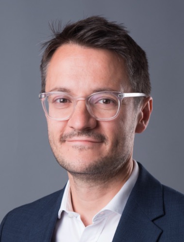 Jean-Emmanuel Bibault合作论文数Radiation Oncology Department, Georges Pompidou European Hospital, Assistance Publique – Hôpitaux de Paris;Jaide;Inserm2
 Maxime Barat合作论文数Service de Radiologie, Hôpital Antoine Béclère2Lorenzo Diane合作论文数2Philippe Soyer合作论文数2Steffen Kunsch合作论文数2




