Sundowning, a common yet poorly understood neuropsychiatric syndrome in Alzheimer’s disease (AD), manifests as evening-specific increases in agitation, confusion, and anxiety. Despite its prevalence and contribution to patient distress, its neural mechanisms remain elusive. Here, we establish a preclinical model of sundowning by characterizing sleep-wake, behavioral, and network-level alterations in an AD mouse model. Aged AD mice exhibit disrupted sleep-wake patterns and reduced slow-wave sleep. Behavioral and pose-tracking analyses revealed motor agitation and a distinct sundowning-like behavioral fingerprint selectively at Sundown. The suprachiasmatic nucleus (SCN) showed disruptions in time-of-day-dependent activation of vasopressin-expressing cells and brain-wide activity-dependent tagging identified hyperconnectivity amongst sensorimotor regions in AD mice at Sundown. Resting-state fMRI data from the Alzheimer’s Disease Neuroimaging Initiative revealed analogous Salience Network alterations in AD subjects. Overall, these cross-species findings define a systems-level framework for sundowning and highlight regions that may be targeted to alleviate a debilitating symptom of AD.
The suprachiasmatic nucleus (SCN) produces diffusible signals sufficient to sustain circadian locomotor rhythms, although the nature of such signals, their targets, and the pathway whereby such signals may travel is unknown. It is possible that the venous portal veins that connect the capillary beds of the SCN to those of the organum vasculosum of the lamina terminalis (OVLT) provide a vascular pathway whereby signals originating in SCN neurons can reach local targets in the OVLT. Given the presence of the blood-brain interface (BBI) within the SCN, it is unclear how diffusible signals originating in SCN neurons might access the capillary vasculature of this nucleus. Estimates of astrocyte coverage of capillary vasculature range widely, from 70 to 100%, and furthermore such coverage can change dynamically. In the present study, we investigated whether three vasoactive peptidergic processes found in the mouse SCN, namely, vasopressin, vasoactive intestinal peptide, and gastrin-releasing peptide, might breach the BBI, thereby accessing capillary vessels. Using widefield and confocal imaging, we found neuron-to-capillary contacts between varicosities bearing each of these vasoactive peptides and capillary basal membranes, pericytes, and the endothelia in the mouse SCN of either sex. The findings suggest that all three vasoactive peptides may functionally breach the BBI of the SCN highlighting the importance of understanding how these peptides act on local vasculature to impact blood flow.
The brain clock located in the suprachiasmatic nucleus (SCN) of the hypothalamus receives direct retinal input, thereby providing the entire body with an internal representation of external solar time. The pathways by which this small nucleus signals so broadly involve co-occurring nervous and diffusible output signals, although the latter are less understood. Portal pathways, such as the well-known pituitary portal pathway, provide a mechanism, whereby signals of neural origin can reach local, specialized targets without suffering dilution in the systemic blood supply. Newly discovered vascular pathways involve direct connections between each of the sensory circumventricular nuclei at its point of attachment to the brain. These nuclei line the brain's ventricles, and their leaky blood vessels and large perivascular spaces represent a route, whereby secretions from the SCN can be relayed and then amplified, providing a pathway to achieve global coordination of circadian clock signaling. This review provides a narrative that incorporates our understanding of SCN neural and diffusible output signals, with particular emphasis on the contribution of brain fluidic compartments and the fluids therein.
Among contributors to diffusible signaling are portal systems which join two capillary beds through connecting veins. Portal systems allow diffusible signals to be transported in high concentrations directly from one capillary bed to the other without dilution in the systemic circulation. Two portal systems have been identified in the brain. The first was discovered almost a century ago and connects the median eminence to the anterior pituitary gland. The second was discovered a few years ago and links the suprachiasmatic nucleus to the organum vasculosum of the lamina terminalis, a sensory circumventricular organ (CVO). Sensory CVOs bear neuronal receptors for sensing signals in the fluid milieu. They line the surface of brain ventricles and bear fenestrated capillaries thereby lacking blood-brain barriers. It is not known whether the other sensory CVOs, namely the subfornical organ (SFO), and area postrema (AP) form portal neurovascular connections with nearby parenchymal tissue. To preserve the integrity of the vasculature of CVOs and their adjacent neuropil, we combined iDISCO clearing and light-sheet microscopy to acquire volumetric images of blood vessels and traced the vasculature in two experiments. In the first, the whole brain vasculature was registered to the Allen Brain Atlas in order to identify the nuclei to which the SFO and AP are attached. In the second study, regionally specified immunolabeling was used to identify the attachment sites and vascular connections between the AP, and the SFO to their respective parenchymal attachment sites. There are venous portal pathways linking the capillary vessels of the SFO and the posterior septal nuclei, namely the septofimbrial nucleus and the triangular nucleus of the septum. Unlike the arrangement of portal vessels, the AP and the nucleus of the solitary tract share a common capillary bed. Taken together, the results reveal that all three sensory CVOs bear direct capillary connections to adjacent neuropil, providing a direct route for diffusible signals to travel from their source to their targets.
The suprachiasmatic nucleus (SCN), locus of a circadian clock, is a small nucleus of approximately 20,000 neurons that oscillate with a period of about 24 hours. While individual neurons produce circadian oscillations even when dispersed in culture, the coherence and robustness of oscillation of the SCN as a whole is dependent on its circuitry. Surprisingly, the individual neurons of the intact SCN do not all oscillate in phase with each other. To understand the oscillatory dynamics across the intact nucleus, we develop a model of the relation of the phase of neurons to their PER2 expression at a particular subjective time (CT1900) using time series data from SCN slice preparations. Next, we use the model, which produces a surprisingly good fit in the SCN slice data, to estimate oscillator phase at a single time point (CT1900) in snapshot data from PER2 expression measurements in intact, unsliced SCN-wide tissue. To monitor temporal changes in phase in time series data, we use PER2::LUC imaging in an ex vivo SCN slice preparation. To study phase in the intact SCN at a fixed time point we use data generated by PER2 staining and a tissue clearing protocol. Because PER2 expression, as measured in the time series slices and the snapshot intact SCN are not directly comparable, the model estimated from time series slices to the snapshot intact SCN data requires a calibrating constant. The results indicate that our model provides a surprisingly good fit to the SCN slice data and is therefore a meaningful method for estimating phase in the intact SCN snapshot data, permitting the study of virtual interventions such as virtual tissue slicing. We next compare oscillation in circuits in the SCN-wide tissue to those that have been disrupted by virtual slicing using a Kuramoto model to simulate the dynamics. The results support prior evidence that the damage done by coronal slicing has the most disruptive impact on SCN oscillation, while horizontal slicing has the least damage. The results point to the importance of connectivity along the caudal-to-rostral axis and indicate that SCN circuit organization depends on the caudal-to-rostral flow of information. In summary, the construction of this model is a major finding of the paper. Our modeling allows us to perform the previously impossible analysis of oscillatory dynamics in static data in an intact SCN captured at a single time point.
It has been 50 years since the suprachiasmatic nucleus (SCN) was first identified as the central circadian clock and 25 years since the last overview of developments in the field was published in the Journal of Biological Rhythms. Here, we explore new mechanisms and concepts that have emerged in the subsequent 25 years. Since 1997, methodological developments, such as luminescent and fluorescent reporter techniques, have revealed intricate relationships between cellular and network-level mechanisms. In particular, specific neuropeptides such as arginine vasopressin, vasoactive intestinal peptide, and gastrin-releasing peptide have been identified as key players in the synchronization of cellular circadian rhythms within the SCN. The discovery of multiple oscillators governing behavioral and physiological rhythms has significantly advanced our understanding of the circadian clock. The interaction between neurons and glial cells has been found to play a crucial role in regulating these circadian rhythms within the SCN. Furthermore, the properties of the SCN network vary across ontogenetic stages. The application of cell type–specific genetic manipulations has revealed components of the functional input-output system of the SCN and their correlation with physiological functions. This review concludes with the high-risk effort of identifying open questions and challenges that lie ahead.
The suprachiasmatic nucleus (SCN) sets the phase of oscillation throughout the brain and body. Anatomical evidence reveals a portal system linking the SCN and the organum vasculosum of the lamina terminalis (OVLT), begging the question of the direction of blood flow and the nature of diffusible signals that flow in this specialized vasculature. Using a combination of anatomical and in vivo two-photon imaging approaches, we unequivocally show that blood flows unidirectionally from the SCN to the OVLT, that blood flow rate displays daily oscillations with a higher rate at night than in the day, and that circulating vasopressin can access portal vessels. These findings highlight a previously unknown central nervous system communication pathway, which, like that of the pituitary portal system, could allow neurosecretions to reach nearby target sites in OVLT, avoiding dilution in the systemic blood. In both of these brain portal pathways, the target sites relay signals broadly to both the brain and the rest of the body.
By transporting products directly from the capillary bed of one region to the capillary bed of another region, vascular portal pathways enable minute amounts of important secretions to reach their specialized targets in high concentrations, without dilution in the systemic circulatory system. For decades there has been only one known portal system in the mammalian brain - that of the pituitary gland, first identified in 1933 (Popa and Fielding, J. Anatomy 1933). This year, we described a second portal pathway in the mouse linking the capillary vessels of the brain's clock suprachiasmatic nucleus (SCN) to those of the organum vasculosum of the lamina terminalis (OVLT), a circumventricular organ (Yao et al., Nat. Comm. 2021). A caveat in this initial work was that the direction of blood flow was unknown. To determine whether the SCN signaled the OVLT or vice-versa, we performed in vivo 2-photon imaging in anesthetized eGFP-vasopressin (VP) rats using a recently developed approach (Roy et al., Cell Report 2021) to study blood flow in this portal system. To delineate the SCN microvasculature in vivo, we intravenously infused fluorescent dextrans in anesthetized rats. The SCN-OVLT portal system was identified as Alexa 633 (an artery/arteriole specific dye)-negative vessels originating from a dense SCN capillary network that run rostrally towards the OVLT. These vessels displayed a mean diameter of ~20 μm. Blood flow in the portal vessels was measured by monitoring red blood cell (RBC) movement after intravenous injections with Rho70 kDa. Using kymographs, we found that in all cases, RBCs flowed rostrally, from the SCN towards the OVLT. Importantly, we found than blood flow was significantly higher at night (ZT17-19) compared to daylight (ZT5-7) (p< 0.001), while directionality remained the same (SCN→OVLT). Taken together, our results support the presence of a functional SCN-OVLT portal system in the rat in which blood flows unidirectionally from the SCN towards the OVLT. Moreover, our studies support the notion that blood flow in this system can be regulated. This clock portal system points to entirely new routes and targets for secreted signals from the SCN, restructuring our understanding of its output pathways. Support: NIH HLBI R01HL162575 to JES, AHA916907 to RKR and NSF 1749500 to RS. This is the full abstract presented at the American Physiology Summit 2023 meeting and is only available in HTML format. There are no additional versions or additional content available for this abstract. Physiology was not involved in the peer review process.
A map of central nervous system organization based on vascular networks provides a layer of organization distinct from familiar neural networks or connectomes. As a well-established example, the capillary networks of the pituitary portal system enable a route for small amounts of neurochemical signals to reach local targets by traveling along specialized pathways, thereby avoiding dilution in the systemic circulation. The first evidence of such a pathway in the brain came from anatomical studies identifying a portal pathway linking the hypothalamus and the pituitary gland. Almost a century later, we demonstrated a vascular portal pathway that joined the capillary beds of the suprachiasmatic nucleus and a circumventricular organ, the organum vasculosum of the lamina terminalis, in a mouse brain. For each of these portal pathways, the anatomical findings opened many new lines of inquiry, including the determination of the direction of flow of information, the identity of the signal that flowed along this pathway, and the function of the signals that linked the two regions. Here, we review landmark steps to these discoveries and highlight the experiments that reveal the significance of portal pathways and more generally, the implications of morphologically distinct nuclei sharing capillary beds.
SUMMARY The suprachiasmatic nucleus (SCN) is the locus of a brain clock that sets the phase of oscillation in cells throughout the brain and body. Anatomical evidence reveals a portal system linking the SCN and the OVLT (here termed SCN-OVLTp). This discovery begs the question of the direction of blood flow and the nature of diffusible signals that flow in this specialized vasculature. Here we show unequivocally that the direction of blood flow is from the SCN to the OVLT, that the rate of flow is under circadian regulation, and that vasopressin (AVP) is present in portal vessels following systemic injection. These findings highlight a previously unknown CNS communication pathway. It is well established that the SCN is required for circadian regulation of AVP in the CSF and that the OVLT bears AVP receptors. Specifically, SCN neurons are necessary for time-stamped signals such as the peptide AVP, that can travel via portal veins to a target in the OVLT. The OVLT, a circumventricular organ offering a “window to the brain,” can relay neural and diffusible signals to broad brain areas via its efferent connections and via the CSF. We conclude that the SCN-OVLTp, like that of the pituitary portal system, discovered almost a century ago, allows neurosecretions to reach nearby specialized target sites, thereby avoiding dilution in the systemic blood. In both of these brain portal pathways, the target site, namely the pituitary and OVLT respectively, relay signals broadly, to both the brain and the rest of the body.
Transplant studies demonstrate unequivocally that the suprachiasmatic nucleus (SCN) produces diffusible signals that can sustain circadian locomotor rhythms. There is a vascular portal pathway between the SCN and the organum vasculosum of the lamina terminalis in mouse brain. Portal pathways enable low concentrations of neurosecretions to reach specialized local targets without dilution in the systemic circulation. To explore the SCN vasculature and the capillary vessels whereby SCN neurosecretions might reach portal vessels, we investigated the blood vessels (BVs) of the core and shell SCN. The arterial supply of the SCN differs among animals, and in some animals, there are differences between the 2 sides. The rostral SCN is supplied by branches from either the superior hypophyseal artery (SHpA) or the anterior cerebral artery or the anterior communicating artery. The caudal SCN is consistently supplied by the SHpA. The rostral SCN is drained by the preoptic vein, while the caudal is drained by the basal vein, with variations in laterality of draining vessels. In addition, several key features of the core and shell SCN regions differ: Median BV diameter is significantly smaller in the shell than the core based on confocal image measurements, and a similar trend occurs in iDISCO-cleared tissue. In the cleared tissue, whole BV length density and surface area density are significantly greater in the shell than the core. Finally, capillary length density is also greater in the shell than the core. The results suggest three hypotheses: First, the distinct arterial and venous systems of the rostral and caudal SCN may contribute to the in vivo variations of metabolic and neural activities observed in SCN networks. Second, the dense capillaries of the SCN shell are well positioned to transport blood-borne signals. Finally, variations in SCN vascular supply and drainage may contribute to inter-animal differences.
Systemic glucose metabolism and insulin activity oscillate in response to diurnal rhythms and nutrient availability with the necessary involvement of adipose tissue to maintain metabolic homeostasis. However, the adipose-intrinsic regulatory mechanism remains elusive. Here, the dynamics of PPARγ acetylation in adipose tissue are shown to orchestrate metabolic oscillation in daily rhythms. Acetylation of PPARγ displays a diurnal rhythm in young healthy mice, with the peak at zeitgeber time 0 (ZT0) and the trough at ZT18. This rhythmic pattern is deranged in pathological conditions such as obesity, aging, and circadian disruption. The adipocyte-specific acetylation-mimetic mutation of PPARγ K293Q (aKQ) restrains adipose plasticity during calorie restriction and diet-induced obesity, associated with proteolysis of a core circadian component BMAL1. Consistently, the rhythmicity in glucose tolerance and insulin sensitivity is altered in aKQ and the complementary PPARγ deacetylation-mimetic K268R/K293R (2KR) mouse models. Furthermore, the PPARγ acetylation-sensitive downstream target adipsin is revealed as a novel diurnal factor that destabilizes BMAL1 and mediates metabolic rhythms. These findings collectively signify that PPARγ acetylation is a hinge connecting adipose plasticity and metabolic rhythms, the two determinants of metabolic health.
Background Steroids are lipid hormones that reach bodily tissues through the systemic circulation, and play a major role in reproduction, metabolism, and homeostasis. All of these functions and steroids themselves are under the regulation of the circadian timing system (CTS) and its cellular/molecular underpinnings. In health, cells throughout the body coordinate their daily activities to optimize responses to signals from the CTS and steroids. Misalignment of responses to these signals produces dysfunction and underlies many pathologies. Questions Addressed To explore relationships between the CTS and circulating steroids, we examine the brain clock located in the suprachiasmatic nucleus (SCN), the daily fluctuations in plasma steroids, the mechanisms producing regularly recurring fluctuations, and the actions of steroids on their receptors within the SCN. The goal is to understand the relationship between temporal control of steroid secretion and how rhythmic changes in steroids impact the SCN, which in turn modulate behavior and physiology. Evidence Surveyed The CTS is a multi-level organization producing recurrent feedback loops that operate on several time scales. We review the evidence showing that the CTS modulates the timing of secretions from the level of the hypothalamus to the steroidogenic gonadal and adrenal glands, and at specific sites within steroidogenic pathways. The SCN determines the timing of steroid hormones that then act on their cognate receptors within the brain clock. In addition, some compartments of the body-wide CTS are impacted by signals derived from food, stress, exercise etc. These in turn act on steroidogenesis to either align or misalign CTS oscillators. Finally this review provides a comprehensive exploration of the broad contribution of steroid receptors in the SCN and how these receptors in turn impact peripheral responses. Conclusion The hypothesis emerging from the recognition of steroid receptors in the SCN is that mutual shaping of responses occurs between the brain clock and fluctuating plasma steroid levels.
EDITORIAL article Front. Neurosci., 09 August 2021Sec. Sleep and Circadian Rhythms https://doi.org/10.3389/fnins.2021.735007
There is only one known portal system in the mammalian brain - that of the pituitary gland, first identified in 1933 by Popa and Fielding. Here we describe a second portal pathway in the mouse linking the capillary vessels of the brain's clock suprachiasmatic nucleus (SCN) to those of the organum vasculosum of the lamina terminalis (OVLT), a circumventricular organ. The localized blood vessels of portal pathways enable small amounts of important secretions to reach their specialized targets in high concentrations without dilution in the general circulatory system. These brain clock portal vessels point to an entirely new route and targets for secreted SCN signals, and potentially restructures our understanding of brain communication pathways.
 Joseph Lesauter合作论文数76Ann-Judith Silverman合作论文数23
 Lance J. Kriegsfeld合作论文数Helen Wills Neuroscience Institute, University of California, Berkeley;Department of Psychology, University of California, Berkeley;Neurobiology Research Laboratory, University of California, Berkeley20Paul Witkovsky合作论文数17
 Michael N Lehman合作论文数17
 Lily Yan合作论文数Joan and Sanford I. Weill Department of Medicine, Weill Cornell Medicine, Cornell University15Yifan Yao合作论文数13
 Ilia N Karatsoreos合作论文数Department of Integrative Physiology and Neuroscience, Washington State University13
 Michael C. Antle合作论文数Department of Psychology, Columbia University12
 Toshiyuki Hamada合作论文数10
 Peter D. Balsam合作论文数Barnard College, Columbia University9
 Xiaoxi Zhuang合作论文数Department of Neurobiology, The University of Chicago9
 Matthew P. Butler合作论文数Department of Integrative Biology, University of California at Berkeley9
 Katherine Nautiyal合作论文数8Mt Romero合作论文数7Harvey H. Feder合作论文数7
 Colin Saldanha合作论文数7Alana Taub合作论文数6
 Nicholas C Foley合作论文数Columbia University6
 Duncan K. Foley合作论文数Department of Economics, New School for Social Research6
 Mario Daniel Caba-Flores合作论文数5
 Claudia Juarez Portilla合作论文数Lab Biomed Integral & Salud, Univ Veracruzana4
 Sato Honma合作论文数Center for Sleep and Circadian Rhythm Disorders, Sapporo Hanazono Hospital4
 Patrick A. Tresco合作论文数Department of Biomedical Engineering, The University of Utah4
 Donald W. Pfaff合作论文数Laboratory of Neurobiology and Behavior, The Rockefeller University4Javier E Stern合作论文数4
 Carl L. Hart合作论文数Department of Psychology, Columbia University4Douglas G. Mcmahon合作论文数Department of Biological Sciences, Vanderbilt University4
 Robert Gábriel合作论文数School of Medicine, The Flinders University of South Australia4
 Ranjan Roy合作论文数Beloit College4Dan Feng Mei合作论文数4C. Reboulleau合作论文数4Isabella K Green合作论文数3
 Rehana Leak合作论文数Div Pharmaceut Sci, Duquesne Univ3
 Kazuyoshi Tsutsui合作论文数Laboratory of Integrative Brain Sciences, Department of Biology, Waseda University;Graduate School of Integrated Sciences for Life, Hiroshima University;Center for Medical Life Science, Waseda University3
 Eric Bittman合作论文数Department of Biology and Program in Neuroscience and Behavior, University of Massachusetts3
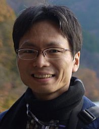 Jihwan Myung合作论文数Brain Sci Inst, RIKEN3
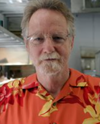 David Crews合作论文数Department of Integrative Biology, College of Natural Sciences, The University of Texas at Austin Austin3
 Matthew Richard Bailey合作论文数3Robotham Margaret合作论文数3
 Sandra Kuhlman合作论文数Department of Physiology and Biophysics, Jacobs School of Medicine & Biomedical Sciences, University at Buffalo3
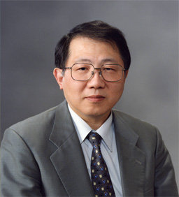 Ken-Ichi Honma (本間研一)合作论文数Division of Physiological Science, Graduate School of Medicine, Hokkaido University3
 Gregory Ball合作论文数Department of Psychology, College of Behavioral and Social Sciences, University of Maryland3
 Pierre Deviche合作论文数Sch Life Sci, Arizona State Univ3
 Eleanor Simpson合作论文数Department of Psychiatry, Columbia University;New York State Psychiatric Institute3Gabriela González-Mariscal合作论文数3Cindy Ramos合作论文数3
 Mona Khalil合作论文数3




