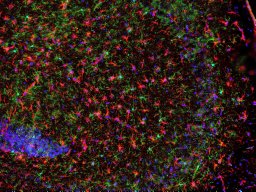
Protein aggregation is a hallmark of several neurodegenerative diseases, including Alzheimer's disease, Parkinson's disease and dementia with Lewy bodies. A common feature of these disorders is the misfolding and aggregation of α-synuclein (α-syn) and amyloid-β (Aβ) proteins into amyloid structures, which disrupt cellular homoeostasis and drive disease progression. While Aβ typically forms extracellular deposits and α-syn accumulates in intracellular inclusions, both ultimately contribute to neuronal damage and neurodegeneration. Increasing in vitro evidence suggests that these proteins can interact, altering their structural properties and, in turn, their biological effects; however, the consequences of their co-occurrence in vivo remain unclear. To address this gap, we examined whether α-syn modulates Aβ deposition and associated neuroinflammation at early stages using a physiologically relevant bigenic mouse model coexpressing human α-syn and APP knock-in Aβ. Using combined histological and biochemical analysis, we characterised Aβ load and microglial responses at early time points. Our results indicate that α-syn expression is associated with altered early Aβ deposition and microglial morphology in vivo. Specifically, while early Aβ deposits were detected in both Aβ/α-syn and Aβ-control mice from 2 months of age, at 4 and 6 months, reduced number and size of Aβ microdeposits was observed in the Aβ/α-syn model. The reduction in Aβ load was accompanied by a more ramified microglial morphology consistent with a less activated microglial state. Whether this delayed response reflects protection or impaired immune surveillance remains unclear. Our findings highlight the complexity of indirect Aβ and α-syn interactions and the need for further studies to clarify their functional impact. The newly generated bigenic mice provide a relevant platform to investigate early co-pathology and its role in disease progression.
Neurodegenerative diseases are characterized by a loss of neuronal function and structure, often in a region-specific manner. Multiple factors contribute to neuronal dysfunction and death, including pathogenic protein buildup, protein mislocalization, and inflammation. Despite extensive research, the common mechanisms driving neurodegeneration remain incompletely understood, partly because pathological processes affect interconnected cellular components such as the cytosol, cytoskeleton, and nucleus. Emerging evidence indicates that changes in nuclear structure and function are not simply secondary to cellular stress but play key roles in aging and neurodegenerative disease progression. This review explores important aspects of nuclear dysfunction, including alterations in nuclear architecture, chromatin organization, nucleocytoplasmic transport, DNA damage responses, and inflammatory signaling. It further discusses how these processes converge to increase neuronal vulnerability and may extend beyond normal aging to promote neurodegeneration. Overall, this review highlights nuclear dysfunction as a significant factor in neuronal impairment and underscores the need for therapies targeting nuclear integrity.
Connexins, fundamental components of gap junctions and hemichannels, regulate intercellular communication and are emerging neurodegeneration regulators. Primary synucleinopathies and co-morbid synuclein pathologies feature pathological α-synuclein (α-Syn) aggregation, yet mechanisms driving pathogenic α-Syn propagation remain unclear. We identify that connexin 50 (Cx50) interacts with α-Syn aggregates in synucleinopathy-affected human brain tissue. Ex vivo dye uptake assays show markedly elevated hemichannel activity in synucleinopathy mouse brain tissue versus wild-type controls, suppressed by selective Cx50 inhibition. Cx50-expressing cell models exhibit strain-dependent brain-derived α-Syn oligomers (BDSOs) uptake, confirmed pharmacologically. In primary neuron-astrocyte co-cultures from mice expressing human wild-type α-Syn, Cx50 knockdown markedly reduced BDSO uptake and α-Syn aggregation. Cx50 knockdown differentially modulates pro-inflammatory cytokines in BDSO-treated conditions, indicating strain-dependent effects and Cx50-mediated neuron-astrocyte crosstalk in regulating neuroinflammation. This identifies Cx50 as a plausible target for modulating initiation and early spread of α-Syn pathology, supporting Cx50-directed interventions for early-stage disease modification.
There is a pressing need to develop novel strategies to ameliorate symptoms and slow the progression of Alzheimer's disease. One of the hallmarks of Alzheimer's disease is the high levels of tau protein, which can form toxic oligomers and characteristic neurofibrillary tangles in the brain. Antitau antibodies can potentially bind tau protein and reduce tau pathology. In order to elicit a powerful antitau antibody response, virus-like-particle bacteriophage Qβ-based conjugate vaccines were developed targeting the microtubule binding region of tau protein. The Qβ-tau vaccines were able to produce a strong antitau antibody response in not only wild-type mice but also in human tau transgenic mice and a llama. The levels of antibody induced were superior to those generated by a corresponding keyhole limpet hemocyanin-based tau conjugate mimicking the one vaccine that successfully completed phase 1/2 human clinical trials. The Qβ-tau vaccine significantly improved the cognitive functions of the immunized mice and reduced the levels of inflammatory cytokines and tau in the brains, suggesting its translational potential.
Tau protein aggregates adopt distinct conformations across tauopathies, yet the protein interactions engaged by disease-specific polymorphs remain poorly characterized. Here, we demonstrate that conformationally distinct tau polymorphs associate with disease-specific interaction networks across Alzheimer’s disease (AD), progressive supranuclear palsy (PSP), and dementia with Lewy bodies (DLB). Interactome profiling of tau aggregates from PBS- and sarkosyl-soluble brain fractions identified 493 high-confidence interactors exhibiting remarkable disease specificity. As an exploratory feature discovery machine learning classification discriminated against diseases using as few as four to six protein features. AD tau selectively engaged glycolytic enzymes, TCA cycle components, and glutamate/GABA cycling machinery, with the astrocytic transporter SLC1A2 showing 27-fold enrichment. PSP tau exhibited extensive interactor depletion alongside selective proteasome enrichment, whereas DLB tau associated with neurogenesis modulators while depleting neuroinflammatory mediators. Interaction patterns were corroborated by parallel reaction monitoring mass spectrometry and proximity ligation assays and corresponded to disease-specific post-translational modification profiles. These findings show that tau polymorph conformations are associated with disease-specific interaction networks, providing molecular insight into tauopathy heterogeneity. Conformationally distinct tau aggregates arise across tauopathies, but the proteins they engage are unknown. This study links tau polymorphs from Alzheimer’s disease (AD), progressive supranuclear palsy (PSP), and dementia with Lewy bodies (DLB) to interaction networks, offering insight into disease heterogeneity. Interactome profiling of tau aggregates from postmortem brain of Alzheimer’s disease (AD), progressive supranuclear palsy (PSP), and dementia with Lewy bodies (DLB) provides new insights into disease heterogeneity.
The pathological misfolding and aggregation of the microtubule associated protein tau (MAPT), a full length Tau2N4R with 441aa, is considered the principal disease relevant constituent in tauopathies including Alzheimer's disease (AD) with an imbalanced ratio in 3R/4R isoforms. The exact cellular fluid composition, properties, and changes that coincide with tau misfolding, seed formation, and propagation events remain obscure. The proteostasis network, along with the associated osmolytes, is responsible for maintaining the presence of tau in its native structure or dealing with misfolding. In this study, for the first time, the roles of natural brain osmolytes are being investigated for their potential effects on regulating the conformational stability of the tau monomer (tauM) and its propensity to aggregate or disaggregate. Herein, the effects of physiological osmolytes myo-inositol, taurine, trimethyl amine oxide (TMAO), betaine, sorbitol, glycerophosphocholine (GPC), and citrulline on tau's aggregation state were investigated. The overall results indicate the ability of sorbitol and GPC to maintain the monomeric form and prevent aggregation of tau, whereas myo-inositol, taurine, TMAO, betaine, and citrulline promote tau aggregation to different degrees, as revealed by protein morphology in atomic force microscopy images. Biochemical and biophysical methods also revealed that tau proteins adopt different conformations under the influence of these osmolytes. TauM in the presence of all osmolytes expressed no toxicity when tested by a lactate dehydrogenase assay. Investigating the conformational stability of tau in the presence of osmolytes may provide a better understanding of the complex nature of tau aggregation in AD and the protective and/or chaotropic nature of osmolytes.
Tau pathology plays a critical role in the onset and progression of multiple neurodegenerative diseases. Although synucleinopathies such as Parkinson’s disease (PD) and dementia with Lewy bodies (DLB) are primarily defined by α-Synuclein (α-Syn) inclusions, they frequently exhibit substantial tau co-pathology. However, whether wild-type (WT) human α-Syn overexpression influences tau pathology in vivo remains poorly defined. Here, we systematically investigated the temporal and regional progression of tau pathology in hSyn transgenic mice overexpressing WT human α-Syn. Using immunohistochemical, biochemical, and structural approaches, we demonstrate progressive, age-dependent accumulation of hyperphosphorylated tau in cortex, hippocampus, and midbrain. Tau pathology was minimal at 5 months, increased substantially by 10 months, and further intensified by 20 months, validated by both phosphorylation-dependent and conformation-specific antibodies. Regional correlation analysis revealed that coordinated accumulation of tau and α-Syn pathology during disease progression (overall r2 = 0.844), with weak association at 5 months and strong, significant correlations by 10 and 20 months. Electron microscopy revealed fibrillar structures in insoluble fractions, and mass spectrometry confirmed the presence of both α-Syn and tau in these fractions. Notably, these pathologies were absent in WT littermates, indicating they arise as a consequence of WT human α-Syn overexpression rather than normal aging. Our findings support tau-targeted therapeutic approaches as a viable strategy for synucleinopathies.
Background Soluble tau oligomers (tauO) are early, synaptotoxic drivers of dysfunction in tauopathies. While selective vulnerability is well documented at the cellular level, emerging evidence suggests that synaptic subtypes may differ in their susceptibility to tau pathology. Still, the key factors that shape synaptic vulnerability to toxic tauO, particularly in humans, remain poorly understood. Objective To define the synaptic compartments and subtypes most vulnerable to tauO and identify molecular correlates underlying this susceptibility. Methods Synaptosomes were isolated from cognitively normal human autopsy specimens and acutely challenged with preformed recombinant tauO. Flow cytometry with multiplexed immunophenotyping resolved tauO engagement across intact pre- and postsynaptic compartments and excitatory versus inhibitory subtypes. Functional effects were assessed by microtransplanting synaptic membranes into Xenopus laevis oocytes and recording ligand-gated GABAergic and glutamatergic responses. Complementary LC-MS/MS proteomics of brain-derived tau oligomers (BDTO) from PBS-soluble hippocampal lysates of primary age-related tauopathy (PART) cases were analyzed using SynGO enrichment to identify molecular correlates. Results TauO preferentially engaged presynaptic compartments and showed elevated association with GABAergic synapses. Functionally, acute tauO exposure selectively enhanced GABAAR-mediated responses, with no effect on AMPAR-mediated currents. The PART BDTO interactome was enriched for presynaptic vesicle-associated proteins involved in vesicle cycling and neurotransmitter release, consistent with a presynaptic axis of vulnerability. Conclusions This integrative analysis identifies a compartment- and subtype-specific vulnerability of human synapses to tauO, highlighting a presynaptic inhibitory bias as a potential driver of synaptic dysfunction and tau propagation in early-stage tauopathies.
Alzheimer's disease (AD) is one of the most common forms of dementia worldwide, making the identification of predictive biomarkers critical for early diagnosis and treatment during the preclinical stage. Recent studies have focused on blood-based biomarkers, particularly plasma brain-derived extracellular vesicles (pl-BDEVs), to detect central nervous system (CNS) alterations. While blood biomarkers like amyloid proteins (Aβ42/Aβ40), tau, and phosphorylated tau show diagnostic promise, identifying predictive biomarkers remains essential. Blood total-tau, originating from non-brain sources, highlights the need to analyze brain-derived tau (BDT) in pl-BDEVs as a more specific biomarker for AD and other neurodegenerative diseases. Longitudinal studies offer great potential for detecting preclinical biomarker patterns in at-risk individuals, yet little attention has been given to oligomers, the most toxic species in AD In this study, we enriched pl-BDEVs from CNS cell types from plasma samples longitudinally collected from participants enrolled in the Texas Alzheimer's Research and Care Consortium (TARCC), who were initially cognitively normal or displayed mild cognitive impairment (MCI), and later either progressed to AD (termed “converters”) or remained cognitively normal/MCI (termed “non-converters”). We evaluated the isolated pl-BDEVs by nanoparticle tracking analysis (size, number, and distribution), western blot (expression of extracellular vesicles markers: CD63, CD9, CD81), and electron microscopy. We immunoprecipitated brain-derived tau oligomers (BDTOs) from pl-BDEVs and characterized them by western blot, proteinase K (PK) digestion, seeding assay, and atomic force microscopy (AFM). Additionally, we treated SH-SY5Y neuroblastoma cells and human synaptosomes isolated from cortex of control samples with these BDTOs and assessed their cytotoxicity and synaptotoxicity, respectively. We demonstrated the successful isolation of pl-BDEVs from plasma samples and the detection of BDTOs within these pl-BDEVs. We identified distinct PK digestion patterns, morphology, and toxicity between BDTOs from converters and non-converters. Additionally, our data revealed different distribution of BDTOs in the pl-BDEVs isolated from the two groups. These findings suggest the presence of two different BDTO strains for converters and non-converters. This study addresses the need for predictive AD biomarkers by analyzing previously unexplored BDTO conformers in pl-BDEVs. Discovering distinct BDTOs in peripheral brain derived extracellular vesicles could enable preclinical forecasting and advance early-stage AD treatments.
Connexins, gap junction components, have been implicated in intercellular connectivity under physiological and pathophysiological conditions, including neurodegeneration. Synucleinopathies comprise a diverse group of neurodegenerative disorders pathologically characterized by α-synuclein (α-Syn) aggregates. However, little is known about connexin-associated α-Syn pathological spread in synucleinopathies Here, we present evidence of connexin-50 (Cx50) directly interacting with α-Syn aggregates in human brains affected by synucleinopathies, including Alzheimer's disease (AD), dementia with Lewy bodies (DLB), and Parkinson's disease (PD). We also observed this interaction in pre-clinical α-Syn mouse models that exhibit Parkinson's disease-related phenotypes. To achieve this, we utilized immunohistochemistry, pharmacological, and genetic manipulation techniques in connexin cell models and primary co-cultures. Utilizing well-characterized α-Syn oligomers (BDSOs) isolated from brains affected by Alzheimer's disease (AD), Lewy body dementia (DLB), and Parkinson's disease (PD), we demonstrate that BDSOs preferentially enter cells expressing connexin 50 (Cx50), a protein found in gap junctions, as confirmed by pharmacological inhibition. In live-cell imaging experiments, we observed a significant reduction in BDSO uptake in primary neurons-astrocytes co-cultured from human wild-type α-Syn transgenic mice expressing genetically modified Cx50. This reduction was accompanied by a decrease in α-Syn aggregates. Moreover, the downregulation of Cx50 was associated with a reduction in the activity of astrocytes, as evidenced by a decrease in pro-inflammatory and an increase in anti-inflammatory cytokines. These findings suggest that Cx50 plays a crucial role in the interplay between neurons and astrocytes. This study presents compelling evidence of the connection between disease-relevant α-Syn aggregates and Cx50 in neurons that regulate astrocyte activity. This insight sheds light on the intricate process of pathogenic α-Syn spread in Synucleiopathies.
Abstract Pathological tau isoforms, including hyperphosphorylated tau at serine 396 (pS396-tau) and tau oligomers (Oligo-tau), are elevated in the retinas of patients with mild cognitive impairment (MCI) due to Alzheimer’s disease (AD) and AD dementia. These patients exhibit significant retinal ganglion cell (RGC) loss, however the presence of tau isoforms in RGCs and their impact on RGC integrity, particularly in early AD, have not been studied. Here, we analyzed retinal superior temporal cross-sections from 25 MCI or AD patients and 16 age- and sex-matched cognitively normal controls. Using the RGC marker ribonucleic acid binding protein with multiple splicing (RBPMS) and Nissl staining, we found a 46–56% reduction in RBPMS+ RGCs and Nissl+ neurons in the ganglion cell layer (GCL) of MCI and AD retinas (P < 0.05–0.001). RGC loss was accompanied by soma hypertrophy (10–50% enlargement, P < 0.05–0.0001), nuclear displacement, apoptosis (30–50% increase, P < 0.05–0.01), and prominent expression of granulovacuolar degeneration (GVD) bodies and GVD-necroptotic markers. Both pS396-tau and Oligo-tau were identified in RGCs, including in hypertrophic cells. PS396-tau+ and Oligo-tau+ RGC counts were significantly increased by 2.1–3.5-fold in MCI and AD retinas versus control retinas (P < 0.05–0.0001). Tauopathy-laden RGCs strongly inter-correlated (r P =0.85, P < 0.0001) and retinal tauopathy associated with RGC reduction (r P =-0.40–(-0.64), P < 0.05–0.01). Their abundance correlated with brain pathology and cognitive deficits, with higher tauopathy-laden RGCs in patients with Braak stages (V–VI), clinical dementia ratings (CDR = 3), and mini-mental state examination (MMSE ≤ 26) scores. PS396-tau+ RGCs in the central and mid-periphery showed the closest associations with disease status, while Oligo-tau+ RGCs in the mid-periphery exhibited the strongest correlations with brain pathology (NFTs, Braak stages, ABC scores; r S =0.78–0.81, P < 0.001–0.0001) and cognitive decline (MMSE; r S =-0.79, P = 0.0019). Overall, these findings identify a link between pathogenic tau in RGCs and RGC degeneration in AD, involving apoptotic and GVD-necroptotic cell death pathways. Future research should validate these results in larger and more diverse cohorts and develop RGC tauopathy as a potential noninvasive biomarker for early detection and monitoring of AD progression.
Apolipoprotein E (APOE), the strongest genetic risk factor for late-onset Alzheimer's disease, exist as 3 isoforms (APOE2, 3, 4) which differentially influence Alzheimer's disease risk and tau pathology in disease. Recent studies have revealed the polymorphic nature of amyloidogenic proteins, including tau, across different diseases, yet there remains a gap in knowledge concerning tau polymorphism across genetic variants such as the APOE isoforms. Here, we address this gap in knowledge by characterizing tau oligomers associated with each APOE isoform. Brain-derived tau oligomers and fibrils were isolated from tissue of patients with various APOE genotypes. Tau oligomers were characterized using proteinase K (PK) digestion and liquid chromatography-tandem mass spectrometry (LC-MS/MS) to investigate proteolytic stability and cleavage site accessibility to PK. Electrophysiology was used to investigate synaptotoxicity of the tau oligomers. Tau oligomers differ in proteolytic stability and cleavage site profiles across the APOE isoforms, indicating conformationally-distinct tau oligomer polymorphs. The tau oligomer polymorphs differentially impair synaptic functioning in an APOE isoform-specific manner, with APOE4-relevant tau oligomers inducing the strongest impairment of synaptic functioning. The APOE isoforms correspond with distinct tau oligomer polymorphs with varying synaptotoxicity. These findings highlight the need for APOE isoform to be considered when generating tau-based therapies for Alzheimer's disease. Specific targeting of APOE isoform-specific tau oligomer polymorphs could provide a novel method of mitigating Alzheimer's Disease pathology and combating disease progression.
Misfolding and aggregation of tau into oligomers and neurofibrillary tangles are associated with Alzheimer's disease and related dementia (ADRD). Misfolded oligomeric species are widely believed to play a critical role in both disrupting cellular functions and propagating protein misfolding between cells. Characterization of the misfolded oligomers is crucial for understanding the mechanisms underlying protein aggregation and its role in disease pathogenesis. However, structural characterization of these misfolded oligomers has proven challenging due to their transient and heterogeneous nature. Here we report structural features of brain-derived tau oligomers extracted from Alzheimer's brains. Initial screening using negative staining transmission electron microscopy (TEM) and atomic force microscopy (AFM) reveal that tau (2N4R) forms a diverse array of pore-like oligomers with a diameter of 5-20 nm and a height of ∼2-8 nm. Higher-resolution structural analyses using cryo-EM on oligomers with diameters of 10-20 nm revealed the presence of two distinct layers within the pore-like structures, resolved at 2.5-4 Å. Our structural studies support the hypothesis that misfolded proteins may function as pore-forming toxins, potentially disrupting cellular membranes.
Tauopathies are a set of neurodegenerative diseases characterized by the pathological accumulation of aggregated tau in the brain. Recent breakthrough evidence has revealed the existence of different strains of tau oligomer (TauO) which direct the different pathological presentation of individual tauopathies. Extensive research efforts have been devoted to search for specific antibodies or drug candidates which target TauO to serve as promising alternatives to treat Alzheimer's disease (AD) in future. To screen for the antibodies which are able to bind with amplified brain derived tau oligomer (aBDTO), we have investigated the binding parameters of the tau oligomer-specific monoclonal antibody-1 (TOMA-1) and toxic tau conformation-specific monoclonal antibody-1 (TTCM-1) with the recombinant tau monomer (rTauM) and aBDTO using isothermal titration calorimetry (ITC). TOMA-1 specifically recognizes the amino acid sequences 367-386, 382-401 and 367-386 and TTCM-1 specifically recognizes the amino acid sequence 307-326 of rTauM and aBDTO, respectively. Our results demonstrated that both TOMA-1 and TTCM-1 have a high binding affinity with aBDTO compared to rTauM. We also observed that higher the binding affinity of the antibody to the aBDTO, lower was the toxicity of the aBDTO and vice versa. Our study taken together presents both TOMA-1 and TTCM-1 to be potential immunotherapeutic agents against AD.
Tau protein aggregates exhibit distinct conformations across tauopathies, but their disease-specific protein interactions remain poorly understood. Here, we demonstrate that disease-specific tau conformations determine unique protein interaction landscapes across Alzheimer's disease (AD), progressive supranuclear palsy (PSP), and dementia with Lewy bodies (DLB). Through comprehensive interactome profiling of misfolded tau aggregates from PBS- and sarkosyl-soluble fractions. We identified 493 high-confidence proteins with remarkable disease specificity-notably, no common interactors overlapping across all three tauopathies. Machine learning classification achieved compelling discrimination between diseases using as few as 4-6 proteins features, demonstrating robust molecular signatures underlying clinical heterogeneity. AD derived tau aggregates uniquely engaged cellular metabolism machinery, including key glycolytic enzymes and TCA cycle proteins, alongside glutamate/GABA neurotransmitter cycling components, with the astrocytic glutamate transporter SLC1A2 showing 27-fold enrichment over other tauopathies. In contrast, PSP tau displayed the most distinctive profile, with extensive protein depletion (52/57 significant proteins) and selective enrichment of proteasome components, particularly PSMB7 showing >3000-fold abundance. DLB tau is associated with neurogenesis modulators while depleting neuroinflammatory mediators. These interaction patterns were validated through proximity ligation assays and correlated with distinct post-translational modification profiles, with PSP tau exhibiting globally elevated ubiquitination, AD showing mixed modification patterns, and DLB displaying minimal ubiquitination. Critically, sarkosyl-soluble fractions revealed reduced interactome complexity across diseases, except for PSP tau which maintained robust interactions with GPCR-ERK signaling and kinetochore proteins, suggesting unique aggregation mechanisms. Our findings establish that conformationally distinct tau strains dictate disease-specific protein interaction networks, providing molecular insight into tauopathy diversity and identifying novel therapeutic targets for precision medicine approaches in neurodegeneration.
The oligomers and fibrils of tau are well known as an indicator of Alzheimer’s disease (AD). Recently, other protein aggregates have been shown to be potentially involved in the development of the disease. One of these proteins is p53, involved in DNA repair. Prior studies in our lab show that p53 will form oligomers and fibrils in AD cases but not healthy controls, and has even been observed as a co-aggregate with tau protein. Owing to the importance of p53 in mitigating damage from diseased cells, it is possible that loss of function or even misfunction of p53 due to aggregation or mislocalisation could be an early contributing factor to AD, having been shown as a potential early biomarker for AD. Because of the co-aggregation of p53 and tau, it is possible that aggregate-specific antibodies will bind to both p53 and tau. Besides giving mechanistic insights into the proteins’ aggregation, this would also suggest that these antibodies could inhibit aggregation in vivo . This study examines this common epitope. • Using recombinant wild-type and mutant p53 and tau proteins, the interaction of p53 and tau specific antibodies with each other was determined based around their immunoreactivity, and compared to amyloid-β. • Co-staining of AD mouse model and AD patient samples for both misfolded tau and p53 was used to determine if this conformation cross-reactivity could also be observed in patient samples. Despite the lack of sequence homology, an in-house conformation and aggregation specific p53 antibody was found to be able to react with tau protein, and vice versa. It was also observed that the p53 mutant proteins had different reactions to these aggregate-specific antibodies. As these antibodies are conformation-specific, this provides information on the similar conformation both proteins must adopt in order to react with MDM2. The common epitope recognised by the p53 and tau antibodies suggests that this is a potential target for antibody therapy, as the ability to prevent the formation of the co-aggregates may permit p53 to continue its role as the ‘guardian of the genome’, a role which, if inhibited, could be a contributing factor to AD.
This study identifies and quantifies diverse pathological tau forms in the retina at both early and advanced stages of Alzheimer’s disease (AD) and assesses their correlation with disease status. In the pathogenesis of AD, the tau protein undergoes post-translational modifications, including hyperphosphorylation (p-tau). As the disease progresses, pathological tau can propagate as oligomers, aggregate into fibrils, and paired helical filaments (PHF), and ultimately form intraneuronal neurofibrillary tangles (NFTs). Previously, increased p-tau and other abnormal forms were detected in postmortem retinas of AD patients; however, their presence and levels at earlier stages – in mild cognitively impaired (MCI due to AD) patients – and their association with brain pathology remain undefined. In this study, we acquired 75 postmortem human eyes and 39 paired brains. Retinal cross-sections in predefined geometric regions were prepared from 34 AD and 11 MCI patients, and 30 age- and sex-matched cognitively normal controls. Histological analyses involved Bielschowsky silver staining and immunostaining of brains and retinas with antibodies for tau (pS396, AT8, AT100, CitR 209 , T22, PHF-1, and MC-1). Stereological examination and quantification were conducted, and correlations were determined with brain pathology and cognition. P-tau forms were also determined by NanoString GeoMx digital spatial profiling. Our immunohistochemical examination revealed significant upregulation of immature hyper-citrullinated (CitR 209 ) and p-tau forms, propagating tau oligomers, as well as mature PHF-tau and NFT forms in the retinas of patients with early-AD (MCI due to AD) and AD-dementia stages. Interestingly, retinal PHF-tau accumulated only in the AD-dementia stage. GeoMx spatial profiling analyses revealed site-specific increases in various p-tau epitopes in AD, particularly at the early MCI stage. Tau oligomers had the largest accumulations in MCI and, moreover, AD retinas, showing a strong correlation to Braak staging, representing the spread of tauopathy across brain regions during disease progression. Strong correlations were also found between retinal tauopathy and brain NFTs, ABC, and MMSE severity scores. Our data demonstrate that most forms of retinal tauopathy are increased in early AD and correlate with one or more AD neuropathology and cognitive parameters, encouraging the development of retinal tauopathy imaging for AD detection and monitoring disease progression.
Pathological tau aggregates cause cognitive decline in neurodegenerative tauopathies, including Alzheimer’s disease (AD), and more abundant in intracellular vs . extracellular compartments. However, current immunotherapies are slow and ineffective at clearing intracellular tau aggregates. We developed toxic tau conformation–specific monoclonal-antibody-2–loaded micelles (TTCM2-ms) that selectively recognize disease-relevant tau aggregates in brain tissues from patients with AD, progressive supranuclear palsy, and dementia with Lewy bodies and potently inhibit tau-seeding activity. A single intranasal dose of TTCM2-ms effectively cleared pathological tau, increased levels of synaptic proteins, and improved cognitive functions in aged tauopathy mice. Mechanistic studies suggest that TTCM2-ms clears intracellular, synaptic, and seed-competent tau aggregates via tripartite motif-containing 21 (TRIM21), an intracellular antibody receptor and E3-ubiquitin ligase. TRIM21 is essential for TTCM2-ms–mediated clearance of tau pathology. Our findings suggest that intranasally administered TTCM2-ms rapidly distributes across the brains of tauopathy mice. Further, TTCM2-ms recognized and cleared pathological tau from the intracellular and synaptic compartments of neuronal cells via TRIM21, thus improving cognitive functions. This study provides insights into the mechanisms of an effective tau immunotherapy strategy against intracellular tau pathology in neurodegenerative tauopathies, including AD.
INTRODUCTION:Pathological tau aggregates form distinct polymorphic species across diseases and even across Alzheimer's disease (AD) patients. However, tau aggregate polymorphism across the apolipoprotein E isoforms (APOE ε2, ε3, ε4), the strongest predictors of late-onset AD development, is unknown. METHODS:This study assessed the conformational and bioactivity properties of tau oligomers from 14 patients with varying APOE genotypes. RESULTS:Tau oligomers differ in proteolytic stability and cleavage site profiles across the APOE isoforms, indicating conformationally distinct polymorphs. APOE isoform-associated tau oligomers affect synaptic plasticity differently, with ε4-associated oligomers having the highest potency and strongest impact on synaptic functioning. Bioactivity assays reveal that ε4-associated oligomers demonstrate particularly high seeding activity. Interestingly, tau oligomer synaptotoxicity and seeding activity are independent characteristics. DISCUSSION:The APOE isoforms are associated with distinct tau oligomer polymorphs with varying bioactivity, underscoring the importance of considering APOE status when generating AD therapies. Polymorph-specific targeting of pathological tau species could provide a novel method of combating AD. HIGHLIGHTS:Conformational and bioactivity distinctions of tau oligomers have not yet been investigated across the APOE isoforms (ε2, ε3, ε4). Tau oligomers differ in conformational properties across the APOE isoforms. APOE ε4-relevant tau oligomers strongly impair synaptic plasticity and demonstrate high tau seeding activity. APOE ε4-relevant tau oligomers exist as a particularly toxic species, making them an ideal target for tau-based AD therapies.
 Urmi Sengupta合作论文数University of Newcastle;University of Hong Kong;University of Newcastle, University of Hong Kong95
 Charles G. Glabe合作论文数School of Biological Sciences, University of California, Irvine;Alzheimer's Disease Research Center, University of California, Irvine65
 Diana L Castillo-Carranza合作论文数UANL), Universidad Autónoma de Nuevo León51
 Nemil Bhatt合作论文数Univ Texas Med Branch, Mitchell Ctr Neurodegenerat Dis, Galveston, TX 77555 USA39
 Julia Gerson合作论文数39Cristian A Lasagna-Reeves合作论文数36
 Marcos Jair Guerrero-Munoz合作论文数Department of Neurology, University of Texas Medical Branch34Giulio Taglialatela合作论文数University of Texas Medical Branch33
 George Jackson合作论文数David Geffen School of Medicine, the University of California33
 Mauro Montalbano合作论文数30
 Puangmalai Nicha合作论文数29Salome McAllen合作论文数Univ Texas Med Branch, Mitchell Ctr Neurodegenerat Dis, Galveston, TX 77555 USA25
 Kelly T Dineley合作论文数University of Texas Medical Branch19Anna Ellsworth合作论文数17
 Balaji Krishnan合作论文数Mitchell Ctr Neurodegenerat Dis, Univ Texas Med Branch15Mariana Carretero-Murillo合作论文数14
 Alice Bittar合作论文数14Cynthia Jerez合作论文数14Wenru Zhang合作论文数14Kathleen Farmer合作论文数13
 Shashirekha Krishnamurthy合作论文数11Michela Marcatti合作论文数Laboratory of Biochemistry, University of Palermo11Filippa Lo Cascio合作论文数10
 Md. Anzarul Haque合作论文数Centre for Interdisciplinary Research in Basic Sciences, Jamia Millia Islamia, New Delhi, 110025, India.10
 Yingxin Zhao合作论文数Department of Internal Medicine, University of Texas Medical Branch at Galveston;Division of Endocrinology, University of Texas Medical Branch at Galveston;Sealy Center for Molecular Medicine, University of Texas Medical Branch at Galveston10
 Alan D. Barrett合作论文数Sealy Institute for Vaccine Sciences, The University of Texas Medical Branch at Galveston;Department of Microbiology & Immunology, The University of Texas Medical Branch at Galveston10
 Sagar Gaikwad合作论文数9
 James E. Hall合作论文数Department of Physiology & Biophysics, School of Medicine, University of California9Wenbo Zhang合作论文数9
 Fracassi Anna合作论文数Department of Science, University Roma Tre9
 Juan C. Troncoso合作论文数School of Medicine, Johns Hopkins University;Brain Resource Center, Johns Hopkins University8
 Keith L. Black合作论文数Department of Neurosurgery, Cedars-Sinai Medical Center8
 Audra L Clos合作论文数George P. and Cynthia Woods Mitchell Center for Neurodegenerative Diseases, The University of Texas8
 Veronica Galvan合作论文数Buck Institute for Age Research8
 Maya Koronyo-Hamaoui合作论文数Cedars-Sinai Medical Center8Yosef Koronyo合作论文数8Saskia Milton合作论文数8Nicole Goertz合作论文数7
 Ashley Nilson合作论文数NIH, Intramural Res Program7Stephanie Garcia合作论文数7
 Tasneem F Hasan合作论文数Dept Neurol, Ochsner Louisiana State Univ7Elizabeth Head合作论文数University of California Irvine7Bayan Aljabari合作论文数7
 Limon Agenor合作论文数Department of Neurobiology and Behavior, University of California7
 Nazanin Mirzaei合作论文数The Centre for Neuroscience, Imperial College London7Fuchs Dieu-Trang合作论文数7
 Michael Bacher合作论文数Department of Neurology, Philipps-University Marburg7
 Stacy A. Hussong合作论文数Department of Biochemistry, Molecular Biology, and Biophysics, University of Minnesota7Peter C Butler合作论文数7




