Abstract MRI fingerprinting (MRF) detects structure-specific relaxometry changes in brain tissue. The purpose of this study is 1) to characterize longitudinal changes in MR Fingerprinting (MRF)-derived T1 and T2 relaxometry within key cognitive structures for brain metastasis patients, and 2) to determine whether MRF changes correlate with the time interval between treatment and post-treatment imaging at the level of individual brain structures. MRF was performed at 1.5 T in 16 patients at pre-treatment baseline, an intermediate scan (128±55 days, n = 3), and post-treatment (157±59 days, range 94–314 days). T1and T2 maps were co-registered to the planning T1Gd using pre-computed rigid-body transforms. CSF-contaminated voxels were excluded (T1 > 2500 ms at 1.5 T). Left and right hippocampal and amygdala structures were combined bilaterally for analysis. Longitudinal MRF changes (PRE→POST) were quantified per structure. At baseline, mean relaxation times were established for the hippocampus (T1=1240±87ms; T2=88±9 ms), amygdala (T1=1204±99 ms; T2=78±8 ms), and corpus callosum (T1=845±88 ms; T2=68±7 ms). While longitudinal changes were observed across all structures—the most prominent being a 2.7% increase in corpus callosum T2 (Δ = 1.79±4.10 ms; d = 0.44)—significant temporal correlations were localized exclusively to the hippocampus (r ≈ -0.65, FDR-corrected p = 0.020), indicating a specific time-dependent decrease in MRF values post-treatment. No significant temporal correlations were observed in the amygdala (p = 0.186) or corpus callosum (p ≥ 0.480). Changes in T1/T2 relaxation times are hypothesis-generating and may represent tissue changes related to cancer burden, radiation therapy, or systemic therapy. These findings support the use of quantitative MRF as a sensitive, regionally resolved biomarker for monitoring brain tissue changes. MRF will be leveraged to develop a cognitive biomarker in the upcoming Athena 3 trial.
BACKGROUND:Magnetic resonance fingerprinting (MRF) is an emerging quantitative imaging technique that enables multiparametric tissue characterization, but its adoption has been hindered by the complexity of data acquisition and post-processing. These technical and implementation challenges have limited its broader clinical deployment. PURPOSE:To develop a modular MRF Development Kit (MRFDK) that enables efficient sequence design, streamlined implementation, and real-time image reconstruction. STUDY TYPE:Prospective. POPULATION:T1 and T2 relaxation phantom, nine volunteers (seven males and two females), five metastatic brain cancer patients. FIELD STRENGTH/SEQUENCE:3 T, MR Fingerprinting. ASSESSMENT:Accuracy of T1 and T2 quantification was estimated from phantom experiments. Manual ROIs were drawn on brain lesions and contralateral white matter for metastatic cancer patients. STATISTICAL TESTS:t-test, in vivo repeatability was calculated with Bland-Altman analysis on healthy volunteer scan-rescan data, significance level p < 0.01. RESULTS:Phantom results showed high accuracy in T1 and T2 assessment, with absolute percentage differences of 3% for T1 and 5% for T2 compared to offline MATLAB reconstruction. In vivo scans of eight healthy subjects further demonstrated excellent repeatability (bias and agreement: 0.95% ± 1.85% for T1; 1.78% ± 5.08% for T2). In patients, metastatic lesions showed significantly higher T1 and T2 values (T1, 1474 ms; T2, 61 ms) compared to normal white matter (T1, 913 ms; T2, 38 ms). With integrated B1 correction, all T1 and T2 maps were available for visualization within 1 min post-MRF scan, enabling immediate image assessment. DATA CONCLUSION:A modular MRF development package enabling efficient 3D acquisition and rapid inline reconstruction was developed and evaluated in this study. LEVEL OF EVIDENCE: 1: TECHNICAL EFFICACY:Stage 2.
PURPOSE:To map temperature via the proton resonance frequency- (PRF-) shift with a high frame rate and high precision using quadratic RF excitation phase-magnetic resonance fingerprinting (qRF-MRF). METHODS:A continuous balanced qRF-MRF sequence was implemented using a constant low-flip-angle excitation with quadratic RF excitation phase increments, which impart sensitivity to resonance frequency changes from heating by repeatedly sweeping the sequence's resonance frequency between - 1 $$ -1 $$ /(2TR) and +1/(2TR) Hz, while minimizing sensitivity to T 1 $$ {T}_1 $$ and T 2 $$ {T}_2 $$ . Temperature maps were reconstructed from sliding windows using the conjugate gradient (CG) algorithm, dictionary matching, and conventional PRF temperature calculations using MRF-synthesized gradient-recalled echo (GRE) images. Monte Carlo simulations were performed to optimize the sequence. qRF-MRF temperature precision was compared to acquisition time-matched 2DFT GRE temperature maps at 3 Tesla in simulations, phantom imaging, and in vivo imaging. The ability to image dynamic temperature changes was validated in a phantom-focused ultrasound (FUS) heating experiment. RESULTS:Compared to 2DFT GRE, the optimized qRF-MRF sequence achieved an 85% reduction in temperature standard deviation in phantom simulation (0.092 vs. 0.014 ∘ C $$ {}^{\circ}\mathrm{C} $$ ), 71% reduction in phantom imaging (0.065 vs. 0.019 ∘ C $$ {}^{\circ}\mathrm{C} $$ ), and 55% reduction in vivo (0.321 vs. 0.147 ∘ C $$ {}^{\circ}\mathrm{C} $$ ). CG MRF reconstruction improved dictionary match inner products ∼ $$ \sim $$ 2 × $$ \times $$ and reduced temperature standard deviation 30% compared to gridding. In FUS heating, qRF-MRF-reconstructed heating pattern and temperature curve closely matched the 2DFT results. qRF-MRF also enabled the reconstruction of linewidth maps. CONCLUSION:Continuous low-flip-angle qRF-MRF is capable of temperature imaging using the PRF shift with similar frame rates but higher precision than conventional GRE thermometry.
Repeatability and reproducibility are imperative for new Magnetic Resonance Imaging (MRI) methods, such as the quantitative technique MR Fingerprinting (MRF), to be clinically adopted for regular patient usage. We tested the repeatability and reproducibility of a new free-breathing (FB) quadratic RF phase Magnetic Resonance Fingerprinting (qRF-MRF) with Pilot Tone (PT) navigator in the abdominal cavity with a focus on liver by performing repeat scan–rescan collection comparisons for 8 healthy volunteers on 2 different Siemens Vida 3T scanners at the same site running different software versions. Using Bland–Altman analysis, our results for T1, T2, and T2* establish the repeatability and reproducibility, via the limits of agreement and bias estimations, of the FB qRF-MRF sequence and compare to its breath-held qRF-MRF and clinical standard counterparts across scanners and scan conditions. Based on the bias and limits of agreement of breath-hold and FB qRF-MRF patients can receive reliable and comparable imaging at different sessions for prognosis and treatment planning.
PURPOSE:Quantitative MRI techniques such as MR fingerprinting (MRF) promise more objective and comparable measurements of tissue properties at the point-of-care than weighted imaging. However, few direct cross-modal comparisons of MRF's repeatability and reproducibility versus weighted acquisitions have been performed. This work proposes a novel fully automated pipeline for quantitatively comparing cross-modal imaging performance in vivo via atlas-based sampling. METHODS:We acquire whole-brain 3D-MRF, turbo spin echo, and MPRAGE sequences three times each on two scanners across 10 subjects, for a total of 60 multimodal datasets. The proposed automated registration and analysis pipeline uses linear and nonlinear registration to align all qualitative and quantitative DICOM stacks to Montreal Neurological Institute (MNI) 152 space, then samples each dataset's native space through transformation inversion to compare performance within atlas regions across subjects, scanners, and repetitions. RESULTS:Voxel values within MRF-derived maps were found to be more repeatable (σT1 = 1.90, σT2 = 3.20) across sessions than vendor-reconstructed MPRAGE (σT1w = 6.04) or turbo spin echo (σT2w = 5.66) images. Additionally, MRF was found to be more reproducible across scanners (σT1 = 2.21, σT2 = 3.89) than either qualitative modality (σT1w = 7.84, σT2w = 7.76). Notably, differences between repeatability and reproducibility of in vivo MRF were insignificant, unlike the weighted images. CONCLUSION:MRF data from many sessions and scanners can potentially be treated as a single dataset for harmonized analysis or longitudinal comparisons without the additional regularization steps needed for qualitative modalities.
This work presents an automated quality control (QC) system within quantitative MRI (qMRI) workflows. By leveraging the ISMRM/NIST quantitative MRI system phantom, we establish an open-source pipeline for rapid, repeatable, and accurate validation and stability tracking of sequence quantification performance across diverse clinical settings. A microservice-based QC system for automated vial segmentation from quantitative maps was developed and tested across various MRF acquisition and protocol designs, with reports generated and returned to the scanner in real time. The system demonstrated consistent and repeatable value segmentation and reporting, successfully extracted all 252 T1 and T2 vial samples tested. Values extracted from the same sequence were found to be repeatable with 0.09
Standard quantitative abdominal MRI techniques are time consuming, require breath-holds, and are susceptible to patient motion artifacts. Magnetic resonance fingerprinting (MRF) is naturally multi-parametric and quantifies multiple tissue properties, including T1 and T2. This work includes T2* and off-resonance mapping into a free-breathing MRF framework utilizing a pilot tone navigator. The new acquisition and reconstruction are compared to current clinical standards. Prospective. Ten volunteers. 3 T scanner, Quadratic-RF MRF, Balanced SSFP, Inversion recovery spin-echo, LiverLab. MRI ROIs were evaluated in the liver, spleen, pancreas, kidney (cortex and medulla), and paravertebral muscle by two abdominal imaging investigators for ten healthy adult volunteers for clinical standard, breath-Hold (BH) qRF-MRF, and free-breathing qRF-MRF with pilot-tone (PT) acquisitions. Bland–Altman analysis as well as Student’s T tests were used to evaluate and compare the respective ROI analyses. Quantitative values between breath-Hold (BH) and free-breathing qRF-MRF with pilot-tone (PT) results show good agreement with clinical standard T1 and T2 quantitative mapping, and Dixon q-VIBE (acquired using the Siemens LiverLAB). In this work, we show free-breathing abdominal MRF (T1, T2) with T2* results that are quantitatively comparable to current breath-hold MRF and clinical techniques.
Purpose For effective optimization of MR fingerprinting (MRF) pulse sequences, estimating and minimizing errors from actual scan conditions are crucial. Although virtual-scan simulations offer an approximation to these errors, their computational demands become expensive for high-dimensional MRF frameworks, where interactions between more than two tissue properties are considered. This complexity makes sequence optimization impractical. We introduce a new mathematical model, the systematic error index (SEI), to address the scalability challenges for high-dimensional MRF sequence design. Methods By eliminating the need to perform dictionary matching, the SEI model approximates quantification errors with low computational costs. The SEI model was validated in comparison with virtual-scan simulations. The SEI model was further applied to optimize three high-dimensional MRF sequences that quantify two to four tissue properties. The optimized scans were examined in simulations and healthy subjects. Results The proposed SEI model closely approximated the virtual-scan simulation outcomes while achieving hundred- to thousand-times acceleration in the computational speed. In both simulation and in vivo experiments, the optimized MRF sequences yield higher measurement accuracy with fewer undersampling artifacts at shorter scan times than the heuristically designed sequences. Conclusion We developed an efficient method for estimating real-world errors in MRF scans with high computational efficiency. Our results illustrate that the SEI model could approximate errors both qualitatively and quantitatively. We also proved the practicality of the SEI model of optimizing sequences for high-dimensional MRF frameworks with manageable computational power. The optimized high-dimensional MRF scans exhibited enhanced robustness against undersampling and system imperfections with faster scan times.
This study aims to quantify the repeatability of a 3D Magnetic Resonance Fingerprinting (MRF) research protocol in the context of a scanner software upgrade. All of MRI assumes consistent hardware performance and raw data pre-processing on the acquisition side. Software upgrades can affect hardware specifications and reconstruction chain parameters. Understanding how vendor-provided software upgrades vary MRF-derived T1 and T2 values is crucial for its application in different settings. Eight healthy volunteers were imaged with an in-house developed 3D MRF pulse sequence using a 3T scanner before and after a software upgrade (VA31A to VA50A, MAGNETOM Vida, Siemens Healthineers). Online MRF reconstruction using Singular Value Decomposition (SVD) timeseries compression and B1+ correction was performed. The study involved test-retest repeatability assessment and a comparison of pre- and post-upgrade data based on automatically extracted T1 and T2 values from MNI-152 Harvard-Oxford Subcortical Structural Atlas regions. Significant mismatches were found directly after the upgrade. However, after an information exchange with the vendor, the 3D-MRF sequence showed consistent repeatability in both intra-version test–retest scenarios and cross-version comparisons: - 1.16 ± 3.18 - 0.54 ± 4.84 - 0.83 ± 3.68 - 0.05 ± 5.81
Magnetic resonance fingerprinting (MRF) is a novel quantitative MR technique that simultaneously provides multiple tissue property maps. When optimizing MRF scans, modeling undersampling errors and field imperfections in cost functions for direct measurement of quantitative errors will make the optimization results more practical and robust. However, optimizing such cost function is computationally expensive and impractical for MRF optimization with tens of thousands of iterations. Here, we introduce a fast MRF simulator to simulate aliased images from actual scan scenarios including undersampling and system imperfections, which substantially reduces computational time and allows for direct error estimation of the quantitative maps and efficient sequence optimization. We evaluate the performance and computational speed of the proposed approach by simulations and in vivo experiments. The simulations from the proposed method closely approximate the signals and MRF maps from in vivo scans, with 158 times shorter processing time than the conventional simulation method using Non-uniform Fourier transform. We also demonstrate the power of applying the fast MRF simulator in MRF sequence optimization. The optimized sequences are validated with in vivo scans to assess the image quality and accuracy. The optimized sequences produce artifact-free T1 and T2 maps in 2D and 3D scans with equivalent mapping accuracy as the human-designed sequence but at shorter scan times. Incorporating the proposed simulator in the MRF optimization framework makes direct estimation of undersampling errors during the optimization process feasible, and provide optimized MRF sequences that are robust against undersampling artifacts and field inhomogeneity.
Objectives To test the feasibility of using 3D MRF maps with radiomics analysis and machine learning in the characterization of adult brain intra-axial neoplasms. Methods 3D MRF acquisition was performed on 78 patients with newly diagnosed brain tumors including 33 glioblastomas (grade IV), 6 grade III gliomas, 12 grade II gliomas, and 27 patients with brain metastases. Regions of enhancing tumor, non-enhancing tumor, and peritumoral edema were segmented and radiomics analysis with gray-level co-occurrence matrices and gray-level run-length matrices was performed. Statistical analysis was performed to identify features capable of differentiating tumors based on type, grade, and isocitrate dehydrogenase (IDH1) status. Receiver operating curve analysis was performed and the area under the curve (AUC) was calculated for tumor classification and grading. For gliomas, Kaplan-Meier analysis for overall survival was performed using MRF T1 features from enhancing tumor region. Results Multiple MRF T1 and T2 features from enhancing tumor region were capable of differentiating glioblastomas from brain metastases. Although no differences were identified between grade 2 and grade 3 gliomas, differentiation between grade 2 and grade 4 gliomas as well as between grade 3 and grade 4 gliomas was achieved. MRF radiomics features were also able to differentiate IDH1 mutant from the wild-type gliomas. Radiomics T1 features for enhancing tumor region in gliomas correlated to overall survival ( p < 0.05). Conclusion Radiomics analysis of 3D MRF maps allows differentiating glioblastomas from metastases and is capable of differentiating glioblastomas from metastases and characterizing gliomas based on grade, IDH1 status, and survival. Key Points • 3D MRF data analysis using radiomics offers novel tissue characterization of brain tumors . • 3D MRF with radiomics offers glioma characterization based on grade, IDH1 status, and overall patient survival .
Background Quantitative T1 and T2 mapping in the abdomen provides valuable information in tissue characterization but is technically challenging due to respiratory motions. The proposed technique integrates magnetic resonance fingerprinting (MRF) and pilot tone (PT) navigator with retrospective gating to provide simultaneous quantification of multiple tissue properties in a single acquisition without breath-holding or patient set-up. Purpose To develop a free-breathing abdominal MRF technique for quantitative mapping in the abdomen. Study Type Prospective. Population Twelve healthy volunteers. Field Strength/Sequence A 3 T, two-dimensional (2D) and three-dimensional (3D) spiral MRF sequence with fast imaging with steady-state free precession (FISP) readout. Assessment The PT navigator was compared to standard respiratory belt performance. The T1 and T2 values acquired using 2D and 3D MRF with and without PT were obtained in a phantom and compared to reference values. Digital phantom simulation was performed to evaluate PT MRF reconstruction with varying breathing patterns. In the in vivo studies, T1 and T2 values derived from PT 2D MRF were compared to 2D breath-hold MRF. T1 and T2 values derived from PT 3D MRF were compared to published values. Statistical Tests Principal component analysis (PCA), linear regression, relative error, Pearson correlation, paired Student's t-test, Bland-Altman Analysis. Results The phantom study showed PT MRF T1 values had a mean difference of 0.2% +/- 0.1%, and T2 values had a mean difference of 0.1% +/- 0.4% when compared to no-PT MRF values. The digital phantom experiment suggested the T1 and T2 maps at both end-exhalation and end-inhalation states resemble the corresponding ground-truth maps. Data conclusion The phantom study showed good agreement between MRF T1 and T2 values and with reference values. In vivo studies demonstrated that 2D and 3D quantitative imaging in the abdomen could be achieved with integration of PT navigation with MRF reconstruction using retrospective gating of respiratory motion. Evidence Level 1 Technical Efficacy Stage 1
Magnetic resonance fingerprinting (MRF) is a method to extract quantitative tissue properties such as [Formula: see text] and [Formula: see text] relaxation rates from arbitrary pulse sequences using conventional MRI hardware. MRF pulse sequences have thousands of tunable parameters, which can be chosen to maximize precision and minimize scan time. Here, we perform de novo automated design of MRF pulse sequences by applying physics-inspired optimization heuristics. Our experimental data suggest that systematic errors dominate over random errors in MRF scans under clinically relevant conditions of high undersampling. Thus, in contrast to prior optimization efforts, which focused on statistical error models, we use a cost function based on explicit first-principles simulation of systematic errors arising from Fourier undersampling and phase variation. The resulting pulse sequences display features qualitatively different from previously used MRF pulse sequences and achieve fourfold shorter scan time than prior human-designed sequences of equivalent precision in [Formula: see text] and [Formula: see text] Furthermore, the optimization algorithm has discovered the existence of MRF pulse sequences with intrinsic robustness against shading artifacts due to phase variation.
PurposeTo implement 3D magnetic resonance fingerprinting (MRF) with quadratic RF phase (qRF‐MRF) for simultaneous quantification of T1, T2, ΔB0, and .Methods3D MRF data with effective undersampling factor of 3 in the slice direction were acquired with quadratic RF phase patterns for T1, T2, and sensitivity. Quadratic RF phase encodes the off‐resonance by modulating the on‐resonance frequency linearly in time. Transition to 3D brings practical limitations for reconstruction and dictionary matching because of increased data and dictionary sizes. Randomized singular value decomposition (rSVD)‐based compression in time and reduction in dictionary size with a quadratic interpolation method are combined to be able to process prohibitively large data sets in feasible reconstruction and matching times.ResultsAccuracy of 3D qRF‐MRF maps in various resolutions and orientations are compared to 3D fast imaging with steady‐state precession (FISP) for T1 and T2 contrast and to 2D qRF‐MRF for contrast and ΔB0. The precision of 3D qRF‐MRF was 1.5‐2 times higher than routine clinical scans. 3D qRF‐MRF ΔB0 maps were further processed to highlight the susceptibility contrast.ConclusionNatively co‐registered 3D whole brain T1, T2, , ΔB0, and QSM maps can be acquired in as short as 5 min with 3D qRF‐MRF.
Magnetic resonance fingerprinting (MRF) is a general framework to quantify multiple MR-sensitive tissue properties with a single acquisition. There have been numerous advances in MRF in the years since its inception. In this work we highlight some of the recent technical developments in MRF, focusing on sequence optimization, modifications for reconstruction and pattern matching, new methods for partial volume analysis, and applications of machine and deep learning. Level of Evidence: 2 Technical Efficacy: Stage 2 J. Magn. Reson. Imaging 2020;51:993-1007.
PurposeTo propose the technique multiband echo‐shifted (MESH) echo planar imaging (EPI), which combines the principles of echo‐shifted acquisition for two‐dimensional multislice EPI, with both in‐plane and multiband acceleration by means of partial parallel imaging techniques.MethodsMESH EPI is suitable for functional MRI (fMRI) in situations where there is sufficient time to insert an additional EPI readout in the dead time between slice selection and the standard EPI readout. In such situations, MESH EPI can further accelerate data acquisition compared with standard multiband techniques. The method is particularly well suited for low static magnetic field strengths and lower spatial resolutions. We compared MESH with multiband and standard EPI with temporal signal‐to‐noise ratio (tSNR) measurements and resting state fMRI data.ResultsResults obtained at 1.5 T from healthy subjects revealed that the additional gradient switching did not additionally affect time course SNR over and above the reduction inherent to multiband imaging. Functional results were qualitatively similar between methods. MESH was not affected by the tSNR reduction and echo shifting gradients. The MESH data were acquired at a factor 2 or 3 faster than corresponding multiband acquisitions for echo shift factors of 1 and 2, respectively.ConclusionMESH can offer further acceleration of image acquisition for fMRI at no loss in sensitivity. Magn Reson Med 77:1981–1986, 2017. © 2016 International Society for Magnetic Resonance in Medicine
 Mark Griswold合作论文数Case Center for Imaging Research, School of Medicine, Case Western Reserve University;Department of Radiology, School of Medicine, Case Western Reserve University;Department of Biomedical Engineering, Case School of Engineering, Case Western Reserve University;Cancer Imaging Program, Case Comprehensive Cancer Center, Case Western Reserve University30
 David G. Norris合作论文数Centre for Cognitive Neuroimaging, Donders Institute for Brain, Cognition and Behaviour, Radboud University;Department of Biophysics, Radboud University14
 Markus Barth合作论文数School of Information Technology and Electrical Engineering, Faculty of Engineering, Architecture and Information Technology, The University of Queensland13
 Andrew Dupuis合作论文数11Peter P. Koopmans合作论文数11
 Dan Ma合作论文数Department of Neurosurgery, Duke University;Department of Biomedical Engineering, Pratt School of Engineering, Duke University9Debra F. McGivney合作论文数7Jenni Schulz合作论文数7
 Yong Chen合作论文数Case Comprehensive Cancer Center, Case Western Reserve University;Department of Radiology, School of Medicine, Case Western Reserve University6Chaitra Badve合作论文数6
 Sherry Huang合作论文数5Siyuan Hu合作论文数4
 Leonardo Kayat Bittencourt合作论文数4Jennifer Bersch合作论文数3
 Reid Bolding合作论文数3
 Eduardo Correia合作论文数3
 B Murat Eyüboğlu合作论文数Dept. of Electr. & Electron. Eng., Middle East Tech. Univ.;c;Dept. of Electr. & Electron. Eng., Middle East Tech. Univ.3
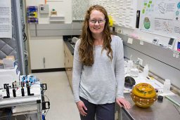 Keenan Kathryn E合作论文数3
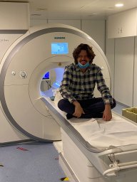 Guido Buonincontri合作论文数School of Medicine, University of California San Diego;Behavioural and Clinical Neuroscience Institute and Department of Psychology, University of Cambridge;Addenbrooke's Hospital, University of Cambridge;Addenbrooke's Hospital, University of Cambridge;Manchester Pharmacy School, University of Manchester;University of California San Diego, University of Manchester3
 William A. Grissom合作论文数Stanford University3mathias nittka合作论文数2
 Vikas Gulani合作论文数Department of Radiology, Medical School, University of Michigan2
 Chris Flask合作论文数Dept Radiol, Case Western Reserve Univ2
 Sun Jessie E P合作论文数Department of Materials Science and Engineering, University of Pennsylvania2Nils C. J. Müller合作论文数2
 Matthias Troyer合作论文数Swiss Joint Research Center, Microsoft;Stellenbosch University2Walter Zhao合作论文数1Gi-Ming Wang合作论文数1Michael E. Beverland合作论文数1stephan kannengiesser合作论文数1Irina Jaeger合作论文数1Wei-Ching Lo合作论文数1Erik S. B. van Oort合作论文数1Charit Tippareddy合作论文数1Louisa Onyewadume合作论文数1Gregor Koerzdoerfer合作论文数1Darryl C. Jacob合作论文数Dept Phys & Astron, Texas A&M Univ1Christina J MacAskill合作论文数1
 Zhilang Qiu合作论文数Chinese Acad Sci, Shenzhen Inst Adv Technol, Paul C Lauterbur Res Ctr Biomed Imaging, Shenzhen 518055, Guangdong, Peoples R China1
 Megan E. Poorman合作论文数Phys Measurement Lab, NIST1
 Nirav Patil合作论文数Dept Biomed Engn, Case Western Reserve Univ1Kelvin Chow合作论文数Department of Radiology, Feinberg School of Medicine, Northwestern University1Yun Jiang合作论文数Department of Biomedical Engineering, College of Engineering, University of Michigan-Ann Arbor1
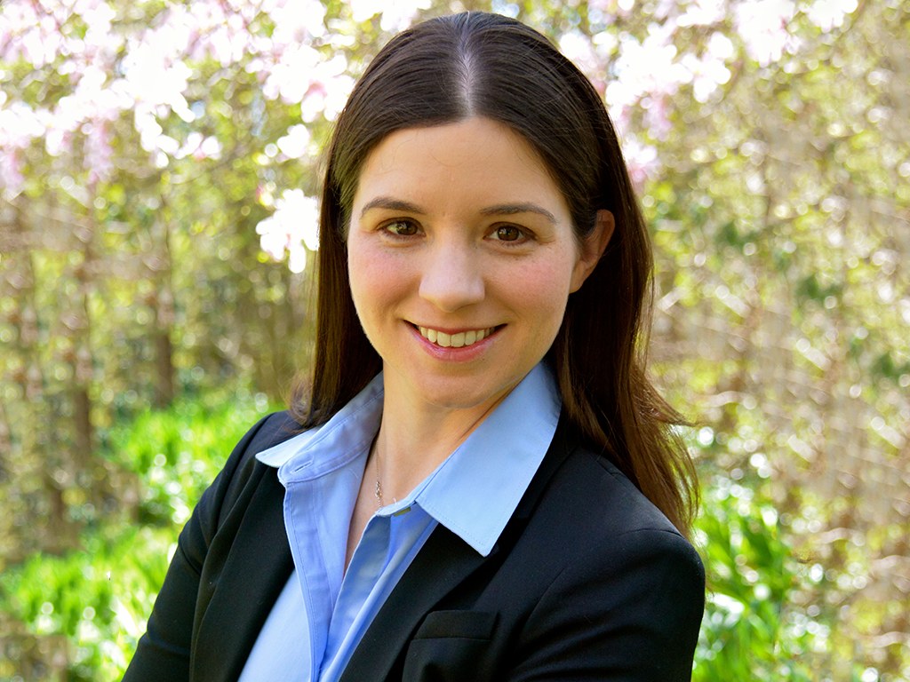 Nicole Seiberlich合作论文数Department of Biomedical Engineering, College of Engineering, University of Michigan-Ann Arbor1
 Evren Degirmenci合作论文数1
 Jill S. Barnholtz-Sloan合作论文数Winship Cancer Institute, Emory University;Department of Biostatistics and Bioinformatics, Rollins School of Public Health, Emory University1Arpinar Volkan Emre合作论文数1Sheyn David合作论文数Department of Obstetrics and Gynecology, University Hospitals Case Medical Center1
 Xin Yu合作论文数Department of Biomedical Engineering, Case School of Engineering, Case Western Reserve University;Department of Physiology and Biophysics, School of Medicine, Case Western Reserve University;Department of Radiology, School of Medicine, Case Western Reserve University1




