Multiple sclerosis (MS) is a chronic inflammatory disease characterized by demyelinating lesions in the central nervous system. Cross-sectional measurements of acute inflammatory lesion activity are typically obtained by detecting the presence of gadolinium enhancement in lesions, which typically lasts 3-6 weeks. We formulate the novel and clinically relevant task of quantification of recent acute lesion activity from the past 24 weeks (6 months) using single-timepoint conventional brain magnetic resonance imaging (MRI). We develop and compare several deep learning (DL) methods for estimating this brain-level acuteness score and show that a 2D-UNet can accurately predict acute disease activity at the patient-level while outperforming transformers and ensemble approaches. In the context of identifying subjects with acute (less than 6 months-old) lesion activity, our 2D-UNet achieves an area under the receiver-operating curve in the range 80-84% on independent relapsing-remitting MS cohorts. When used in conjunction with measurements of gadolinium-enhancing lesion activity, our model significantly improves the prognostication of future acute lesion activity (over the next 6 months). This model could thus be leveraged for population recruitment in clinical trials to identify a higher number of patients with acute inflammatory activity than current standard approaches (e.g., gadolinium positivity) with a predictable precision/recall trade-off.
Evaluate the performance of an automated detection and quantification tool for identification and severity assessment of amyloid-related imaging abnormalities (ARIA).
In addition to focal lesions, diffusely abnormal white matter (DAWM) is seen on brain MRI of multiple sclerosis (MS) patients and may represent early or distinct disease processes. The role of MRI-observed DAWM is understudied due to a lack of automated assessment methods. Supervised deep learning (DL) methods are highly capable in this domain, but require large sets of labeled data. To overcome this challenge, a DL-based network (DAWM-Net) was trained using semi-supervised learning on a limited set of labeled data for segmentation of DAWM, focal lesions, and normal-appearing brain tissues on multiparametric MRI. DAWM-Net segmentation performance was compared to a previous intensity thresholding-based method on an independent test set from expert consensus (N = 25). Segmentation overlap by Dice Similarity Coefficient (DSC) and Spearman correlation of DAWM volumes were assessed. DAWM-Net showed DSC > 0.93 for normal-appearing brain tissues and DSC > 0.81 for focal lesions. For DAWM-Net, the DAWM DSC was 0.49 +/- 0.12 with a moderate volume correlation (rho = 0.52, p < 0.01). The previous method showed lower DAWM DSC of 0.26 +/- 0.08 and lacked a significant volume correlation (rho = 0.23, p = 0.27). These results demonstrate the feasibility of DL-based DAWM auto-segmentation with semi-supervised learning. This tool may facilitate future investigation of the role of DAWM in MS.
Purpose T-1 mapping is a widely used quantitative MRI technique, but its tissue-specific values remain inconsistent across protocols, sites, and vendors. The ISMRM Reproducible Research and Quantitative MR study groups jointly launched a challenge to assess the reproducibility of a well-established inversion-recovery T-1 mapping technique, using acquisition details from a seminal T-1 mapping paper on a standardized phantom and in human brains. Methods The challenge used the acquisition protocol from Barral et al. (2010). Researchers collected T-1 mapping data on the ISMRM/NIST phantom and/or in human brains. Data submission, pipeline development, and analysis were conducted using open-source platforms. Intersubmission and intrasubmission comparisons were performed. Results Eighteen submissions (39 phantom and 56 human datasets) on scanners by three MRI vendors were collected at 3 T (except one, at 0.35 T). The mean coefficient of variation was 6.1% for intersubmission phantom measurements, and 2.9% for intrasubmission measurements. For humans, the intersubmission/intrasubmission coefficient of variation was 5.9/3.2% in the genu and 16/6.9% in the cortex. An interactive dashboard for data visualization was also eveloped: https://rrsg2020.dashboards.neurolibre.org. Conclusion The T-1 intersubmission variability was twice as high as the intrasubmission variability in both phantoms and human brains, indicating that the acquisition details in the original paper were insufficient to reproduce a quantitative MRI protocol. This study reports the inherent uncertainty in T-1 measures across independent research groups, bringing us one step closer to a practical clinical baseline of T-1 variations in vivo.
Background Tremor affects up to 45% of patients with Multiple Sclerosis (PwMS). Current understanding is based on insights from other neurological disorders, thus, not fully addressing the distinctive aspects of MS pathology. Objective To characterize the brain white matter (WM) correlates of MS-related tremor using diffusion tensor imaging (DTI). Methods In a prospective case-control study, PwMS with tremor were assessed for tremor severity and underwent MRI scans including DTI. PwMS without tremor served as matched controls. After tract selection and segmentation, the resulting diffusivity measures were used to calculate group differences and correlations with tremor severity. Results This study included 72 PwMS. The tremor group (n = 36) exhibited significant changes in several pathways, notably in the right inferior longitudinal fasciculus (Cohen's d = 1.53, q < 0.001) and left corticospinal tract ( d = 1.32, q < 0.001), compared to controls (n = 36). Furthermore, specific tracts showed a significant correlation with tremor severity, notably in the left medial lemniscus (Spearman's coefficient [ r s p] = −0.56, p < 0.001), and forceps minor of corpus callosum ( r s p = -0.45, p < 0.01). Conclusion MS-related tremor is associated with widespread diffusivity changes in WM pathways and its severity correlates with commissural and sensory projection pathways, which suggests a role for proprioception or involvement of the dentato-rubro-olivary circuit.
This dataset includes both raw data and processed T1 maps obtained from the 2020 challenge on inversion recovery T1 mapping organized by the International Society in Magnetic Resonance in Medicine (ISMRM) Reproducible Research Study Group (RRSG). For a comprehensive overview of the data distribution submitted for this challenge, please visit https://rrsg2020.db.neurolibre.org. It's important to note that this dataset exclusively comprises ISMRM-NIST system phantom data. Dataset provided for NeuroLibre preprint. Author repo: https://github.com/rrsg2020/paper NeuroLibre fork:https://github.com/roboneurolibre/paper For details, please visit the corresponding NeuroLibre technical screening. https://neurolibre.org
Introduction: Left ventricular ejection fraction (LVEF) improvement in HFrEF patients is classified as heart failure with improved ejection fraction (HFimpEF) and is associated with better long-term outcomes than persistent HFrEF. HFimpEF is difficult to predict and underlying mechanisms remain poorly understood. Hypothesis: Better myocardial high-energy phosphate metabolism at baseline predicts future LVEF recovery and outcomes in a general HFrEF population Methods: We measured cardiac energetics, the ratio of the two main high-energy phosphates, phosphocreatine and ATP (PCr/ATP), and the rate of ATP synthesis through the primary cardiac energy reserve reaction creatine kinase (“CK flux”), in 37 nonischemic HFrEF patients (LVEF ≤40%) with 31 P cardiac magnetic resonance spectroscopy (MRS) at 3T. Baseline transthoracic echocardiogram (TTE) was obtained prior to 31 P MRS and follow-up consecutive TTEs with the two highest LVEFs meeting HFimpEF criteria (LVEF >40% and ≥10% increase over baseline LVEF) over a median follow-up of 6.9 years were averaged and used to classify HFimpEF. Results: There were no significant differences in demographics, HF etiology, baseline TTE metrics, or medications between persistent HFrEF (n=22) and eventual HFimpEF (n=15). Cardiac CK flux was significantly higher at baseline in patients who later recovered to HFimpEF than in those who remained HFrEF (p=0.031). Better baseline cardiac CK flux was associated with increases in LVEF (p=0.025) and decreases in LV end-diastolic diameter (p=0.016) over time. By both dichotomous and continuous metrics, better preserved CK flux was associated with future HFimpEF. Fourteen subjects (38%), all persistent HFrEF, experienced cardiac death, transplant, or LVAD and had significantly lower baseline CK flux than outcome-free patients (p=0.017). Cardiac PCr/ATP was not associated with eventual HFimpEF, outcomes, or changes in TTE indices. Conclusion: Better preserved cardiac CK flux is associated with future LVEF recovery and LV reverse remodeling while lower CK flux is associated with worse cardiac outcomes in HFrEF patients. Interventions restoring CK flux, and possibly other energy-generating metabolic reactions, may elicit myocardial recovery in HFrEF patients.
Objective: The purpose of this study is to recover 3D-T1 weighted brain MRI scans from Multiple Sclerosis (MS) patients to reproduce a homologous representation of their lesion-free brain. Background: In population-level studies, the comparison of MS brain MRIs with their synthetic lesion-free counterpart may highlight disease-specific variability which may provide insights into MS pathophysiology. Design/Methods: A generative adversarial network (GAN) based on StyleGAN2 was trained to generate synthetic healthy brain scans from a 512-dimensional latent variable. Then, each MS brain was projected to this latent-space to recover the latent variable that generates the synthetic 'healthy' brain which is the most similar to the MS brain. For model training, 3500 3D T1-weighted brain MRIs from healthy adults were pooled across the HCP, OASIS2 and IXI datasets. For evaluation, scans from 2398 MS patients from ADVANCE (NCT00906399) and ASCEND (NCT01416181) trials were used. All scans were skull-stripped, resampled to 2-mm isotropic voxel spacing, and intensity-normalized via min-max mapping to the range [−1, 1]. Model evaluation included visual inspection and quantitative measures: Mean Squared Error (MSE), Structural Similarity Metric (SSIM) and Peak Signal to Noise Ratio (PSNR) between the original MS brain scans and their synthetic lesion-free counterpart, excluding lesion mask areas. Results: We applied our method to 3D T1-weighted MRIs of MS patients to recover lesion-free brains with an isotropic resolution of 2mm. The generated lesion-free brain scans showed no MS lesions and displayed great similarity to the diseased brain outside of the MS lesion areas, as highlighted by the following metrics: SSIM = 0.99, PSNR = 34.5, MSE = 1.49e-3. Conclusions: Our method reconstructed realistic lesion-free representations of 3D T1-weighted brain MRI scans of MS patients. This method could be generalizable to other neurological diseases. Disclosure: Mr. SPINAT has received personal compensation for serving as an employee of Therapanacea. Mr. Caba has received personal compensation for serving as an employee of Therapanacea. Mr. Caba has received personal compensation for serving as an employee of Biogen. Mr. Caba has received personal compensation in the range of $50,000-$99,999 for serving as a Research Engineer with Therapanacea. Mr. Caba has received personal compensation in the range of $100,000-$499,999 for serving as a Scientist II, ML/AI with Biogen. Dr. Ioannidou has received personal compensation for serving as an employee of Therapanacea. Dr. Gafson has stock in Biogen. Dr. Teboul has nothing to disclose. Xiaotong Jiang has nothing to disclose. Dr. Lepetit has received personal compensation in the range of $500-$4,999 for serving on a Scientific Advisory or Data Safety Monitoring board for Therapanacea. Dr. Arnold has received personal compensation in the range of $5,000-$9,999 for serving as a Consultant for Biogen. Dr. Arnold has received personal compensation in the range of $5,000-$9,999 for serving as a Consultant for Celgene. Dr. Arnold has received personal compensation in the range of $500-$4,999 for serving as a Consultant for Frequency Therapeutics. Dr. Arnold has received personal compensation in the range of $5,000-$9,999 for serving as a Consultant for Merck. Dr. Arnold has received personal compensation in the range of $10,000-$49,999 for serving as a Consultant for Novartis. Dr. Arnold has received personal compensation in the range of $10,000-$49,999 for serving as a Consultant for Roche. Dr. Arnold has received personal compensation in the range of $5,000-$9,999 for serving as a Consultant for Sanofi. Dr. Arnold has received personal compensation in the range of $500-$4,999 for serving as a Consultant for Shionogi. Dr. Arnold has received personal compensation in the range of $500-$4,999 for serving as a Consultant for Xfacto communications. Dr. Arnold has received personal compensation in the range of $500-$4,999 for serving as a Consultant for Eli Lilly. Dr. Arnold has received personal compensation in the range of $500-$4,999 for serving as a Consultant for EMD Serono. Dr. Arnold has stock in NeuroRx. Dr. Gabr has received personal compensation for serving as an employee of Biogen. Dr. Gabr has stock in Biogen. Enrica Cavedo has nothing to disclose. Colm Elliott has received personal compensation for serving as an employee of NeuroRx Research. Nikos Paragios has received personal compensation in the range of $500-$4,999 for serving on a Speakers Bureau for AstraZeneca. Nikos Paragios has received personal compensation in the range of $500-$4,999 for serving on a Speakers Bureau for Ipsen. Nikos Paragios has received personal compensation in the range of $500-$4,999 for serving as an officer or member of the Board of Directors for ArteDrone. Nikos Paragios has received personal compensation in the range of $10,000-$49,999 for serving as an Editor, Associate Editor, or Editorial Advisory Board Member for Elsevier. Nikos Paragios has received intellectual property interests from a discovery or technology relating to health care. Nikos Paragios has received personal compensation in the range of $100,000-$499,999 for serving as a CEO with TheraPanacea. Dr. Belachew has received personal compensation for serving as an employee of Biogen Inc. Dr. Belachew has received stock or an ownership interest from Biogen Inc.
Abstract Background and purpose The discovery of glymphatic function in the human brain has generated interest in waste clearance mechanisms in neurological disorders such as multiple sclerosis (MS). However, noninvasive in vivo functional assessment is currently lacking. This work studies the feasibility of a novel intravenous dynamic contrast MRI method to assess the dural lymphatics, a purported pathway contributing to glymphatic clearance. Methods This prospective study included 20 patients with MS (17 women; age = 46.4 [27, 65] years; disease duration = 13.6 [2.1, 38.0] years, expanded disability status score (EDSS) = 2.0 [0, 6.5]). Patients were scanned on a 3.0T MRI system using intravenous contrast‐enhanced fluid‐attenuated inversion recovery MRI. Signal in the dural lymphatic vessel along the superior sagittal sinus was measured to calculate peak enhancement, time to maximum enhancement, wash‐in and washout slopes, and the area under the time‐intensity curve (AUC). Correlation analysis was performed to examine the relationship between the lymphatic dynamic parameters and the demographic and clinical characteristics, including the lesion load and the brain parenchymal fraction (BPF). Results Contrast enhancement was detected in the dural lymphatics in most patients 2–3 min after contrast administration. BPF had a significant correlation with AUC (p < .03), peak enhancement (p < .01), and wash‐in slope (p = .01). Lymphatic dynamic parameters did not correlate with age, BMI, disease duration, EDSS, or lesion load. Moderate trends were observed for correlation between patient age and AUC (p = .062), BMI and peak enhancement (p = .059), and BMI and AUC (p = .093). Conclusion Intravenous dynamic contrast MRI of the dural lymphatics is feasible and may be useful in characterizing its hydrodynamics in neurological diseases.
HomeCirculationVol. 148, No. 24Energetic Basis of Recovered Ejection Fraction in Human Heart Failure No AccessResearch ArticleRequest AccessFull TextAboutView Full TextView PDFView EPUBSections ToolsAdd to favoritesDownload citationsTrack citationsPermissions ShareShare onFacebookTwitterLinked InMendeleyReddit Jump toNo AccessResearch ArticleRequest AccessFull TextEnergetic Basis of Recovered Ejection Fraction in Human Heart Failure Joseph R. Goldenberg, Allison G. Hays, Refaat E. Gabr, Michael Schӓr, T. Jake Samuel, Lisa R. Yanek, Gary Gerstenblith, Paul A. Bottomley and Robert G. Weiss Joseph R. GoldenbergJoseph R. Goldenberg https://orcid.org/0000-0002-9451-8235 Department of Medicine, Cardiology Division (J.R.G., A.G.H., T.J.S., G.G., R.G.W.), Johns Hopkins University School of Medicine, Baltimore, MD. , Allison G. HaysAllison G. Hays https://orcid.org/0000-0003-2138-1589 Department of Medicine, Cardiology Division (J.R.G., A.G.H., T.J.S., G.G., R.G.W.), Johns Hopkins University School of Medicine, Baltimore, MD. , Refaat E. GabrRefaat E. Gabr https://orcid.org/0000-0002-8802-3201 Department of Diagnostic and Interventional Imaging, University of Texas Health Science Center at Houston (R.E.G.). , Michael SchӓrMichael Schӓr Department of Radiology, Division of MR Research (M.S., P.A.B., R.G.W.), Johns Hopkins University School of Medicine, Baltimore, MD. , T. Jake SamuelT. Jake Samuel https://orcid.org/0000-0001-7638-1864 Department of Medicine, Cardiology Division (J.R.G., A.G.H., T.J.S., G.G., R.G.W.), Johns Hopkins University School of Medicine, Baltimore, MD. , Lisa R. YanekLisa R. Yanek https://orcid.org/0000-0001-7117-1075 Division of General Internal Medicine (L.R.Y.), Johns Hopkins University School of Medicine, Baltimore, MD. , Gary GerstenblithGary Gerstenblith https://orcid.org/0000-0001-6046-6812 Department of Medicine, Cardiology Division (J.R.G., A.G.H., T.J.S., G.G., R.G.W.), Johns Hopkins University School of Medicine, Baltimore, MD. , Paul A. BottomleyPaul A. Bottomley Department of Radiology, Division of MR Research (M.S., P.A.B., R.G.W.), Johns Hopkins University School of Medicine, Baltimore, MD. and Robert G. WeissRobert G. Weiss Correspondence to: Robert G. Weiss, MD, Johns Hopkins University School of Medicine, Blalock 544, Johns Hopkins Hospital, 600 N. Wolfe St, Baltimore, MD 21287-6568. Email E-mail Address: [email protected] https://orcid.org/0000-0002-1844-8677 Department of Medicine, Cardiology Division (J.R.G., A.G.H., T.J.S., G.G., R.G.W.), Johns Hopkins University School of Medicine, Baltimore, MD. Department of Radiology, Division of MR Research (M.S., P.A.B., R.G.W.), Johns Hopkins University School of Medicine, Baltimore, MD. Originally published11 Dec 2023https://doi.org/10.1161/CIRCULATIONAHA.123.065217Circulation. 2023;148:1976–1978FootnotesFor Sources of Funding and Disclosures, see page 1978.Circulation is available at www.ahajournals.org/journal/circCorrespondence to: Robert G. Weiss, MD, Johns Hopkins University School of Medicine, Blalock 544, Johns Hopkins Hospital, 600 N. Wolfe St, Baltimore, MD 21287-6568. Email rweiss@jhmi.eduREFERENCES1. Heidenreich PA, Bozkurt B, Aguilar D, Allen LA, Byun JJ, Colvin MM, Deswal A, Drazner MH, Dunlay SM, Evers LR, et al. 2022 AHA/ACC/HFSA guideline for the management of heart failure: a report of the American College of Cardiology/American Heart Association Joint Committee on Clinical Practice Guidelines.Circulation. 2022; 145:e895–e1032. doi: 10.1161/CIR.0000000000001063LinkGoogle Scholar2. Wilcox JE, Fang JC, Margulies KB, Mann DL. Heart failure with recovered left ventricular ejection fraction: JACC scientific expert panel.J Am Coll Cardiol. 2020; 76:719–734. doi: 10.1016/j.jacc.2020.05.075CrossrefMedlineGoogle Scholar3. Gabr RE, El-Sharkawy A-MM, Schär M, Panjrath GS, Gerstenblith G, Weiss RG, Bottomley PA. Cardiac work is related to creatine kinase energy supply in human heart failure: a cardiovascular magnetic resonance spectroscopy study.J Cardiovasc Magn Reson. 2018; 20:81. doi: 10.1186/s12968-018-0491-6CrossrefMedlineGoogle Scholar4. Weiss RG, Gerstenblith G, Bottomley PA. ATP flux through creatine kinase in the normal, stressed, and failing human heart.Proc Natl Acad Sci U S A. 2005; 102:808–813. doi: 10.1073/pnas.0408962102CrossrefMedlineGoogle Scholar5. Bottomley PA, Panjrath GS, Lai S, Hirsch GA, Wu K, Najjar SS, Steinberg A, Gerstenblith G, Weiss RG. Metabolic rates of ATP transfer through creatine kinase (CK flux) predict clinical heart failure events and death.Sci Transl Med. 2013; 5:215re3. doi: 10.1126/scitranslmed.3007328CrossrefMedlineGoogle Scholar eLetters(0)eLetters should relate to an article recently published in the journal and are not a forum for providing unpublished data. Comments are reviewed for appropriate use of tone and language. Comments are not peer-reviewed. Acceptable comments are posted to the journal website only. Comments are not published in an issue and are not indexed in PubMed. Comments should be no longer than 500 words and will only be posted online. References are limited to 10. Authors of the article cited in the comment will be invited to reply, as appropriate.Comments and feedback on AHA/ASA Scientific Statements and Guidelines should be directed to the AHA/ASA Manuscript Oversight Committee via its Correspondence page.Sign In to Submit a Response to This Article Previous Back to top Next FiguresReferencesRelatedDetails December 12, 2023Vol 148, Issue 24 Advertisement Article InformationMetrics © 2023 American Heart Association, Inc.https://doi.org/10.1161/CIRCULATIONAHA.123.065217PMID: 38079486 Originally publishedDecember 11, 2023 Keywordsadenosine triphosphatecardiac failureenergy metabolismmagnetic resonance spectroscopyphosphocreatinePDF download Advertisement SubjectsHeart FailureMagnetic Resonance Imaging (MRI)Metabolism
We developed an end-to-end deep learning-based motion correction technique that utilized the time-segmented nature of MRI data acquisition. Results of computer simulations and in vivo studies show the network to be highly effective in correcting motion artifacts.
Background: Abnormalities in cardiac energy metabolism occur in heart failure (HF) and contribute to contractile dysfunction, but their role, if any, in HF-related pathologic remodeling is much less established. CK (creatine kinase), the primary muscle energy reserve reaction which rapidly provides ATP at the myofibrils and regenerates mitochondrial ADP, is down-regulated in experimental and human HF. To test the hypotheses that pathologic remodeling in human HF is related to impaired cardiac CK energy metabolism and that rescuing CK attenuates maladaptive hypertrophy in experimental HF. Methods: First, in 27 HF patients and 14 healthy subjects, we measured cardiac energetics and left ventricular remodeling using noninvasive magnetic resonance 31P spectroscopy and magnetic resonance imaging, respectively. Second, we tested the impact of metabolic rescue with cardiac-specific overexpression of either Ckmyofib (myofibrillar CK) or Ckmito (mitochondrial CK) on HF-related maladaptive hypertrophy in mice. Results: In people, pathologic left ventricular hypertrophy and dilatation correlate closely with reduced myocardial ATP levels and rates of ATP synthesis through CK. In mice, transverse aortic constriction-induced left ventricular hypertrophy and dilatation are attenuated by overexpression of CKmito, but not by overexpression of CKmyofib. CKmito overexpression also attenuates hypertrophy after chronic isoproterenol stimulation. CKmito lowers mitochondrial reactive oxygen species, tissue reactive oxygen species levels, and upregulates antioxidants and their promoters. When the CK capacity of CKmito-overexpressing mice is limited by creatine substrate depletion, the protection against pathologic remodeling is lost, suggesting the ADP regenerating capacity of the CKmito reaction rather than CK protein per se is critical in limiting adverse HF remodeling. Conclusions: In the failing human heart, pathologic hypertrophy and adverse remodeling are closely related to deficits in ATP levels and in the CK energy reserve reaction. CKmito, sitting at the intersection of cardiac energetics and redox balance, plays a crucial role in attenuating pathologic remodeling in HF. REGISTRATION: URL: https://www.clinicaltrials.gov ; Unique identifier: NCT00181259.
Changes in cerebral perfusion occur early in relapsing and progressive multiple sclerosis (MS) patients, though whether cerebral blood flow (CBF) can be altered by therapy is unknown. We sought to characterize the time course of change in CBF (cerebral vascular reactivity [CVR]), following intravenous (IV) acetazolamide (ACZ) in whole brain and within various gray and white matter brain regions in MS patients.
Background Optic disc edema develops in most astronauts during long-duration spaceflight. It is hypothesized to result from weightlessness-induced venous congestion of the head and neck and is an unresolved health risk of space travel. Purpose Determine if short-term application of lower body negative pressure (LBNP) could reduce internal jugular vein (IJV) expansion associated with the supine posture without negatively impacting cerebral perfusion or causing IJV flow stasis. Study Type Prospective. Subjects Nine healthy volunteers (six women). Field Strength/Sequence 3T/cine two-dimensional phase-contrast gradient echo; pseudo-continuous arterial spin labeling single-shot gradient echo echo-planar. Assessment The study was performed with two sequential conditions in randomized order: supine posture and supine posture with 25 mmHg LBNP (LBNP25). LBNP was achieved by enclosing the lower extremities in a semi-airtight acrylic chamber connected to a vacuum. Heart rate, bulk cerebrovasculature flow, IJV cross-sectional area, fractional IJV outflow relative to arterial inflow, and cerebral perfusion were assessed in each condition. Statistical Tests Paired t-tests were used to compare measurement means across conditions. Significance was defined as P < 0.05. Results LBNP25 significantly increased heart rate from 64 +/- 9 to 71 +/- 8 beats per minute and significantly decreased IJV cross-sectional area, IJV outflow fraction, cerebral arterial flow rate, and cerebral arterial stroke volume from 1.28 +/- 0.64 to 0.56 +/- 0.31 cm(2), 0.75 +/- 0.20 to 0.66 +/- 0.28, 780 +/- 154 to 708 +/- 137 mL/min and 12.2 +/- 2.8 to 9.7 +/- 1.7 mL/cycle, respectively. During LBNP25, there was no significant change in gray or white matter cerebral perfusion (P = 0.26 and P = 0.24 respectively) and IJV absolute mean peak flow velocity remained >= 4 cm/sec in all subjects. Data Conclusion Short-term application of LBNP25 reduced IJV expansion without decreasing cerebral perfusion or inducing IJV flow stasis. Level of Evidence 1 Technical Efficacy Stage 1
Editorial for “A Multi-Modality Fusion Deep Learning Model Based on DCE-MRI for Preoperative Prediction of Microvascular Invasion in Intrahepatic Cholangiocarcinoma” Intrahepatic cholangiocarcinoma (ICC) is relatively rare and accounts for 5%–20% among all liver cancers. However, the incidence of ICC is increasing at an alarming rate. For example, between 2004 and 2015, the annual increase in the incidence of ICC is 4.16%. Only 60%–70% ICC patients are eligible for surgical resection, currently the most effective treatment. Majority of the patients who underwent surgical resection experiences recurrence. For nonsurgical patients, the median survival rate is 12–15 months. ICC with microvascular invasion (MVI) is a risk factor for early recurrence and poor survival following surgical resection. Published studies also suggest that adjuvant treatments might improve the survival rate in advanced or unresectable ICC patients with MVI. Thus, there is a need to confirm the presence of MVI in ICC prior to surgery for treatment customization. Currently, the presence of MVI is confirmed on histology, which is invasive. A few studies based on radiomic analysis to predict the MVI in mass-forming ICC were reported (see, eg Reference 8). However, this approach requires accurate tumor segmentation that may not always be possible. Thus, there is a need for a noninvasive technique for automatically predicting MVI in ICC. In the current study, the authors have applied deep learning (DL) for predicting MVI in ICC patients . DL is based on neural networks and is being increasingly adapted by the imaging community for image analysis, generating images with multiple contrasts, creating very high-resolution images from routine clinical images, and many other applications. In this study, the authors used multimodality fusion CNN (MMFCNN) network that accepts multicontrast MRI as input. The multicontrast MRI data in this study include fat-suppressed T2-weighted, diffusion-weighted imaging (DWI), apparent diffusion coefficient (ADC) maps, 3D T1-weighted dynamic contrast imaging in the precontrast, arterial, venous, and delayed phases. MRI data were acquired in 451 surgically treated patients with and without MVI from one center at 1.5 T and 3 T for training and validation of the network. In addition, 68 patients from two other centers served as the test cohort. This study uses three models for predicting MVI on the presurgical MRI: mono-modality, later fusion model, and MMFCNN. In the mono-modality model, seven networks are trained with a single contrast image. In the later fusion model, the seven mono-modality features in the last layer were fused. In contrast, the MMFCNN captures the complimentary information from the multicontrast images at the middle layers. These three models were trained to predict MVI from the volume-of-interest (VOI) that includes both tumor and peri-tumor areas. Based on the area under the receiver operating characteristics curve (AUC) analysis of these three models, the authors conclude that MMFCNN has the best prediction. This manuscript addresses an important clinical question and the methodology is based on state-of-the-art neural network approach. The relatively high AUC value of 0.888 on a separate test cohort is quite promising in predicting MVI in ICC. Unlike radiomic analysis, this methodology does not require manual feature extraction since the networks learn features from the data. The sample size appears to be adequate for training, validating, and testing the network. A common problem with the network models is the poor generalizability of the results. The multicenter MRI acquisition at two different field strengths improves the generalizability of the results. Additional testing on independent data from other centers adds to the rigor of this work. Neural networks are generally considered to be a black box. However, using gradient-weighted class activation mapping (Grad-CAM), this study produced visualization of the spatial locations that are important for predicting MVI. This is a clinically useful tool for spatial localization of MVI.
 Ponnada Narayana合作论文数Department of Diagnostic and Interventional Imaging, McGovern Medical School, Health Science Center at Houston, The University of Texas;Graduate School of Biomedical Sciences, MD Anderson Cancer Center, The University of Texas31
 Paul A Bottomley合作论文数Department of Radiology, School of Medicine, Johns Hopkins University25
 Khader M. Hasan合作论文数18
 Michael Schar合作论文数Department of Otolaryngology-Head and Neck Surgery, School of Medicine, Johns Hopkins University17
 Robert Weiss合作论文数Department of Human Genetics, School of Medicine, The University of Utah16
 Jerry S. Wolinsky合作论文数Department of Neurology, McGovern Medical School, The University of Texas Health Science Center at Houston14
 John Lincoln合作论文数Doctors Office Center, UMDNJ-New Jersey Medical School13
 Ivan Coronado合作论文数Dept Diagnost & Intervent Imaging, Univ Texas Hlth Sci Ctr Houston UTHlth10
 Sheeba J Sujit合作论文数Sch Biomed Informat, Univ Texas Hlth Sci Ctr Houston10
 Abdel-Monem M. El-Sharkawy合作论文数Department of Electrical and Computer Engineering, Johns Hopkins University8
 Arash Kamali合作论文数Department of Diagnostic and Interventional Imaging, University of Texas Medical School at Houston8Xiaojun Sun合作论文数7Haque Muhammad合作论文数7
 Yi Zhang (张祎)合作论文数College of Biomedical Engineering and Instrument Science, Zhejiang University;Children's Hospital, School of Medicine, Zhejiang University7
 Amol Pednekar合作论文数Philips Healthcare7P. Narayana合作论文数6Sean I. Savitz合作论文数6
 Octavio Arevalo合作论文数Health Sciences Center, Louisiana State University5
 Gary Gerstenblith合作论文数Department of Otolaryngology-Head and Neck Surgery, School of Medicine, Johns Hopkins University5
 Flavia Nelson合作论文数Department of Neurology, University of Texas Medical School at Houston5
 Yasser Kadah合作论文数medical Engineering Department, Bio;Cairo University;Biomedical Engineering Department, Cairo University5
 Shibeshih Belachew合作论文数Department of Neurology, University of Liège5
 B. Caba合作论文数4Arie Gafson合作论文数4
 Melvin Deloyd Robinson合作论文数Coll Sci & Engn, Houston Baptist Univ4
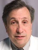 Fred Lublin合作论文数Icahn School of Medicine, Mount Sinai;Corinne Goldsmith Dickinson Center for Multiple Sclerosis, Mount Sinai4
 Sarah George合作论文数Faculty of Health Sciences, University of Bristol4
 Douglas Arnold合作论文数Department of Neurology and Neurosurgery, Faculty of Medicine and Health Sciences, McGill University;NeuroRx Research4Xu Zhang合作论文数Computer and Information Science and Engineering, University of Florida Gainesville, FL 32611, USA
4Jaroslaw Aronowski合作论文数4
Douglas Arnold合作论文数Department of Neurology and Neurosurgery, Faculty of Medicine and Health Sciences, McGill University;NeuroRx Research4Xu Zhang合作论文数Computer and Information Science and Engineering, University of Florida Gainesville, FL 32611, USA
4Jaroslaw Aronowski合作论文数4 Abou-Bakr M. Youssef合作论文数Biomedical Enginerring Department, Cairo University4Sushmita Datta合作论文数4
 Colm Elliott合作论文数LMS Medical Systems4Walter R Lowe合作论文数3
 Mayank Rao合作论文数McGovern Medical School, The University of Texas Health Science Ctr at Houston3
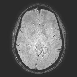 Seth Boren合作论文数McGovern Medical School, The University of Texas Health Science Center at Houston3Cassady Christopher I合作论文数Texas Children's Fetal Center, Texas Children's Hospital3
 Manickam Kumaravel合作论文数Health Science Center, University of Texas3Alderman Susan合作论文数3
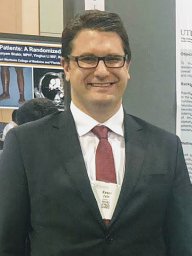 Keser Zafer合作论文数Department of Physical Medicine and Rehabilitation and TIRR Memorial Hermann Neuro-Recovery Research Center, University of Texas Health Science Center3
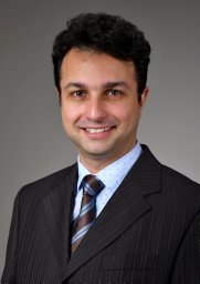 Payam Zandiyeh合作论文数Faculty of Kinesiology, University of Calgary3
 Christopher D. Harner合作论文数The University of Texas Health Science Center at Houston3
 Larry A. Kramer合作论文数Department of Diagnostic and Interventional Imaging, McGovern Medical School, The University of Texas Health Science Center at Houston3Jinyuan Zhou合作论文数3Scott Tashman合作论文数3
 William J. Allen合作论文数Ellis and Moore Consulting Engineers, Station House, Ashley Avenue, Bath BA1 3DS, UK3Clark W. Sitton合作论文数3
 Xiurong Zhao合作论文数3K. Hasan合作论文数2




