Impaired wound healing in type 2 diabetes (T2D) is associated with microvascular dysfunction and remains a significant clinical challenge. We aimed to determine whether primary human dermal microvascular endothelial cells (HDMVECs) from individuals with T2D exhibit abnormal cellular functions, and whether exposure to T2D serum impacts healthy endothelial function. In Experiment 1, T2D-HDMVECs displayed paradoxically higher migratory and angiogenic capacities than their healthy counterparts, despite markedly reduced eNOS expression and disrupted endothelial-identity gene expression. In Experiments 2 and 3, healthy HDMVECs showed decreased tube formation, nitric oxide production, and Notch/angiogenesis-related gene expression after exposure to both healthy and T2D serum, suggesting the presence of serum-derived factors that suppress these pathways. However, T2D-HDMVECs remained largely unresponsive to these serum-driven effects, reinforcing an intrinsic reprogramming of T2D endothelial cells. Additional analyses revealed selective alterations in redox and angiogenic signaling pathways (e.g., NOX4, FLT1), whereas canonical regulators such as VEGFA and PFKB3 were not affected by serum exposure. Overall, our data reveal a complex interplay between cell-autonomous alterations and extrinsic signals in diabetic endothelial dysfunction. Therapeutic strategies targeting both intrinsic cellular programs (e.g., eNOS, Notch signaling) and the circulating milieu may represent promising avenues for enhancing wound repair in patients with T2D.
IntroductionPulmonary arterial hypertension (PAH) is distinguished by elevated blood pressure and vascular resistance in the arteries of the lungs. Patients with PAH demonstrate pulmonary vascular remodeling, wall thickening, and a high rate of morbidity due to right heart failure. Notably, while female patients are more likely to develop PAH, male patients suffer from higher morbidity rates after diagnosis. The molecular mechanism(s) underlying PAH development is poorly understood, though heritable PAH linked to mutations in bone morphogenic protein receptor 2 (Bmpr2) and caveolin-1 (Cav1) may provide novel insights into the disease’s pathophysiology.MethodsTo interrogate this dynamic, we utilized a global Cav1 knockout (Cav1-KO) mouse model (Cav1-/-) in conjunction with chronic hypoxia to induce symptoms of PAH as demonstrated by hemodynamic and ECHO cardiography recordings.ResultsBoth female and male Cav1-/- mice in chronic hypoxia demonstrated elevated right ventricular systolic pressure (RVSP) of 48.49 mmHg and 47.78 mmHg respectively. Female knockout mice began dying earlier in hypoxic conditions (4 wks), though male mice showed greater total mortality by the end of the 8 wks of hypoxia. In addition to wildtype controls, we compared this knockout mouse to endothelial-specific Cav1 reconstituted (Cav1-RC) knockouts and found that restoration of Cav1 expression only in endothelial cells (ECs) is sufficient to ameliorate PAH symptoms, highlighting the importance of vascular Cav1 in maintaining pulmonary artery function. RNA-sequencing of the lungs revealed that Cav1-/- is associated with downregulation of biological process gene pathways involved in cilium assembly in normoxic conditions for both sexes. In hypoxic conditions, Cav1 knockout in females leads to downregulation of bone morphogenetic protein (BMP) signaling, while male hypoxic Cav1-/- led to a significant increase in muscle cell development genes. Reconstitution of Cav1 in ECs leads to upregulation of immune signaling pathways, muscle cell development, and various cell differentiation pathways in both sexes; females showed a unique upregulation of cilia-related pathways, while males demonstrated increased BMP signaling.DiscussionThese data indicate that muscle cell development, angiogenesis, cilia assembly, immune response, and BMP signaling pathways undergo sex-specific transcriptional regulation during PAH development that may underlie sex differences in PAH patient outcome.
Natural killer (NK) cells contribute to tumour immunosurveillance. They detect and eliminate circulating tumour cells by degranulation, releasing perforin and granzyme. Also involved in NK-cell-mediated tumour cell defense are cytokines with proapoptotic and anti-apoptotic characteristics as well as extracellular vesicles containing perforin. Recent clinical studies suggest that volatile anesthetics might negatively impact cancer outcomes compared with intravenous agents. A possible factor could be the impairment of NK cell activity by volatile anesthetics. In this study, we aimed to investigate the effects of sevoflurane on NK-mediated tumour cell defense in a cell model in vitro. In an in vitro laboratory study, we exposed a NK cell line to 2.2
BACKGROUND:During obesity, endothelial cells (ECs) become lipid laden, leading to endothelial dysfunction. We tested posttranslational modification on cluster of differentiation 36 (CD36) that may regulate EC lipid accumulation. METHODS:We used an EC-specific Cav1 (caveolin-1) knockout mouse, nitrosation and palmitoylation assays, and whole animal Nγ-nitro-l-arginine methyl ester administration to examine blood lipids. RESULTS:EC-specific Cav1 knockout male mice are hyperlipidemic regardless of diet but retain endothelial cell function. We found these mice have significantly increased NO in response to the lack of Cav1, and the presence or absence of NO toggled inversely EC lipid content and plasma lipid in mice. The NO nitrosated the fatty acid translocase CD36 at the same cysteines that are palmitoylated on CD36. The nitrosation of CD36 prevented its trafficking to the plasma membrane and decreased lipid accumulation. The physiological effect of this mechanism was a reliance on NO for endothelial function and not dilation. CONCLUSIONS:This work suggests that CD36 nitrosation occurs as a protective mechanism to prevent EC lipotoxicity.
Dynamic protein distribution within and across the plasma membrane is pivotal in regulating cell communication. However, rapid, high-density labeling methods for multiplexed live imaging across diverse cell types remain scarce. Here, we demonstrate N-hydroxysuccinimide (NHS)-ester-based amine crosslinking of fluorescent dyes to uniformly label live mammalian cell surface proteins. Using model cell systems, we capture previously elusive membrane topology and cell-cell interactions. Live imaging shows transient membrane protein accumulation at cell-cell contacts and bidirectional migration patterns guided by membrane fibers in DC2.4 dendritic cells. Multiplexed superresolution imaging reveals the biogenesis of membrane tunneling nanotubes that facilitate intercellular transfer in DC2.4 cells, and caveolin 1-dependent endocytosis of insulin receptors in HEK293T cells. 3D superresolution imaging reveals membrane topology remodeling in response to stimulation, generation of microvesicles, and phagocytic activities in Jurkat T cells. Furthermore, NHS-labeling remains stable in vivo, enabling visualization of intercellular transfer among splenocytes using a T cell lymphoma mouse model.
ABSTRACT Progressive aging is known to negatively affect cardiopulmonary function, which increases the risk of developing cardiac/respiratory diseases. However, the relationship between the decline in pulmonary function and aortic arch stiffness in aging is not well understood. This study investigated the correlation between lung function impairment and aortic arch stiffness in male and female C57BL/6 mice from 2 to 52 weeks of age. Lung function was assessed using forced oscillation ventilation and aortic arch stiffness was measured through echocardiography. Our results show a progressive decline in lung function markers such as respiratory system resistance (Rrs) and tissue elastance (H) with age, alongside an increase in inspiratory capacity (IC) and compliance of the respiratory system (Crs). Aortic arch stiffness significantly increased with age, particularly during early adulthood, and was found to be a strong predictor of lung function impairment in both sexes. While linear regression models indicated that body weight was a more accurate predictor of lung function variability, aortic arch stiffness emerged as a reliable marker for the decline in pulmonary function. These findings suggest that aortic arch stiffness can serve as an early indicator of declining lung function and therefore provide a noninvasive method to assess cardiopulmonary health in aging populations. Future studies should explore the molecular mechanisms underlying these changes and extend the investigation to older mice to fully understand the long‐term impacts of aging on cardiopulmonary function.
Background The intraluminal middle cerebral artery occlusion (MCAO) model is widely used in preclinical stroke research, but outcomes remain inconsistent because of variable occlusion times and inadvertent filament placement into the pterygopalatine artery (PPA). Methods We applied a modified version of the Koizumi method to directly visualize the internal carotid artery (ICA)–PPA bifurcation and systematically compare targeted ICA versus PPA occlusion. We combined multiple complementary methods - survival studies, neurological scoring, 2,3,5-Triphenyltetrazolium chloride (TTC) infarct staining, Evans blue blood brain barrier (BBB) permeability assays, laser speckle cerebral blood flow imaging, small-animal magnetic resonance imaging (MRI), and evaluation of inflammatory gene expression through qPCR - to rigorously characterize injury patterns, survival, and inflammatory responses. Results ICA occlusion approximately caused a rapid 70% reduction in cerebral blood flow, progressive infarction (15 min: smaller infarct with approximately 50% 7-day survival; 30 min: moderate infarct but >50% mortality), and cortical edema. In contrast, PPA occlusion produced only a gradual 44% decline in blood flow, small infarcts at 60 min) and preserved survival (80% at 7 days). Finally, magnetic resonance imaging in 15-minute ICA occluded mice identified the development of cortical edema which was not observed in sham, 15-minute PPA, or 60-minute PPA mice. Conclusion These findings identify 15-minute ICA occlusion as the most practical balance between reproducible infarction and survival for long-term studies, while 30 minutes is best reserved for short-term infarct induction when survival is less critical. By resolving two critical sources of MCAO variability, occlusion duration and vascular targeting, this study provides a standardized protocol and evidence-based occlusion benchmarks that will improve reproducibility in preclinical stroke studies ### Competing Interest Statement The authors have declared no competing interest. * BBB : blood-brain barrier CBF : Cerebral Blood Flow CCA : Common carotid artery HG : Hypoglossal ICA : Internal carotid artery IL-6 : Interleukin 6 LCCA : Left common artery LECA : Left external carotid artery LICA : Left internal carotid artery MCA : Middle cerebral artery MCAO : middle cerebral artery occlusion MRI : Magnetic Resonance Imaging PPA : pterygopalatine artery rCBF : relative cerebral blood flow TGF-β : Tumor Growth Factor Beta TTC : 2,3,5-Triphenyltetrazolium chloride UIC College of Medicine seed fund
Vaso-occlusive episodes (VOEs) in the setting of hyperhemolysis can rapidly evolve into multiorgan failure in sickle cell disease (SCD). Although the mechanisms for rapid progression to multiorgan failure are unclear, a systemic vasculopathy with thrombotic microangiopathy-type features has been described. Reduced thrombomodulin (TM) function is implicated in some thrombotic microangiopathy syndromes. We observed a greater decline in platelet count and hemoglobin concentration and an increase in vascular injury biomarkers within 24 hours of admission for a VOE in 12 patients with SCD with multiorgan failure versus 12 patients without multiorgan failure. We observed decreased TM expression on the lung and kidney vasculature of 3 additional patients with SCD with multiorgan failure compared with a control patient without SCD. Transgenic SCD mice challenged with cell-free hemoglobin had reduced TM function, increased vascular injury biomarkers, and reduced renal cortical blood flow. Infusion of recombinant TM 2 or 24 hours after the challenge restored cortical blood flow and mitigated increases in vascular injury, complement activation, and tubular injury biomarkers, and protected against acute kidney and lung injury. We demonstrated that impaired TM function may be involved in the systemic vasculopathy of SCD-related multiorgan failure, and infusion of recombinant TM may restore vascular function and protect against acute organ damage.
Acute chest syndrome in sickle cell disease (SCD) carries high morbidity and mortality, with up to 15% of patients requiring invasive mechanical ventilation. However, the rates and mechanisms of ventilator-induced lung injury (VILI) in SCD remain poorly understood. We hypothesized individuals with SCD are protected from VILI in a caveolin-1 (Cav-1) dependent manner. SCD mice and control littermates underwent either spontaneous tidal breathing or high tidal volume mechanical ventilation for 4 h (VILI model), or received intratracheal Escherichia coli-derived lipopolysaccharide (LPS) or sterile phosphate-buffered saline and recovered for 16 h (LPS model). Bronchoalveolar lavage (BAL) samples were analyzed for inflammatory cytokine profiles and lung tissues were used for histology and Western blot. SCD mice were protected from VILI but were more susceptible to LPS-induced lung injury, as evidenced by higher BAL fluid total protein concentrations, polymorphonuclear cell infiltration and total cell count. Inflammatory cytokine profiles differed significantly in BAL fluid: IL-6, KC and MIP-2 levels were attenuated in the SCD-VILI model, while TNF-α levels were significantly increased after LPS exposure. Cav-1 expression was reduced at baseline in SCD mice and further decreased after exposure to VILI when compared to control animals. Phosphorylated Cav-1 expression increased, leading to depletion of total Cav-1 in the SCD-VILI model. These data suggest SCD mice are protected from VILI, but not LPS-induced lung injury. These differences appear to be mediated by distinct inflammatory cytokine profiles and expression of Cav-1. Further studies are needed to explore differences in lung injury patterns in patients with SCD.
The mechanisms that govern whether T cells cross blood-brain barrier (BBB) endothelium by transcellular versus paracellular routes are unclear. Caveolin-1 is a membrane scaffolding and signaling protein associated with transcellular transmigration through the endothelial cytoplasm. Here, we report that the neuroinflammatory chemokine CXCL10 induced transcellular, caveolar transmigration of CXCR3+ CD4+ T cells. Specifically, data revealed that CXCL10-induced transcellular transmigration requires expression of Caveolin-1 and ICAM-1 in brain endothelial cells and of the CXCL10 receptor, CXCR3, and LFA-1 in T cells. Moreover, Caveolin-1 promoted CXCL10 aggregation into brain endothelial cytoplasmic stores, providing a mechanism for activation and recruitment of CXCR3+ T cells to migrate at cytoplasmic locations, distal to cell-cell junctions. Consistent with our in vitro data, genetic ablation of Caveolin-1 reduces infiltration of CXCR3+ CD4+ T cells into the CNS in experimental autoimmune encephalomyelitis. Our findings establish a novel mechanism by which brain endothelial cells utilize Caveolin-1 dependent CXCL10 intracellular stores to license T cells for transcellular migration across the blood-brain barrier.
Heart failure (HF) is a common clinical syndrome marked by reduced cardiac output, elevated intracardiac pressures, and heart dysfunction. Chronic HF (CHF) is a syndrome characterized by a lack of blood flow and impaired pumping ability to the heart over time, while acute HF (AHF) arises suddenly due to incidents like myocardial infarction or cardiac arrest. HF has a significant impact on pulmonary health and function, leading to conditions such as pulmonary edema and restrictive lung patterns. Clinical evidence highlights the bidirectional relationship between HF and lung dysfunction. Declining lung function serves as a predictor for HF progression and severity, while HF contributes to worsening lung health. Animal models that induce HF through surgical methods further demonstrate the connection between heart and lung pathology. The main mechanisms linking HF and lung dysfunction are pressure overload and chronic systemic inflammation, with changes in the extracellular matrix (ECM) also playing a role. Additionally, environmental factors like air pollution exacerbate lung inflammation, increasing the risk of both HF and chronic obstructive pulmonary disease (COPD) incidence. Combined treatment approaches involving pharmaceutical drugs such as statins, Angiotensin-converting enzyme (ACE) inhibitors, and Angiotensin receptor blockers (ARBs) may benefit by reducing inflammation. This review will explore the complex interplay between HF and lung function, emphasizing their interconnected pathophysiology and potential integrated treatment strategies.
This review explores the complex relationship between social determinants of health and the biology of chronic wounds associated with diabetes mellitus, with an emphasis on racial/ethnic disparities. Chronic wounds pose significant healthcare challenges, often leading to severe complications for millions of people in the United States, and disproportionally affect African American, Hispanic, and Native American individuals. Social determinants of health, including economic stability, access to healthcare, education, and environmental conditions, likely influence stress, weathering, and nutrition, collectively shaping vulnerability to chronic diseases, such as obesity and DM, and an elevated risk of chronic wounds and subsequent lower extremity amputations. Here, we review these issues and discuss the urgent need for further research focusing on understanding the mechanisms underlying racial/ethnic disparities in chronic wounds, particularly social deprivation, weathering, and nutrition, to inform interventions to address these disparities.
Blood-brain barrier (BBB) permeability can cause neuroinflammation and cognitive impairment. Caveolin-1 (Cav-1) critically regulates BBB permeability, but its influence on the BBB and consequent neurological outcomes in respiratory viral infections is unknown. We used Cav-1-deficient mice with genetically encoded fluorescent endothelial tight junctions to determine how Cav-1 influences BBB permeability, neuroinflammation, and cognitive impairment following respiratory infection with mouse adapted (MA10) SARS-CoV-2 as a model for COVID-19. We found that SARS-CoV-2 infection increased brain endothelial Cav-1 and increased transcellular BBB permeability to albumin, decreased paracellular BBB Claudin-5 tight junctions, and caused T lymphocyte infiltration in the hippocampus, a region important for learning and memory. Concordantly, we observed learning and memory deficits in SARS-CoV-2 infected mice. Importantly, genetic deficiency in Cav-1 attenuated transcellular BBB permeability and paracellular BBB tight junction losses, T lymphocyte infiltration, and gliosis induced by SARS-CoV-2 infection. Moreover, Cav-1 KO mice were protected from the learning and memory deficits caused by SARS-CoV-2 infection. These results establish the contribution of Cav-1 to BBB permeability and behavioral dysfunction induced by SARS-CoV-2 neuroinflammation.
Autophagy serves as a critical regulator of immune responses in sepsis. Macrophages are vital constituents of both innate and adaptive immunity. In this study, we delved into the intricate role of p120-catenin (p120) in orchestrating autophagy in macrophages in response to endotoxin stimulation. Depletion of p120 effectively suppressed LPS-induced autophagy in both J774A.1 macrophages and murine bone marrow- derived macrophages. LPS not only elevated the interaction between p120 and L chain 3 (LC3) I/II but also facilitated the association of p120 with mammalian target of rapamycin (mTOR). p120 depletion in macrophages by small interfering RNA reduced LPS-induced dissociation of mTOR and Unc-51-like kinase 1 (ULK1), leading to an increase in the phosphorylation of ULK1. p120 depletion also enhanced LPS-triggered macrophage apoptosis, as evidenced by increased levels of cleaved caspase 3, 7-aminoactinomycin D staining, and TUNEL assay. Notably, inhibiting autophagy reversed the decrease in apoptosis caused by LPS stimulation in macrophages overexpressing p120. Additionally, the ablation of p120 inhibited autophagy and accentuated apoptosis in alveolar macrophages in LPS-challenged mice. Collectively, our findings strongly suggest that p120 plays a pivotal role in fostering autophagy while concurrently hindering apoptosis in macrophages, achieved through modulation of the mTOR/ULK1 signaling pathway in sepsis. This underscores the potential of targeting macrophage p120 as an innovative therapeutic avenue for treating inflammatory disorders. The Journal of Immunology, 2024, 213:1666-1675.
Rationale: α-SNAP is part of a large 20S complex composed of 3 SNARE, 4 α-SNAP, and 6 N-ethylmaleimide sensitive factor proteins that together regulate endothelial cell Weibel Palade body fusion and exocytosis of von Willebrand factor (vWF). We previously showed that Gα12 is essential for basal and thrombin-induced vWF secretion, that GST-α-SNAP pulldown of Gα12 is dependent on N-terminal domain aa 10-15 in Gα12, and that a myristoylated 6 aa α-SNAP Binding Domain Gα12 peptide (Myr-SBD6) reduces vWF secretion, thrombo-inflammation, and lethality in septic mice. Here, using direct binding assays (Surface Plasmon Resonance (SPR), AlphaLISA, and mass spectrometry (MS)), we assessed the molecular underpinnings of SBD6 and modified higher affinity peptide (SBD15) binding directly to α-SNAP. Methods: GST-α-SNAP was incubated alone or with SBD6, SBD15-PEG-biotin, or SBD6 with C to S mutation and then evaluated by Maldi MS. SPR was performed for both SBD6 and SBD15 to assess binding affinity ( K D ) and kinetics ( k a and k d ). After demonstrating the covalent linkage of α-SNAP with SBD15-PEG-biotin, cysteine mapping was performed to identify the SBD binding site on α-SNAP. Free cysteines in SBD15-biotin conjugated α-SNAP were capped with N-ethylmaleimide, disulfide bonds were reduced with DTT, reduced Cys residues were capped with iodoacetamide (IAA), and tryptic peptide digests were analyzed by LC/MS/MS. Results: Maldi MS studies showed that SBD15-PEG-biotin exhibited covalent binding to GST-α-SNAP. Further, proteomic analysis of Cys mapped fragments identified a peptide containing C103, KADPQEAINC 103 LMR, that was modified by NEM in control samples and shifted in the presence of SBD15, indicating modification by IAA in α-SNAP - SBD15 conjugates. Further, the C103S α-SNAP mutant was unable to bind to SBD6, and C11S-SBD15-PEG-biotin was unable to bind to α-SNAP. Conclusion: We propose that amino-terminal Gα12 Cys11 covalently binds to α-SNAP Cys103 and that this plays a critical role in the mechanism regulating vWF secretion. The discovery of this disulfide binding mechanism between Gα12 and α-SNAP opens up new avenues for therapeutic intervention in thrombotic disorders associated with elevated plasma vWF.
Sepsis is a life-threatening clinical condition caused by infection and transposition of pathogens and pathogen-associated molecular patterns (PAMPs) into the host bloodstream. During sepsis, activation of toll-like receptors (TLRs) on immune cells triggers the release of pro-inflammatory cytokines and overstimulates the production of vasodilatory mediators such as nitric oxide (NO). These vascular changes lead to widespread inflammation, tissue damage, multiple organ failure, and often death. New therapeutic options are urgently needed. To this end, thiostrepton (TST) has emerged as a candidate for sepsis treatment due to its action as an antibiotic and anti-inflammatory molecule (TLR7-9 inhibitor). Reports in the literature suggest that TLR9 inhibition substantially suppresses the excessive host inflammatory response and attenuates sepsis-induced mortality in the cecal ligation and puncture (CLP) murine model of sepsis. However, to the best of our knowledge, TST has never been directly tested as a therapeutic option for the management of sepsis, possibly due to its low water solubility and drug delivery issues. These facts prompted us to test the central hypothesis that TST encapsulated in phospholipid sterically stabilized micelles (TST-SSM) could be developed into a novel treatment for sepsis. Thus, using our published method of encapsulating the hydrophobic antibiotic TST-SSM, we evaluated the in vivo efficacy of TST-SSM nanomedicine in the murine model of polymicrobial sepsis. We found that TST-SSM increased the median survival of CLP-induced septic mice from 31 to 44 hr by reducing the bacterial burden in the blood and peritoneal lavage. Moreover, plasma levels of pro-inflammatory cytokines (interleukin 6 and tumor necrosis factor-alpha) and NO derivatives were also reduced, whereas renal and hepatic function biomarkers creatinine and aspartate transferase were significantly improved. In conclusion, we identified that TST-SSM nanomedicine has significant potential as a therapeutic agent for sepsis management, primarily due to its anti-inflammatory and antibiotic properties.
Leukocyte infiltration of the CNS can contribute to neuroinflammation and cognitive impairment. Brain endothelial cells regulate adhesion, activation, and diapedesis of T cells across the blood-brain barrier (BBB) in inflammatory diseases. The integral membrane protein Caveolin-1 (Cav-1) critically regulates BBB permeability, but its influence on T cell CNS infiltration in respiratory viral infections is unknown. In this study, we sought to determine the role of Cav-1 at the BBB in neuroinflammation in a COVID-19 mouse model. We used mice genetically deficient in Cav-1 to test the role of this protein in T cell infiltration and cognitive impairment. We found that SARS-CoV-2 infection upregulated brain endothelial Cav-1. Moreover, SARS-CoV-2 infection increased brain endothelial cell vascular cell adhesion molecule-1 (VCAM-1) and CD3+ T cell infiltration of the hippocampus, a region important for short term learning and memory. Concordantly, we observed learning and memory deficits. Importantly, genetic deficiency in Cav-1 attenuated brain endothelial VCAM-1 expression and T cell infiltration in the hippocampus of mice with SARS-CoV-2 infection. Moreover, Cav-1 KO mice were protected from the learning and memory deficits caused by SARS-CoV-2 infection. These results indicate the importance of BBB permeability in COVID-19 neuroinflammation and suggest potential therapeutic value of targeting Cav-1 to improve disease outcomes.
Glycosphingolipids (GSLs) are products of lipid glycosylation that have been implicated in the development of cardiovascular diseases. In diabetes, the adipocyte microenvironment is characterized by hyperglycemia and inflammation, resulting in high levels of GSLs. Therefore, we sought to assess the GSL content in extracellular vesicles derived from the adipose tissues (adiposomes) of obese-diabetic (OB-T2D) subjects and their impact on endothelial cell function. To this end, endothelial cells were exposed to adiposomes isolated from OB-T2D versus healthy subjects. Cells were assessed for caveolar integrity and related signaling, such as Src-kinase and caveolin-1 (cav-1) phosphorylation, and functional pathways, such as endothelial nitric oxide synthase (eNOS) activity. Compared with adiposomes from healthy subjects, OB-T2D adiposomes had higher levels of GSLs, especially LacCer and GM3; they promoted cav-1 phosphorylation coupled to an obvious loss of endothelial surface caveolae and induced eNOS-uncoupling, peroxynitrite generation, and cav-1 nitrosylation. These effects were abolished by Src kinase inhibition and were not observed in GSL-depleted adiposomes. At the functional levels, OB-T2D adiposomes reduced nitric oxide production, shear response, and albumin intake in endothelial cells and impaired flow-induced dilation in healthy arterioles. In conclusion, OB-T2D adiposomes carried a detrimental GSL cargo that disturbed endothelial caveolae and the associated signaling.
Thrombomodulin (TM) is a transmembrane protein that plays a pivotal role in maintaining microvascular health. Functions of TM include 1) binding thrombin and inhibiting its interaction with fibrinogen, 2) augmenting protein C function, and 3) down-regulating complement activation. Reduced TM function has been implicated in several thrombotic microangiopathies, including atypical hemolytic uremic syndrome and disseminated intravascular coagulation (DIC) (PMID 29866818). Previous in vitro studies have demonstrated that cell-free hemoglobin (Hb) mediates loss of TM from the endothelial surface which, in turn, is associated with a higher risk of kidney injury in patients with sickle cell disease (PMID 34929051). Infusion of intact, soluble TM may restore TM's function in the microvasculature, as observed in critically ill patients with DIC (PMID 17059423). In transgenic sickle mice (Townes model, Jackson Laboratory; Bar Harbor, Maine), we investigated whether 1) cell-free Hb reduces TM function leading to glomerular endothelial and kidney injury and 2) if infusion of intact, soluble TM (Asahi Kasei Pharma ©; Tokyo, Japan) can restore TM function and reduce the toxic effects of cell-free Hb in the kidney. Sickle mice (4 male, 4 females per condition) were challenged with cell-free Hb (0.24g/kg intravenous [iv]) or normal saline followed 2 hours later by either infusion of TM (5mg/kg subcutaneous, 1mg/kg iv) or normal saline. Blood and urine samples were collected 24 hours after the challenge and the mice were sacrificed for histopathologic evaluation. In a separate set of similar experiments, sickle mice (3 males, 3 females per condition) received iv microbubbles (Vevo MicroMarker TM Contrast) 24 hours after the challenge to assess renal cortical blood flow by contrast-enhanced ultrasound (Vevo2100 system). Mean ± standard error of mean values are provided. Comparisons between Hb-only and Hb+TM conditions were made using the Mann-Whitney test. Vasculopathy: Infusion of cell-free Hb resulted in decreased TM antithrombotic function within the glomerulus; infusion of TM 2 hours after the challenge restored these functions (fibrinogen relative fluorescent intensity [RFI]: control, 24.2 ± 0.9; Hb-only, 40.6 ± 1.7; Hb+TM: 26.8 ± 1.6) and anti-complement function (C3 RFI: control, 12.9 ± 0.4; Hb-only, 19.3 ± 1.4; Hb+TM, 14.1 ± 0.5) (P≤0.007). Circulating vascular injury biomarkers rose in the cell-free Hb challenged sickle mice and this was abated with TM rescue for p-selectin (control, 271 ± 25 ng/mL; Hb-only, 533 ± 47 ng/mL; Hb+TM, 356 ± 34 ng/mL), VEGF (control, 59 ± 4 pg/mL; Hb-only, 93 ± 7 pg/mL; Hb+TM 69 ± 3 pg/mL), and vWF (control: 91 ± 9 ng/mL, Hb-only: 299 ± 31 ng/mL, Hb+TM: 183 ± 22 ng/mL) (P≤0.01). Kidney Damage: Urine biomarkers of tubular injury increased after the cell-free Hb challenge and improved with TM (KIM-1: control, 317 ± 66 pg/day; Hb-only, 1201 ± 142 pg/day; Hb+TM, 638 ± 78 pg/day; P=0.003) (NGAL: control, 234 ± 34 ng/day; Hb-only, 1624 ± 145 ng/day; Hb+TM, 767 ± 118 ng/day; P=0.003). An elevation in serum creatinine concentration is used to define acute kidney injury. The cell-free Hb challenge led to an acute rise in serum creatinine while the TM rescued mice had serum creatinine concentrations that were significantly lower than the Hb-only challenged mice (P=0.03) and similar to the control conditions (Figure 1A). Preliminary testing of TM administered 24 hours after the cell-free Hb challenge has also led to kidney protective effects as assessed by serum creatinine, p-selectin, and vWF (P < 0.05). Cortical Blood Flow: Peak enhancement, a measure of relative blood volume, was reduced in the cell-free Hb challenged mice compared to control conditions by contrast-enhanced ultrasound. The TM-rescued mice had improved peak enhancement compared to the Hb-only treated mice (P=0.03) and similar to control conditions (Figure 1B). In conclusion, cell-free Hb reduces TM function resulting in increased vascular and kidney injury and decreased cortical blood flow. Infusion of soluble TM 2 hours after the cell-free Hb challenge restores TM function, protects against vascular and kidney injury, and preserves cortical blood flow. Our data highlights TM as a potential candidate therapy to preserve vascular function and abate kidney injury in patients with sickle cell disease during severe vaso-occlusive or hyperhemolytic episodes, when concentrations of cell-free Hb increase several fold.
 Asrar B. Malik合作论文数Department of Pharmacology and Regenerative Medicine, College of Medicine, University of Illinois at Chicago106
 Stephen M. Vogel合作论文数51
 Marcelo G Bonini合作论文数Center for Human Immunobiology, Feinberg School of Medicine, Northwestern University;Department of Medicine, Feinberg School of Medicine, Northwestern University;Department of Biochemistry and Molecular Genetics, Feinberg School of Medicine, Northwestern University48Chinnaswamy Tiruppathi合作论文数46Guochang Hu合作论文数37
 Zhenlong Chen合作论文数Division of Endocrinology, Diabetes and Metabolism, Beth Israel Deaconess Medical Center Boston32Maricela Castellon合作论文数31
 Suellen Darc Oliveira合作论文数Institute of Biophysics Carlos Chagas Filho, Federal University of Rio de Janeiro;Institute of Biomedical Sciences, Federal University of Rio de Janeiro;National Institute of Translational Research in Health and Environment in the Amazon Region;Federal University of Rio de Janeiro, Federal University of Rio de Janeiro29
 David Schwartz合作论文数Department of Biochemistry, Rush-Presbyterian St Luke's Medical Center27
 Beatrice Beck-Schimmer合作论文数Institute of Anesthesiology, University of Zurich;Institute of Physiology, University of Zurich;University of Zurich, University of Zurich26Farnaz R Bakhshi合作论文数20
 Jiwang Chen合作论文数University of Illinois at Chicago19
 Randal O. Dull合作论文数Department of Anesthesiology, The University of Utah18Piegeler Tobias合作论文数16Aaron T. Place合作论文数14
 Vasily Shinin合作论文数13Sf Rabito合作论文数12
 Xiaoping Du合作论文数Du Lab, Department of Pharmacology & Regenerative Medicine, College of Medicine, University of Illinois12Alain Borgeat合作论文数12
 Irena Levitan合作论文数Department of Medicine, College of Medicine, University of Illinois at Chicago12
 Ayesha N. Shajahan-Haq合作论文数12Guoquan Liu合作论文数11Rusu Luiza合作论文数11
 Roberto F Machado合作论文数11DJ MILETICH合作论文数10
 Peter C Hart合作论文数Coll Sci Hlth & Pharm, Roosevelt Univ10
 Richard Ye (叶德全)合作论文数School of Life and Health Sciences, The Chinese University of Hong Kong, Shenzhen9Xiao-Pei Gao合作论文数9
 Orly Lazarov合作论文数University of Illinois at Chicago9Dolly Mehta合作论文数Department of Pharmacology, University of Illinois at Chicago9Viktor Brovkovych合作论文数9An Shajahan合作论文数8Gina Votta-Velis合作论文数8Olga Chernaya合作论文数8
 Martin Schläpfer合作论文数Institute of Physiology, University of Zurich8
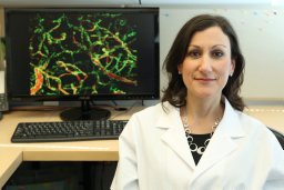 Sarah E Lutz合作论文数Department of Anatomy and Neurobiology, University of Kentucky College of Medicine8
 Peter P. Toth合作论文数Medicine Division of Cardiology, Johns Hopkins University School of Medicine;Community Medicine University of Illinois College of Medicine;Preventative Cardiology CGH Medical Center8RF ALBRECHT合作论文数7
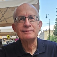 Randal Skidgel合作论文数Dupage Medical Technology, Inc.7
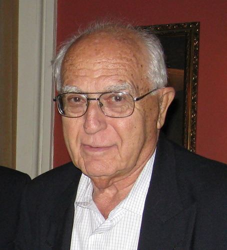 Ervin G. Erdös合作论文数Department of Pharmacology and Anesthesiology, University of Illinois7
 Guy Weinberg合作论文数University of Illinois7
 Sergei M. Danilov合作论文数Institute of Experimental Cardiology, Cardiology Research Center of Russia7John W. Christman合作论文数7
 Arshad Rahman合作论文数Department of Pharmacology;University of Illinois;College of Medicine;College of Medicine, University of Illinois7
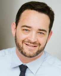 Brant Isakson合作论文数School of Medicine, University of Virginia7Jalees Rehman合作论文数7Ta John合作论文数6R Sandoval合作论文数Dept Pharmacol, Univ Illinois6Elizabeth LeMaster合作论文数6




