OBJECTIVE Hearing outcomes following Gamma Knife radiosurgery (GKRS) for vestibular schwannoma (VS) are multifactorial and poorly characterized in prior literature. In this study the authors evaluated hearing outcomes chronologically to identify prognostic factors of serviceable hearing preservation (HP) rates following GKRS for VS. METHODS Six medical databases were queried according to Preferred Reporting Items for Systematic Reviews and Meta-Analyses (PRISMA) guidelines. Eligible studies reported VS treated with single-fraction GKRS and included the HP rate following GKRS. HP was defined as a postoperative Gardner-Robertson score ≤ 2 among patients with preoperative serviceable hearing. A meta-analysis with random-effects modeling was performed for variables of interest. RESULTS Data from 42 articles with a total of 6582 patients were analyzed; the average age of patients was 54 years and the average follow-up time was 68 months. The pooled proportion of preoperative serviceable hearing was 76%, and the pooled HP rate was 60% at the last follow-up visit. At < 5 years after GKRS, age was significantly correlated with HP on both continuous and categorical analyses (p = 0.001 and p = 0.011, respectively). Between 5 and < 10 years of follow-up, HP was associated with a radiation dose of 12.5 Gy, but not with age or tumor volume. At ≥ 10 years after radiosurgery, a significant negative correlation was seen between marginal radiation dose and HP on both continuous and categorical analyses (p = 0.001 and p = 0.021, respectively). CONCLUSIONS This meta-analysis identifies age and radiation dose as independent prognostic factors for HP. Age-related hearing deterioration appears to be concentrated in the first 5 years after GKRS, whereas radiation dose was associated with HP at last follow-up, between 5 and < 10 years, and at ≥ 10 years after radiosurgery. This meta-analysis offers an objective overview of the literature and a framework for clinical decision-making, with applications for treatment planning and patient counseling.
Cystic vestibular schwannoma (cVS) has been shown to have high rates of recurrence and nerve damage after both surgery and stereotactic radiosurgery (SRS). However, cVS studies are few and contradictory, and there is no consensus on optimal treatment strategies. Six medical databases were queried according to PRISMA guidelines for articles with primary surgery or SRS for cVS. Clinical outcomes were compared using Wilcoxon and Student’s t-tests. A meta-analysis was performed using random-effects modeling to account for interstudy heterogeneity. 1,345 total patients from 28 studies were included and stratified into two cohorts: 547 SRS and 798 surgical cases. For SRS and surgery, respectively, mean follow-up periods were 65 and 30 months (p=0.01), and pooled tumor control (TC) rates were 92% and 97% (p=0.16). Greater facial nerve (FN) preservation was seen after SRS (98.9%) relative to surgical resection for cVS (84.7%, p<0.01). Hearing preservation was higher after surgery (78.2%) compared to SRS (58.5%), although not significant (p=0.50). Pooled rates of postoperative hydrocephalus requiring a shunt were 2.9% after SRS and 4.9% after surgery (p=0.73). Postoperative mortality was minimal and similar between the cohorts (0.0% SRS, 0.3% surgery). Pooled rates of salvage surgery and SRS were all <3%, with no significant difference between the cohorts (p=0.90, p=0.64, respectively). Both SRS and surgery are effective treatment modalities for cVS with a low risk of complications. While TC rates are similar between SRS and surgery, SRS may permit greater FN preservation in cVS.
High grade meningiomas provide challenges to treatment as these neoplasms are more likely to recur, exhibit invasion of brain parenchyma, and result in decreased long term survival. Recent works demonstrate the epigenetic modifier, EZH2, to be increased in higher grade meningiomas and to correlate with poor survival, along with loss of SMARCB1, a known antagonizer of EZH2. The selective targeting of EZH2 has shown promise in other malignancies with overexpression of or gain-of-function mutations in EZH2, leading to promising findings. Here we sought to investigate the effect of EZH2 inhibition on meningioma cell growth and viability, using a panel of in vitro and in vivo models. Human immortalized meningioma cell lines, mouse meningioma cell lines and a panel of human primary cell cultures with varying baseline protein expression of NF2, SMARCB1, and EZH2, were selected for investigation. Cells were treated with several small molecule inhibitors of EZH2 including the anti-viral agent ribavirin. Cell proliferation, cell cycle, and cell death processes were assessed. RNAseq and Western blots were used to assess downstream target effects after EZH2 inhibition. In vivo orthotopic xenograft models were used to assess survival. EZH2 inhibition via a panel of inhibitors significantly impairs meningioma cell proliferation, at varying IC50s, in all cell lines and primary cultures tested. Furthermore, EZH2 inhibition results in a reduction of Ki67+ cells, induction of cell cycle arrest, and apoptosis. Ribavirin (a known EZH2 inhibitor) -treated animals exhibited a significantly increased median survival (67 days) compared to controls (41 days; p=0.0009;) in an orthotopic convexity meningioma xenograft model. Furthermore, ribavirin treatment resulted in increased survival in an orthotopic skull base meningioma xenograft model. Our work demonstrates that EZH2 inhibition in meningioma results in decreased proliferation, induced cell cycle arrest and cell death, in vitro, while ribavirin treatment resulted in increased survival, in vivo, potentially via inhibition of EZH2. This work provides the framework for further investigation of EZH2 inhibition as targeted therapy in meningioma.
Abstract INTRODUCTION High grade meningiomas provide challenges to treatment as these neoplasms are more likely to recur, exhibit invasion of brain parenchyma, and result in decreased long term survival. Recent works demonstrate the epigenetic modifier, EZH2, to be increased in higher grade meningiomas, and to correlate with poor survival. The selective targeting of EZH2 has shown promise in other malignancies with overexpression of or gain-of-function mutations in EZH2, leading to promising findings. Here we sought to investigate the effect of EZH2 inhibition on meningioma cell growth and viability, using a panel of in vitro models. METHODS Human immortalized meningioma cell lines, mouse meningioma cell lines and a panel of human primary cell cultures with varying baseline protein expression of NF2, SMARCB1, and EZH2, were selected for investigation. Cells were treated with several selective small molecule inhibitors of EZH2. Cell proliferation, cell cycle and cell death processes were assessed. RNAseq was used to assess transcriptional changes in response to EZH2 inhibition. Western blots were used to assess protein changes in response to EZH2 inhibition. RESULTS EZH2 inhibition significantly impairs meningioma cell proliferation, at varying IC50s, in all cell lines and primary cultures tested, utilizing a panel of EZH2 inhibitors. EZH2 inhibition resulted in significantly decreased colony formation in a high-grade cell line. Furthermore, EZH2 inhibition results in a reduction of Ki67+ cells, induction of cell cycle arrest, and apoptosis. Similar grouping of differential gene expression in immortalized cell lines and primary cell cultures is observed across cell lines and primary cell cultures, after EZH2 inhibition. CONCLUSION Our work demonstrates that EZH2 inhibition results in growth inhibition, cell cycle arrest, and cell death, across all cell lines, and cell cultures tested. Futhermore, modulation of protein levels of the EZH2 target H3K27 is observed upon treatment. RNA changes are broad and involve cell proliferative and migratory processes. This work provides the framework for further investigation of EZH2 inhibition in meningioma.
Abstract INTRODUCTION High grade meningiomas provide significant challenges to treatment. Recent works have demonstrated meningiomas to express higher levels of the epigenetic regulator enhancer of zeste homolog 2 (EZH2), as well as the protein eIF4E, a key player in translation. These two molecules are demonstrated targets of the well tolerated antiviral drug, ribavirin, which has recently shown promise as an anti-neoplastic agent. Based on these findings we sought to investigate the effect of ribavirin on meningioma tumorigenesis utilizing in vitro and in vivo meningioma models. METHODS Several immortalized human meningioma cell lines, three immortalized mouse meningioma cell lines, and a panel of human primary cell cultures were assessed. Cell proliferation, cell colony formation, clonogenic assay, and cell death were assessed. Furthermore, we tested ribavirin efficacy in vivo utilizing two aggressive high grade syngeneic orthotopic allograft meningioma models in FVB WT mice. RESULTS We provide evidence that ribavirin significantly impairs meningioma cell growth and proliferation, and induces cell death in vitro. Most importantly, we demonstrate that ribavirin alone significantly improves the survival of mice orthotopically implanted with MGS2 cells. Ribavirin-treated animals exhibited a significantly increased median survival (67 days) compared to controls (41 days; p=0.0009;) in an orthotopic convexity meningioma xenograft model. Furthermore, ribavirin treatment resulted in increased survival in an orthotopic skull base meningioma xenograft model. CONCLUSION Our work establishes that ribavirin is effective against meningioma in vitro and in vivo. Ribavirin treatment reduced proliferation in all cell lines tested, in both high and low grade models, induced cell death, and prolonged survival in two aggressive in vivo models, potentially via modulation of the EZH2 and eIF4E pathways. Given the lack of medical therapy for high grade meningiomas, these findings provide a framework for further investigation into ribavirin’s effects on meningioma tumorigenesis, and demonstrate ribavirin as a potential new therapeutic option in the treatment of meningioma.
INTRODUCTION:Although meningiomas are the most common primary brain tumor, there are limited treatment options for recurrent or aggressive lesions. Compared to other brain tumors, meningiomas may be uniquely amenable to immunotherapy by virtue of their location outside the blood-brain barrier. AREAS COVERED:This review describes our current understanding of the immunology of the meninges, as well as immune cell infiltration and immune signaling in meningioma. Current literature on meningioma immunology and immunotherapy was comprehensively reviewed and summarized by a comprehensive search of MEDLINE (1/1/1990-6/1/2024). Further, we describe the current state of immunotherapeutic approaches, as well as potential future targets. Potential immunotherapeutic approaches include immune checkpoint inhibition, CAR-T approaches, tumor vaccine therapy, and immunogenic molecular markers. EXPERT OPINION:Meningioma immunotherapy is in early stages, as no immunotherapies are currently included in treatment guidelines. There is substantial heterogeneity in immune cell infiltration, immunogenicity, and immune escape across tumors, even within tumor grade. Furthering our understanding of meningioma immunology and tumor classification will allow for careful selection of tumors and patient populations that may benefit from primary or adjunctive immunotherapy for meningioma.
Introduction: Adjuvant therapy for recurrent and progressive meningioma remains a challenge. Radiotherapy shows a varied clinical response, particularly among WHO grade II-III meningiomas. Mammalian target of rapamycin (mTOR)-inhibition has been suggested as a potential medical adjuvant agent for growth arrest. There is a paucity of data on the efficacy and mechanism of radiation and mTOR inhibition combination therapy.
Introduction: Radiotherapy remains the primary adjunctive treatment for recurrent or progressive meningioma. Nevertheless, varied clinical response to radiotherapy in meningioma remains a challenge, particularly in higher-grade tumors. There is a paucity of predictors of radiosensitivity. Basal BCL-2-interacting mediator of cell death (BIM) peptide is an intrinsic mediator of apoptosis, and BIM-responsiveness has been postulated as a metric of general sensitivity to apoptotic stress. Herein we investigated its utility as a metric of radiosensitivity in meningioma.
Abstract INTRODUCTION Radiotherapy remains the primary adjunctive treatment for recurrent or progressive meningioma. Nevertheless, varied clinical response to radiotherapy in meningioma remains a challenge, particularly in higher grade tumors. Currently there is a paucity of predictors of radio-sensitivity. Bim peptide is an intrinsic mediator of apoptosis, and Bim-responsiveness has been postulated as a metric of general sensitivity to apoptotic stress. Herein we investigated its utility as a metric of radio-sensitivity in meningioma. METHODS In an in vitro model of meningioma, a number of immortalized human and murine meningioma cell lines and patient-derived primary cultures (WHO grades 1 + 2), were exposed to ionizing radiation at range of doses (0, 2, 4, 8, 16Gy); cells were counted (Celigo, Nexelcom) to assess for growth. Separately, cultures were irradiated at 4Gy and apoptosis was analyzed 24H later using Annexin/PI staining by flow cytometry (Attune NxT). Lastly, untreated cultures were exposed to Bim peptide as a range of doses (0.01uM-10uM); induction of apoptosis was assessed via cytochrome C staining by flow cytometry. RESULTS We were able to recapitulate a wide range of radio-sensitivities both by growth metrics and radiation-induced apoptosis. Immortalized lines showed more radiation-induced growth inhibition relative to primary cultures (p = 0.03). Activation of apoptosis in response to Bim exposure demonstrated a similarly wide range of basal apoptotic sensitivities, akin to that seen for radio-sensitivity. In fact, there was a significant correlation (Pearson, p < 0.01) between Bim-responsiveness and levels of radiation-induced apoptosis. CONCLUSIONS We identify basal state Bim-responsiveness as a potential predictor of radio-sensitivity in meningioma. Further work in this vein may shed light on mechanisms of radio-resistance in meningioma, particularly with respect to the interaction of Bim with the anti-apoptotic members of the Bcl-2 family.
Introduction: Genomic instability near the NF2 locus (22q12.1) has been linked with meningioma oncogenesis and is a prognosticator of poorer outcomes; many have attributed this to the antiapoptotic effects of the NF2 gene product—merlin. Herein, we applied a novel technique (BH3 profiling) to assess the intrinsic sensitivity of meningioma cell lines to apoptotic stress as a unifying endpoint of adjuvant therapies including ionizing radiation. Correlating this assessment across grades and NF2 status promises to identify functional differences between meningioma lines to uncover differential vulnerabilities.
Purpose: Ganglion cysts are benign soft tissue lesions found in joints, most commonly wrists. The incidence for juxtafacet cysts, the condition under which spinal ganglion cysts are categorized, is between 0.06% and 5.8%. Spinal ganglion cysts often arise in the most mobile segment of the lumbar spine, L4-L5. Patients commonly present with pain, radiculopathy, and weakness. Conservative management is used, but surgical resection is the most common treatment modality. We aim to review the literature and present a rare case of an L2-L3 situated spinal ganglion cyst, treated with maximal safe resection. Methods: A systematic review of literature was conducted in accordance with PRISMA guidelines. PubMed, Web of Science, and Cochrane databases were queried using Boolean operators and search terms, "spinal ganglion cyst, lumbar ganglion cyst, and lumbar juxtafacet cyst". Presentation, surgical management, and postoperative course of a 29-year-old male with an L2-L3 spinal ganglion cyst are also described. Results: The search yielded 824 articles; 23 met inclusion criteria. These papers consisted of 27 spinal ganglion cyst cases with disaggregated patient data. 63.0% of patients were male, and 53.4 years (range: 23-86) was the average age at presentation. Mean symptom duration was 1.9 years (range: 3 days-12 years). 70.4% of patients reported complete symptom resolution. 14.8% of cases noted neural foramen involvement. Conclusions: Spinal ganglion cysts are benign lesions typically presenting with radiculopathy. Maximal safe resection is an effective treatment modality with low complication rates. Future studies are needed to understand if neural foramen involvement leads to increased symptom severity.
OBJECTIVE Medulloblastoma, the most common pediatric brain malignancy, has Sonic Hedgehog (SHH) and group 3 (Myc driven) subtypes that are associated with the activity of eukaryotic initiation factor 4E (eIF4E), a critical mediator of translation, and enhancer of zeste homolog 2 (EZH2), a histone methyltransferase and master regulator of transcription. Recent drug repurposing efforts in multiple solid and hematologic malignancies have demonstrated that eIF4E and EZH2 are both pharmacologically inhibited by the FDA-approved antiviral drug ribavirin. Given the molecular overlap between medulloblastoma biology and known ribavirin activity, the authors investigated the preclinical efficacy of repur- posing ribavirin as a targeted therapeutic in cell and animal models of medulloblastoma. METHODS Multiple in vitro assays were performed using human ONS-76 (a primitive SHH model) and D425 (an aggressive group 3 model) cells. The impacts of ribavirin on cellular growth, death, migration, and invasion were quantified using proliferation and Cell Counting Kit-8 (CCK-8) assays, flow cytometry with annexin V (AnnV) staining, scratch wound assays, and Matrigel invasion chambers, respectively. Survival following daily ribavirin treatment (100 mg/kg) was assessed in vivo in immunodeficient mice intracranially implanted with D425 cells. RESULTS Compared to controls, ribavirin treatment led to a significant reduction in medulloblastoma cell growth (ONS-76 proliferation assay, p = 0.0001; D425 CCK-8 assay, p < 0.0001) and a significant increase in cell death (flow cytometry for AnnV, ONS-76, p = 0.0010; D425, p = 0.0284). In ONS-76 cells, compared to controls, ribavirin significantly decreased cell migration and invasion (Matrigel invasion chamber assay, p = 0.0012). In vivo, ribavirin significantly extended survival in an aggressive group 3 medulloblastoma mouse model compared to vehicle-treated controls (p = 0.0004). CONCLUSIONS The authors demonstrate that ribavirin, a clinically used drug known to inhibit eIF4E and EZH2, has significant antitumor effects in multiple preclinical models of medulloblastoma, including an aggressive group 3 animal model. Ribavirin may represent a promising targeted therapeutic in medulloblastoma.
OBJECTIVE Atypical teratoid rhabdoid tumors (ATRTs) are aggressive pediatric brain tumors with no current standard of care and an estimated median patient survival of 12 to 18 months. Previous genetic analyses have implicated cyclin D1 and enhancer of zeste homolog 2 (EZH2), a histone methyltransferase that is implicated in many cancers, as key drivers of tumorigenicity in ATRTs. Since the effects of EZH2 and cyclin D1 are facilitated by a host of cyclin-dependent kinases (CDKs), the authors sought to investigate the potential therapeutic effects of targeting CDKs in ATRTs with the multi-CDK inhibitor, TG02. METHODS Human ATRT cell lines BT12, BT37, CHLA05, and CHLA06 were selected for investigation. The effects of TG02 on cell viability, proliferation, clonogenicity, and apoptosis were assessed via Cell Counting Kit-8 assays, cell counting, clonogenic assays, and flow cytometry, respectively. Similar methods were used to determine the effects of TG02 combined with radiation therapy (RT) or cisplatin. Synergism indices for TG02-cisplatin combination therapy were calculated using CompuSyn software. RESULTS TG02 was observed to significantly impair ATRT cell growth in vitro by limiting cell proliferation and clonogenicity, and by inducing apoptosis. TG02 inhibited ATRT cell proliferation and decreased cell viability in a dose-dependent manner with nanomolar half maximal effective concentration (EC50) values (BT12, 207.0 nM; BT37, 127.8 nM; CHLA05, 29.7 nM; CHLA06, 18.7 nM). TG02 (150 nM) dramatically increased the proportion of apoptotic ATRT cells 72 hours posttreatment (TG02 8.50% vs control 1.52% apoptotic cells in BT12, p < 0.0001; TG02 70.07% vs control 15.36%, p < 0.0001). Combination therapy studies revealed that TG02 acted as a potent radiosensitizer in ATRT cells (BT12 surviving fraction, RT 51.2% vs RT + TG02 21.7%). Finally, CompuSyn analysis demonstrated that TG02 acted synergistically with cisplatin against ATRT cells at virtually all therapeutic doses. These findings were consistent in cell lines that cover all three molecular subgroups of ATRTs. CONCLUSIONS The results of this investigation have established that TG02 is an effective therapeutic against ATRTs in vitro. Given the lack of standard therapy for ATRTs, these findings help fill an unmet need and support further study of TG02 as a potential therapeutic option for patients with this deadly disease.
BackgroundMedulloblastoma (MB) is the most common brain malignancy in children, and is still responsible for significant mortality and morbidity. The aim of this study was to assess the safety and efficacy of Disulfiram (DSF), an FDA-approved inhibitor of Aldehyde-Dehydrogenase (ALDH), and Copper (Cu++) in human SSH-driven and Group 3 MB. The molecular mechanisms, effect on cancer-stem-cells (CSC) and DNA damage were investigated in xenograft models.MethodsThe cytotoxic and anti-CSC effects of DSF/Cu++ were evaluated with clonogenic assays, flow-cytometry, immunofluorescence, western-blotting. ONS76, UW228 (SHH-driven with Tp53m), D425med, D283 and D341 (Group 3) cell-lines were used. In vivo survival and nuclear protein localization protein-4 (NPL4), Ki67, Cleaved-Caspase-3, GFAP and NeuN expression were assessed in two Group 3 MB xenografts with immunohistochemistry and western-blotting.ResultsSignificant in vitro cytotoxicity was demonstrated at nanomolar concentrations. DSF/Cu++ induced cell-death through NPL4 accumulation in cell-nucleus and buildup of poly-ubiquitylated proteins. Flow-cytometry demonstrated a significant decrease in ALDH+, Nestin+ and CD133+ following treatment, anti-CSC effect was confirmed in vitro and in vivo. DSF/Cu++ prolonged survival, and increased nuclear NPL4 expression in vivo.ConclusionsOur data suggest that this combination may serve as a novel treatment, as monotherapy or in combination with existing therapies, for aggressive subtypes of pediatric MB.
BACKGROUND:Captopril is a well-characterized, FDA-approved drug that has demonstrated promise as a repurposed oncology therapeutic. Captopril's known anti-cancer effects include inhibition of Matrix Metalloproteinase-2 (MMP-2), an endopeptidase which selectively breaks down the extracellular matrix to promote cell migration. MMP-2 is a known therapeutic target in gliomas, tumors with significant clinical need. Using an aggressive gliosarcoma model, we assessed captopril's effects on MMP-2 expression in vitro and in vivo as well as its efficacy as an adjuvant in combination therapy regimens in vivo. METHODS:Following captopril treatment, MMP-2 protein expression and migratory capabilities of 9 L gliosarcoma cells were assessed in vitro via western blots and scratch wound assays, respectively. Rats were intracranially implanted with 9 L gliosarcoma tumors, and survival was assessed in the following groups: control; captopril (30 mg/kg/day); temozolomide (TMZ) (50 mg/kg/day), and captopril+TMZ. In vivo experiments were accompanied by immunohistochemistry for MMP-2 from brain tissue. RESULTS:In vitro, captopril decreased MMP-2 protein expression and reduced migratory capacity in 9 L gliosarcoma cells. In a gliosarcoma animal model, captopril decreased MMP-2 protein expression and extended survival as a TMZ adjuvant relative to untreated controls, captopril monotherapy, and TMZ monotherapy groups (27.5 versus 14 (p < 0.001), 16 (p < 0.001), and 23 (p = 0.018) days, respectively). CONCLUSIONS:Captopril decreases gliosarcoma cell migration, which may be mediated by reduction in MMP-2 protein expression. Captopril provided a survival advantage as a TMZ adjuvant in a rat intracranial gliosarcoma model. Captopril may represent a promising potential adjuvant to TMZ therapy in gliosarcoma as a modulator of the MMP-2 pathway.
Abstract Nasopharyngeal carcinoma (NPC) is a squamous cell carcinoma with a propensity for metastasis, leading many patients to fail available treatments and/or present with advanced-stage disease. There is currently a scarcity of targeted therapies for NPC, despite working knowledge of several proteins with key roles in NPC cancer biology. These include EZH2, Snail, eIF4E, and IMPDH, which are all overexpressed in NPC and correlated with poor patient prognosis. These proteins are known to be targeted by ribavirin, a well-characterized anti-viral drug that has recently been repurposed as an anti-cancer agent in several solid and hematologic malignancies. In the present study, we investigated the potential of ribavirin as a targeted therapy and radiosensitizing agent in five human NPC cell lines. We show in vitro, using cellular growth assays, flow cytometry, BrdU cell proliferation assays, scratch wound assays, and invasion assays, that ribavirin decreases NPC cellular proliferation, migration, and invasion and promotes cell cycle arrest and cell death. Modulation of EZH2, Snail, eIF4E, IMPDH, and mTOR were observed in western blots and enzymatic activity assays in response to ribavirin treatment. In vivo, monotherapy with ribavirin reduced flank tumor growth in multiple NPC xenograft models. Given that radiation therapy is a mainstay of NPC treatment, we next investigated the effects of combining ribavirin with radiation. Using clonogenic assays and flow cytometry, we demonstrate that ribavirin enhanced the cytotoxic effects of radiation on NPC cells in vitro. Most importantly, using a flank tumor xenograft model, we show that pre-treatment with ribavirin potentiated the effects of radiation therapy in vivo. Our work suggests that NPC responds to ribavirin-mediated EZH2, Snail, eIF4E, IMPDH, and mTOR modulation and positions ribavirin for clinical evaluation as a targeted therapy and radiosensitizing agent in this cancer. Citation Format: Sakibul Huq, Joshua Casaos, Riccardo Serra, Michael Peters, Yuanxuan Xia, Andy Ding, Jeffrey Ehresman, Jayanidhi Kedda, Manuel Morales, Noah Gorelick, Tianna Zhao, Wataru Ishida, Alexander Perdomo-Pantoja, Arba Cecia, Chenchen Ji, Ian Suk, David Sidransky, Mariana Brait, Henry Brem, Nicolas Skuli, Betty Tyler. Use of the anti-viral drug ribavirin as a radiosensitizing agent in nasopharyngeal carcinoma [abstract]. In: Proceedings of the Annual Meeting of the American Association for Cancer Research 2020; 2020 Apr 27-28 and Jun 22-24. Philadelphia (PA): AACR; Cancer Res 2020;80(16 Suppl):Abstract nr 6268.
BACKGROUND:Sacrificing the superior petrosal vein (SPV) is controversial during a microvascular decompression (MVD). There have been multiple reports of complications including life-threatening brainstem infarction and cerebellar edema.OBJECTIVE:To analyze the potential for vascular complications when the SPV is sacrificed during an MVD.METHODS:Retrospective chart review was performed to identify all MVDs for trigeminal neuralgia and hemifacial spasm from 2007 to 2018 at 1 institution. Cases with ≥1 mo of follow-up were included and SPV sacrifice was noted. The primary outcome was complications related to SPV sacrifice including sinus thrombosis, cerebellar edema, and midbrain or pontine infarction. Imaging was used to confirm all potential vascular complications noted in medical records. Fisher's exact test and unpaired t-tests were used to compare between groups.RESULTS:A total of 732 MVD cases were identified and 592 met inclusion criteria with an average follow-up of 11.8 ± 16.4 mo and a male-to-female ratio of 1:2.2. The SPV was sacrificed in 217 cases and retained in 375 cases. No SPV-related vascular complications were found in this study. Two unrelated cases of vascular complications were identified and both were in the nonsacrificed group. One case involved cerebellar bleeding while the other was an ipsilateral transverse sinus thrombosis that was present preoperatively.CONCLUSION:In MVDs, there is no difference in the rate of vascular complications when the SPV is sacrificed compared to preserved. To best visualize a cranial nerve and optimize safe decompression, surgeons should feel free to sacrifice the SPV.
Abstract BACKGROUND Medulloblastoma (MB), the most common brain malignancy among children, is classified into four molecular subtypes, WNT-driven, Shh-driven, Group 3 and 4. The aim of this study was to assess the effects of the combination of Disulfiram, an inhibitor of Aldehyde Dehydrogenase, and Copper in Shh-driven and Group 3 MB, in vitro and in vivo. METHODS The mechanisms of action and anti-cancer stem-cell effects of disulfiram were evaluated with clonogenic assays, flow-cytometry (FC), western blotting (WB), and immunofluorescence (IF) using established MB cell-lines - ONS76, UW228, D425med, D341 and D283- representing the main molecular subtypes. Survival, tumor volume, nuclear protein localization protein-4-expression (NPL4) and markers of proliferation/apoptosis were assessed in multiple models of Group 3 MB in vivo and ex vivo with WB and immunohistochemistry (IHC). RESULTS Significant in vitro cytotoxicity was demonstrated at nanomolar concentrations of DSF in all lines. DSF/Cu++ induced cell death (increased AnnV/PI, cleaved-Poly(ADP-ribose)polymerase fraction, and Apoptosis Inducing Factor on WB/FC/IF, in vitro and ex vivo) through NPL4 accumulation in cell nucleus and intracellular buildup of poly-ubiquitylated proteins. DNA damage was also detected with WB and H2AX foci on immunofluorescence. Flow-cytometry analysis demonstrated a significant reduction in ALDH, Nestin- and CD133-positive cells in Group 3 lines, confirmed with WB in vitro and ex vivo. DSF/Cu++-toxicity was tested in vivo in multiple dose-escalating trials, and the combination significantly prolonged survival and reduced tumor volume in D425 and D341 xenografts. IHC showed lower Ki67 and increased Cleaved-Caspase-3 expression, higher NPL4-positive cells and no difference in NeuN and GFAP-positive cells after treatment. CONCLUSIONS DSF/Cu++ demonstrated a potent therapeutic effect on Shh-driven and Group 3 MB cell lines by determining apoptosis, targeting cancer stem cells and inducing DNA damage. Our data suggest that this combination may serve as a novel treatment, alone or with existing therapies, for pediatric MB.
 Betty Tyler合作论文数Departments of Neurology and Neurosurgery, School of Medicine, Biomedical Engineering18
 Henry Brem合作论文数Department of Neurosurgery, School of Medicine, The Johns Hopkins University17
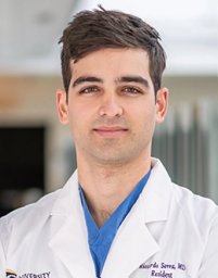 Riccardo Serra合作论文数Department of Neurosurgery, University of Maryland12
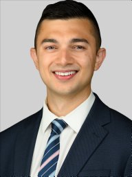 Sakibul Huq合作论文数Dept Neurosurg, Johns Hopkins Univ12
 Noah Gorelick合作论文数12
 Alexander Perdomo-Pantoja合作论文数9
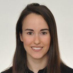 Maya Harary合作论文数Department of Neurosurgery, University of California, Los Angeles9Nicolas Skuli合作论文数8
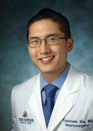 Yuanxuan Xia合作论文数Dept Neurosurg, Johns Hopkins Univ8
 Marco Giovannini合作论文数Ludwig Institute for Cancer Research and Department of Clinical Genetics, Karolinska Hospital;Molecular Genetics Laboratory, The Salk Institute;and Laboratory of Tumor Genetics, Institut Curie;The Salk Institute, Institut Curie8
 Richard G Everson合作论文数Department of Surgery, Duke University Medical Center8
 Jeremie Vitte合作论文数Department of Human Genetics, Hadassah University Hospital8Arba Cecia合作论文数7Tianna Zhao合作论文数Johns Hopkins Univ, Sch Med, Dept Neurosurg, Baltimore, MD 21205 USA7Antonella Mangraviti合作论文数Sant'Andrea University Hospital;Department of Neuroscience, Mental Health and Sense Organs, Sapienza University of Rome7
 Andy S Ding合作论文数Sch Med, Johns Hopkins Univ6
 Wataru Ishida合作论文数Dept Neurosurg, Johns Hopkins Univ6Kelly Blaine合作论文数6Hrusch Cara L合作论文数6
 Anne I. Sperling合作论文数School of Medicine, University of Virginia6
 Brian Na合作论文数5
 Witham Timothy F合作论文数Johns Hopkins University School of Medicine5
 Imre Noth合作论文数Univ Chicago5Alessandro Olivi合作论文数4Stephenie M. Takahashi合作论文数4
 M. Jaffery合作论文数Sect Pulm & Crit Care, Univ Chicago4Elizabeth Fernandez合作论文数Dept Mol & Med Pharmacol, Univ Calif Los Angeles4
 Robert M. Prins合作论文数Department of Neurosurgery, David Geffen School of Medicine, University of California, Los Angeles;Brain Research Institute, University of California, Los Angeles4
 David Nathanson合作论文数Molecular Biology Institute, University of California, Los Angeles4Lo Sheng-fu L合作论文数4
 Bonham Catherine合作论文数4
 Isaac Yang合作论文数University of California4Seba Ramhmdani合作论文数3Tarik Lott合作论文数3Raphael Felder合作论文数3
 Jeff Ehresman合作论文数Sch Med, Johns Hopkins Univ3Manuel Morales合作论文数3Chenchen Ji合作论文数3
 Michael Lim合作论文数School of Medicine, Johns Hopkins University;School of Medicine, Stanford University3
 Nicholas Theodore合作论文数St. Joseph’s Hospital and Medical Center, Barrow Neurological Institute3
 John S. Choi合作论文数jet propulsion laboratory3Alejandro Chara合作论文数2
 Nivedha V Kannapadi合作论文数2Noah L Gorelick合作论文数2
 Miguel A Ruiz-Cardozo合作论文数Department of Neurological Surgery, Washington University2
 Christina Jackson合作论文数Dept Neurosurg, Johns Hopkins Univ2Ravi Medikonda合作论文数2Timothy Kim合作论文数2Alice Hung合作论文数2




