Multiple systems have been developed for grading the adverse effects (AEs) of cancer treatment. The National Cancer Institute Common Toxicity Criteria (CTC) system has substantially evolved since its inception in 1983. The most recent version, CTCAE v3.0 (Common Terminology Criteria for Adverse Events version 3.0) represents the first comprehensive, multimodality grading system for reporting the acute and late effects of cancer treatment. The new CTC requires changes in the application of AE criteria including new guidelines regarding late effects, surgical and pediatric effects, multimodality issues, and for reporting the duration of an effect. It builds on the strengths of previous systems, represents a considerable effort among hundreds of participants, and signifies an international collaboration and consensus of the oncology research community. This article updates recent progress in the evolution of adverse effects grading systems and reviews the development of CTCAE v3.0.
A one and one-half day workshop to assess the current state of the science in brachytherapy was convened at the request of the Radiation Research Program, Division of Cancer Treatment and Diagnosis, National Cancer Institute. The advances in the knowledge of the molecular biology of the radiation response, and in biologic imaging, image fusion, and computer technology made this an appropriate time to address the state of the science and research opportunities for brachytherapy. The aspects of brachytherapy that make it unique include the ability to choose a wide range of dose rates and energies not available with teletherapy, and the ability to precisely localize the placement of radiation sources within tumor, or portions of the tumor. The agenda included both the biology and physics related to brachytherapy. Much of the first day was devoted to overviews of clinical and research topics. The participants then separated into two breakout sessions. Appendix I includes the workshop participants. The following morning session included a presentation and discussion of each breakout session and recommendations for future research. Some research recommendations are specific for brachytherapy and others are more general, as noted. ErratumInternational Journal of Radiation Oncology, Biology, PhysicsVol. 55Issue 5Preview Full-Text PDF
To the Editor: We appreciated the spirited comments from Dr. Schulz and Dr. Kagan regarding the medical physics workshop entitled “Research in Medical Physics.” The workshop, sponsored by the Radiation Research Program, was held “to define the current state of the science in medical physics as it may be applied to radiation oncology, and to discuss potential directions for future research” ( 1 Cumberlin R.L. Coleman C.N. Research in medical physics. Int J Radiat Oncol Biol Phys. 2000; 49: 891-895 Abstract Full Text Full Text PDF Scopus (4) Google Scholar ). Breakout Group A identified “eight discrete areas of research. These represent not only areas of immediate interest but also include areas that, while somewhat speculative now, may quickly develop into important research topics as more is learned about the molecular effects of ionizing radiation on biologic systems.” Breakout Group B “identified four specific areas of research. These represent not only areas of research interest, but they describe some of the most important problems that need to be solved to improve our capability to plan, deliver, and verify conformal dose distributions to patients” ( 1 Cumberlin R.L. Coleman C.N. Research in medical physics. Int J Radiat Oncol Biol Phys. 2000; 49: 891-895 Abstract Full Text Full Text PDF Scopus (4) Google Scholar ). As noted in the accompanying editorial, a central goal of the Radiation Oncology Sciences Program is to advance the field of radiation oncology ( 2 Coleman C.N. Cumberlin R. We’re from the government and we’re here to help you. Int J Radiat Oncol Biol Phys. 2001; 49: 617-618 Abstract Full Text Full Text PDF PubMed Scopus (1) Google Scholar ). The National Cancer Institute supports research primarily through grants. Workshops are “designed to bring together experts to help stimulate research, collaboration and the generation of new ideas” ( 2 Coleman C.N. Cumberlin R. We’re from the government and we’re here to help you. Int J Radiat Oncol Biol Phys. 2001; 49: 617-618 Abstract Full Text Full Text PDF PubMed Scopus (1) Google Scholar ).
A 1.5-day workshop to assess the current state of the science in neutron capture therapy (NCT) was convened at the request of the Radiation Research Program, Division of Cancer Treatment and Diagnosis, National Cancer Institute and the U.S. Department of Energy. The topics were primarily clinical with physics, chemistry, and biology relevant to the immediate trials discussed. The morning of the first day was directed toward updates on epithermal neutron sources, the chemistry of medicinal boron compounds, and preclinical studies. In the afternoon, participants from Europe, Asia, and North America were invited to present their clinical experience with NCT. The participants then separated into breakout sessions. The following morning session included presentations and discussions of each breakout session. These are presented below. NCT was first proposed in 1936, just 4 years after the neutron itself was discovered. NCT is a unique form of radiotherapy that carries a potential for a significant improvement in therapeutic gain. The classification of neutrons into energy categories is somewhat arbitrary, but for the present purpose “thermal neutrons” have an energy 1 eV, “epithermal neutrons” have an energy between 1 eV and 10 KeV, and “fast neutrons” may be considered, for therapeutic purposes, to be in the megavoltage range. NCT is a form of binary therapy, similar to photodynamic therapy, in which neither the thermal neutrons nor the boron carrier molecule has significant cytotoxic effect but produces highly radiobiologically effective particles when the two interact. This is in contrast to the more familiar “fast neutrons,” which are highly radiobiologically effective by themselves. These reactions between a neutron carrier molecule such as boron-10 (B-10) and thermal neutrons produce He-4 and Li-7 ions of very high linear energy transfer (LET) but very short ( 10 m) range. This therapy was first studied in glioblastoma in 1951 using crude thermal neutron beams and B-10–enriched boric acid. Since then, NCT has been studied in several countries, usually with glioblastoma. The difficulties with NCT were, and continue to be, difficulty in finding appropriate neutron beams, a scarcity of suitable boron carrier agents, uncertain dosimetry, and a lack of rigorous and reproducible clinical trials. This workshop was convened to address these issues. These issues were divided into three broad topics: carrier agent development, preclinical studies, and clinical studies.
A 1.5 day workshop was convened at the request of the Radiation Research Program, Division of Cancer Treatment and Diagnosis, National Cancer Institute (RRP/DCTDNCI) for the purpose of defining the current state of the science in tumor biology as it may be applied to treatment with ionizing radiation and to discuss potential clinical applications. The first morning session included overview presentations of seven research areas that were felt to be potential areas of new investigation: Opening Remarks (C. N. Coleman, R. Cumberlin); Presentations: Normal Tissue (P. Okunieff), Signal Transduction (R. Schmidt-Ullrich, A. Dritschilo), Angiogenesis (R. Weichselbaum), Apoptosis (Z. Fuks), Microenvironmental Effects (J. M. Brown), Chemotherapy–Radiotherapy Interactions (T. Lawrence, D. Brizel, J. Mitchell), and Radiation Induced Gene Expression, (R. Weichselbaum). Appendix I includes the list of attendees.
This issue of the Journal inaugurates a special section, “NCI Radiation Research Program Meeting Reports.” We are most appreciative of the offer by Editor-in-Chief Dr. Jim Cox for including this new series. The Radiation Research Program (RRP) is one of the three major programs/branches within the new Radiation Oncology Sciences Program (ROSP) within the National Cancer Institute. The RRP is within the extramural Division of Cancer Treatment and Diagnosis (DCTD), Dr. Robert Wittes, Director. The other two components of ROSP are the Radiation Biology Branch (RBB), Dr. James Mitchell, Branch Chief, and the Radiation Oncology Branch (ROB), which are within the intramural Division of Clinical Sciences (DCS), Dr. Edison Liu, Director. The current members of the RRP are Richard Cumberlin, Frank Mahoney, Helen Stone, Rosemary Wong, Frank Govern, and Norman Coleman.
A one and one-half day Medical Physics Workshop was convened at the request of the Radiation Research Program, Division of Cancer Treatment and Diagnosis, NCI, for the purpose of defining the current state of the science in medical physics as it may be applied to radiation oncology, and to discuss potential directions for future research.
Patients with basal cell skin cancer can be treated with afterloading 192Ir surface molds constructed from dental wax. High activity 192Ir seeds in ribbons are distributed in the mold to uniformly irradiate the target volume. The treatment is preplanned with a treatment planning system to achieve a uniform dose distribution in the planned target volume. The implant parameters optimized include the seed strength and number of seeds, inter- and intracatheter spacing between 192Ir seeds, and the distance between the implant and treatment planes. The radioactive 192Ir strands are afterloaded in the catheters embedded in the wax mold and the position secured with buttons. The treatment area drawn on the patient surface is visually overlapped with the uniformly irradiated area sketched on the mold surface. The mold is taped to the head to secure this position. The dose rate on the surface of the mold in contact with the patient skin is measured with calibrated LiF TLD chips and is within +5% of the computer preplanned dose rate value. This technique is a viable alternative to external beam treatments when daily treatments are not feasible and a dose distribution conforming to the treatment area is desirable.
Ionizing radiation has long been recognized as a weak carcinogen, and the risk of developing a radiation induced neoplasm after exposure to therapeutic radiation has been established. In the case of therapeutic radiation for treatment of existing malignancies, concomitant risk factors for second malignancies can confound the effect of radiation alone. This study presents a model for evaluating the isolated contribution of ionizing radiation to the induction of second malignancies in cancer patients, and presents estimates of the expected number of second malignancies induced in selected sensitive sites by scatter radiation during radiotherapy for cancer. The study focused on the year 1987, during which it was estimated that 192,761 new cancer patients received radiotherapy as part of their initial treatment plan. The model predicted that radiation may induce 63-84 secondary breast cancers, 64-72 secondary thyroid cancers, 94-157 secondary lung cancers, and 489-707 secondary leukemias over the remaining lifetime of this patient population. This represents a lifetime incidence of 0.7% for leukemia, and 0.3% for the solid tumors. This incidence must be placed in perspective with the current concepts of cancer management, such as combined modality therapy that may carry a risk of carcinogenesis greater than either modality alone, and when the alternatives to radiotherapy may be nonexistent or may be cosmetically or functionally undesirable. The information presented may be used in weighing the risks and benefits of alternative treatments for cancer.
Thirty-three children under age 20 with medulloblastoma, treated between 1962 and 1976, at the University of California and the Claire Zellerback Saroni Tumor Institute of Mount Zion Hospital, San Francisco, were retrospectively studied. A relationship between dose and local control rate was suggested by an improved five-year survival in those patients receiving doses greater than 5000 rads to the posterior fossa. The posterior fossa, either alone or with the spinal cord, was the most frequent site of failure. Results of re-irradiation for failure were encouraging and no significant complications were noted. A study of the effects of craniospinal irradiation on the hematopoietic and immune system demonstrated a marked decrease in the peripheral lymphocyte population at the completion of therapy and suggested a functional impairment of the remaining lymphocytes. Other side effects of irradiation included suppression of the hypothalamic-pituitary axis and one instance of brain necrosis. Current treatment policy and proposals for future modifications are discussed.
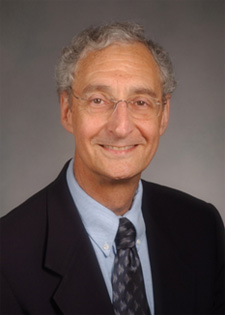 C. Norman Coleman合作论文数Radiation Oncology Branch, Center for Cancer Research, National Cancer Institute, National Institutes of Health, U.S. Department of Health and Human Services6
 Volker Budach合作论文数1
 TA STRIKE合作论文数1Robert Dowsett合作论文数1Ann Setser合作论文数1
 Indra J Das合作论文数Radiation Oncology, Feinberg School of Medicine, Northwestern University1
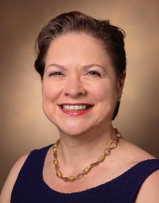 Barbara A. Murphy合作论文数Vanderbilt-Ingram Cancer Center, Vanderbilt University Medical Center1
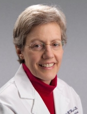 Valerie W. Rusch合作论文数Department of Surgery, Memorial Sloan-Kettering Cancer Center;Weill Cornell Medical College1
 William M. Wara合作论文数Department of Radiation Oncology, University of California, San Francisco;Kaiser Permanente1Mossman K L合作论文数1
 Jeffrey F. Williamson合作论文数Department of Radiation Oncology, School of Medicine, Washington University in St. Louis;Virginia Commonwealth University1
 Anatoly Dritschilo合作论文数1
 Charles Wilson合作论文数Department of Neurological Surgery, Weill Institute for Neurosciences, University of California, San Francisco1Katharina Schad合作论文数1Philip Rubin合作论文数1
 James Deye合作论文数1James Easley合作论文数1
 Andy Trotti合作论文数Center for Infection Research in Cancer, Moffitt Cancer Center1Michael Dattoli合作论文数1
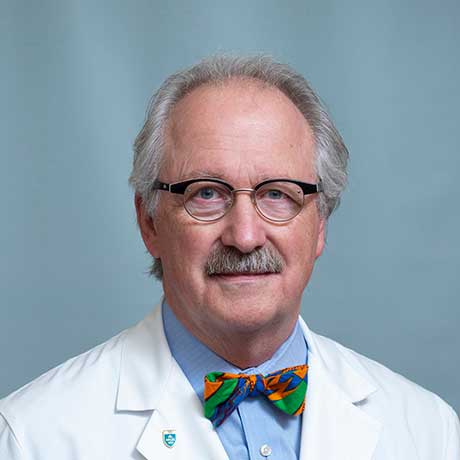 Paul Busse合作论文数Mass General Research Institute, Massachusetts General Hospital;Radiation Oncology Department, Massachusetts General Hospital;Harvard Medical School1David Jaques合作论文数1
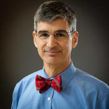 Alexander Dimitrios Colevas合作论文数Medicine Department, Stanford University1
 Corey J. Langer合作论文数Perelman School Of Medicine, University of Pennsylvania1Juan Carlos合作论文数1Kenneth H. Luk合作论文数1
 George Callender合作论文数School of Electronics and Computer Science, Faculty of Engineering and Physical Sciences, University of Southampton1Surendra N. Rustgi合作论文数1




