Lung cancer is the commonest fatal malignant disease worldwide causing more than 37 000 deaths in the UK per annum.1 The five year survival rate of 5% in the UK compares poorly with rates of up to 14% in some European countries.2 Serious concerns have also been raised about inequalities in patient access to oncology services related to the geographical location and age of the patients.3–6 The Calmann-Hine Committee report recommended a review of lung cancer services with increased emphasis on a multidisciplinary approach to care.7 Initial reports show that such multiprofessional teamwork improves coordination and patient support and reduces delays in diagnosis and treatment.8 In the British Thoracic Society (BTS) recommendations to respiratory physicians for organising the care of patients with lung cancer, emphasis was placed on reducing the delay in arranging investigations and the necessary treatment.9 In the new NHS Cancer Plan,10 the Department of Health has recommended that the maximum wait from urgent general practitioner referral to first treatment should be 62 days with a maximum wait of 31 days between diagnosis and first treatment for all cancers. Although there is little evidence to suggest that management delays adversely affect the clinical outcomes, anecdotal reports suggest that borderline resectable tumours at presentation may become unresectable if there is an inordinate delay in treatment planning. A multidisciplinary working group was established within our hospital trust to review the diagnostic journey of patients attending rapid access lung cancer clinics (RAC). The aim of the group was to rationalise this journey, to reduce waiting times, and improve the patients’ experience of this often stressful process. Our multidisciplinary team (MDT) coordinator visited Papworth Hospital, Cambridge, UK to review their tertiary diagnostic service, as described by Laroche et al .8 An away day …
Many sleep centres employ a preliminary screening test in order to reduce the number of polysomnographies required in the routine diagnosis of the sleep apnoea/hypopnoea syndrome (SAHS). We investigated the combination of heart rate and oximetry information as a means of performing this test. A retrospective study of 100 patients with suspected SAHS was made. All patients had in-hospital polysomnography on one night. We estimated the number of respiratory event-related arousals by counting the number of autonomic arousals (assessed on the basis of changes in the heart interbeat interval) that were coincident with a rise in oximetry. The hourly index of such events was denoted the 'cardiac-oximetry disturbance index' (CODI). The median apnoea/hypopnoea index (AHI) was 16.5 (range 1.0-93.6) h(-1) . The CODI correlated significantly with the AHI (Spearman correlation coefficient r (s) = 0.88, P < 0.01), and the area (+/- standard error) under the receiver operating characteristic (ROC) was 0.94 +/- 0.05. Oximetry alone (based on 4% dips) was a less effective screening test (r (s) = 0.80, P < 0.01; area under ROC 0.83 +/- 0.06). Using 2% dips in oximetry offered comparable performance with the CODI (r (s) = 0.91, P < 0.01; area under ROC 0.93 +/- 0.04). The CODI was better correlated with the electroencephalograph arousal index (r (s) = 0.84, P < 0.01) than was oximetry (2% dips, r (s) = 0.57, P < 0.01). The CODI algorithm also offers an informal measure of self-validation: a large discrepancy between the number of autonomic arousals and the number of rises in oximetry indicates the presence of autonomic arousals without changes in oximetry (or vice versa). This self-validation mechanism identified several patients in this study, and may be useful in identifying sleep disruption due to chronic pain or other causes.
Formulating a rational health policy necessitates the ability to compare between different healthcare interventions and disease scenarios. Continuous positive airway pressure (CPAP) therapy with a conservative lifestyle strategy in sleep apnoea/hypopnoea syndrome (SAHS) was evaluated using health utility and quality-adjusted life years (QALYs) as outcome measures. A total 71 SAHS (apnoea/hypopnoea index ≥15 h−1) patients completed a randomised, parallel group study over 3 months using utilities derived by the standard gamble approach (Usg) and European quality of life questionnaire (Euroqol) (Ueq). The severely impaired health status at baseline improved by 23% (Usg 0.32 to 0.55) adding 8 QALYs in the CPAP group, compared to a 4% improvement with 4.7 QALYs added in the lifestyle group (Usg 0.31 to 0.35). The Ueq showed a marginal change with CPAP (0.73 to 0.77) but did not demonstrate any improvement with lifestyle intervention. The health status impairment in sleep apnoea/hypopnoea syndrome patients is markedly improved by continuous positive airway pressure compared to a modest improvement with conservative lifestyle strategies using the standard gamble utility, which may be incorporated in effectiveness and economic analyses. The European quality of life questionnaire did not reflect a similar degree of impact and is probably not useful in this population.
The modest daytime hypertension and sympathetic upregulation associated with the sleep apnoea/hypopnoea syndrome (SAHS), does not explain the relatively large increased risk of cardiac morbidity and mortality in the SAHS patients population. Therefore, efferent vagal and sympathetic activity was evaluated during wakefulness in SAHS subjects and matched healthy controls, in order to determine if vagal downregulation may play a role in the aetiology of cardiac disease in the SAHS. The awake autonomic nervous system function of 15 male subjects, with mild-to-moderate SAHS was compared to that of 14 healthy controls matched for age, body mass index, gender and blood pressure. All subjects were free from comorbidity. Vagal activity was estimated from measurements of heart rate variability high frequency power (HF) and sympathetic activity was measured from urine catecholamine excretion. The %HF power was significantly (p < 0.03) reduced in SAHS patients (10+/-1.6 (mean+/-SEM)) as compared to controls (17 +/- 3). In addition, HF power correlated with the apnoea/hypopnoea index in the SAHS subjects (R = -0.592, p = 0.02). There was no statistically significant difference in the daytime excretion of nonadrenaline between control (242 +/- 30 nmol x collection(-1)) and SAHS (316 +/- 46 nmol x collection(-1)) subjects (p = 0.38). In these sleep apnoea/hypopnoea syndrome patients there was limited evidence of increased waking levels of urine catecholamines. The principal component altering waking autonomic nervous system function, in the sleep apnoea/hypopnoea syndrome subjects, was a reduced daytime efferent vagal tone.
The authors present a method of screening for obstructive sleep apnoea based on the electrocardiogram (ECG). The algorithm combines information from the ECG-derived respiration (EDR) signal and the RR interval tachogram. Power spectral features from the EDR signal were computed using the discrete harmonic wavelet transform, considering the power at the respiratory frequency and at frequencies below 0.1 Hz. Cycles of tachy/bradycardia (consistent with an arousal from sleep, as would be expected at the end of an episode of apnoea) were identified front the RR interval tachogram. Features were collated into minute-by-minute vectors and passed to a classifier. The algorithm correctly classified 81% of all minutes in the test database, with 29/30 patients correctly identified as apnoea or normal. Visual classification produced 92% correct classification, with all 30 patients correct
The sleep apnoea/hypopnoea syndrome (SAHS) elicits a unique heart rate rhythm that may provide the basis for an effective screening tool. The study uses the receiver operator characteristic (ROC) to assess the diagnostic potential of spectral analysis of heart rate variability (HRV) using two methods, the discrete Fourier transform (DFT) and the discrete harmonic wavelet transform (DHWT). These two methods are compared over different sleep stages and spectral frequency bands. The HRV results are subsequently compared with those of the current screening method of oximetry. For both the DFT and the DHWT, the most diagnostically accurate frequency range for HRV spectral power calculations is found to be 0.019–0.036 Hz (denoted by AB2). Using AB2, 15 min sections of non-REM sleep data in 40 subjects produce ROC areas, for the DFT, DHWT and oximetry, of 0.94, 0.97 and 0.67, respectively. In REM sleep, ROC areas are 0.78, 0.79 and 0.71, respectively. In non-REM sleep, spectral analysis of HRV appears to be a significantly better indicator of the SAHS than the current screening method of oximetry, and, in REM sleep, it is comparable with oximetry. The advantage of the DHWT over the DFT is that it produces a greater time resolution and is computationally more efficient. The DHWT does not require the precondition of stationarity or interpolation of raw HRV data.
The unique heart rate rhythm in the sleep apnoea/hypopnoea syndrome (SAHS) may provide the basis for an effective screening tool. This paper assesses the variability (HRV), using the Discrete Fourier Transform (DHWT). Spectral HRV results are compared to those of the current screening method of oximetry. Diagnostic indicator of the SAHS in both NREM and REM sleep. The DHWT offers advantages over the DFT in that it supplies greater time resolution and is computationally more efficient.
In patients with cystic fibrosis (CF), nasal intermittent positive pressure ventilation (NIPPV) is currently used as a short-term bridge to transplantation but its precise role has yet to be determined. Patients were offered a therapeutic trial of NIPPV when candidates for lung transplantation, with respiratory failure unresponsive to medical treatment. Twelve patients, six male of mean age of 26 +/- 1.4 years, had a trial of NIPPV. At recruitment the mean percentage predicted forced expired volume in one second (FEV1) was 15.1% +/- 1.2%, arterial carbon dioxide (PaCO2) 8.7 +/- 0.6 kPa, arterial oxygen (PaO2) with variable FiO2 7.4 +/- 0.6 kPa and arterial bicarbonate (HCO3-) 40.1 +/- 1.6 mmol l-1. Ten cases tolerated NIPPV for 1-15 months, mean 5.1 +/- 1.4 months, with subjective improvement in headache and quality of sleep. At 3 months, there was significant improvement in forced vital capacity, PaCO2 and arterial HCO3- and there was a reduction in the number of hospital inpatient days (P < 0.05). Subsequently three cases had lung transplantation, four died on the active list and three are awaiting organs. Two patients failed to tolerate NIPPV owing to abdominal bloating and increasing hypercapnia. In conclusion, NIPPV, if tolerated, was a useful adjunct in the treatment of CF patients with hypercapnic respiratory failure awaiting transplantation. Further prospective studies are required to determine the optimum time to commence NIPPV and to clarify its precise role.
A 47 year old man presented with a two year history of increasing cervical dysphagia, dyspnoea, and cutaneous signs. He had been diagnosed 27 years previously with Wilson's disease and was treated with penicillamine (1.5 g daily). Systemic abnormality of elastic fibres was confirmed by light and electron microscopy following biopsy of skin, lung, oesophageal muscle, gum, pharyngeal tissue, and cervical connective tissue. Dysphagia was relieved by cricopharyngeal myotomy. Substitution of trientene dihydrochloride for penicillamine relieved cutaneous and systemic manifestations. This is possibly the first case demonstrating an association between prolonged penicillamine use and biopsy proved systemic pseudo-pseudoxanthoma elasticum. The presenting symptoms may have resulted from the abnormal numbers and properties of elastic fibres, and the changes were caused by penicillamine use, rather than by idiopathic, inherited pseudoxanthoma elasticum.
Precise relationships between symptoms and diseases are rarely documented in the literature, and yet it is essential that the physician establishes a diagnostic label that will lead to the appropriate therapy. In obstructive sleep apnoea (a breathing disorder during sleep which is characterised by intermittent pauses in respiration) the borderline between normal and pathological is arbitrary. As such, fuzzy sets provide an intuitively appealing framework for representing the medical knowledge. In this paper, the expert system CADOSA for decision support in a hospital sleep clinic is presented. The medical knowledge in the system is stored in the form of fuzzy logical relationships between symptoms and diseases, between symptoms themselves and between symptom combinations and diseases. The symptoms present in the patient are confirmed by an interview, a physical examination, a questionnaire and laboratory tests, including an overnight oximetry study which measures oxygen saturation and heart rate. The fuzzy inference in the system is performed using the max-min compositional rule to calculate indication relations expressing occurrence and confirmability. These lead to confirmed and excluded diagnoses as well as diagnostic hypotheses. In an evaluation of CADOSA using 21 patients, the proportion diagnosed correctly as confirmed (excluded) obstructive sleep apnoea was 0.95 (1.00).
BACKGROUND:An association between psychological morbidity and asthma is well recognized and an increase in negative psychosocial factors in brittle asthma has been previously reported. Such factors, apart from affecting patient perceptions, may alter patients' self-management of their condition.METHODS:We have undertaken a case-control study on 29 well characterized brittle and non-brittle asthmatics in the West Midlands Region to assess their level of psychological morbidity, using the General Health Questionnaire (GHQ) and the living with asthma questionnaire, and their responses to changes in asthma symptoms, using the Asthma Symptom Checklist and a taped interview.RESULTS:Significant differences in GHQ (mean score brittle 19.5 vs non-brittle 7.2, P = 0.0002) and living with asthma (mean score brittle 1.30 vs non-brittle 1.00, P = 0.002) reinforced the presence of psychological factors in this group of patients with severe asthma. Interviews regarding responses to hypothetical asthma attacks showed that patients with brittle asthma delayed seeking medical attention more often than those with non-brittle asthma and instead preferred to self-administer beta-agonist medication. Sixteen (55.2%) of the 29 patients with brittle asthma would have delayed 7 days before seeking medical attention in a slow onset attack compared with 6/29 (20.7%) in the non-brittle group. In a fast onset attack 14 (48.3%) patients with brittle asthma would not have summoned help, despite finding it difficult to walk to the kitchen for a drink whereas in the non-brittle group 24 out of 29 (82.8%) would have summoned help. Levels of family support tended to be lower in patients with brittle asthma (mean family APGAR 7.3 vs 8.65 P = 0.09).CONCLUSIONS:Brittle asthma is associated with greater psychological morbidity and altered strategies for coping with deteriorating asthma symptoms. Attention should be paid to the presence of such factors in the management of patients with brittle asthma.
Guidelines for the spectral analysis of electrocardiographic R-R intervals have recently been published. However, little infomation is given on the effect of the electrocardiogram sampling frequency with respect to spectral analysis results. Developing modelled R-R data, using the Integral Pulse Frequency Modulated model at differing sampling frequencies, we have produced a methodology for determining if a recorded R-R signal has a high enough signal to noise ratio to perform accurate spectral analysis
Background - Patient self-management plans for asthma use peak expiratory flow (PEF) meter readings for decisions on adjusting asthma treatment. PEF meters have been shown to be inaccurate and the effect of this inaccuracy on such treatment plans has been determined.Methods - PEF measurements were made by 127 severe asthmatic patients at least twice a day for at least two weeks using a mini-Wright meter. The daily variation from ''best'' PEF and the within day PEF variability were calculated before and after correction for the meter's known inaccuracy. The effect of this data correction on the number of days when trigger points were reached for changing asthma therapy was then determined.Results - Continuous PEF readings were available from 114 subjects with a median of 157.5 days of data per subject (range 15-489 days). Correction of the PEF data led to the number of days of satisfactory asthma control being reduced in 72% of subjects with just one subject showing an increase in satisfactory control. Data correction reduced the percentage of total days of satisfactory control from 46% to 36% of days, and increased the days requiring more inhaled steroids from 33% to 36%. The days on which a course of oral corticosteroids was required increased from 16% to 23%.Conclusions - The accuracy of PEF meters significantly influences the interpretation of currently used asthma self-management plans. Managing asthma with the corrected PEF data would have increased the amount of treatment received by these patients since the severity of the asthma was underestimated by the raw data.
BACKGROUND--The British Thoracic Society report on the diagnosis and treatment of the sleep apnoea/hypopnoea syndrome (SAHS) suggests that, if the pulse oximetry baseline oxygen saturation is above 90%, then 15 4% oxygen desaturation/hour in bed will diagnose SAHS requiring treatment. The diagnostic outcome of applying these guidelines has been studied. METHODS--One hundred patients referred to a district general hospital sleep clinic were recruited. After initial clinical assessment, overnight pulse oximetry measurements were performed, followed by full polysomnography at the regional laboratory. RESULTS--Sixty nine patients underwent both pulse oximetry and polysomnography. All 10 patients with more than 15 4% desaturations/hour on pulse oximetry had SAHS confirmed on polysomnography (specificity = 100%). Twenty two patients with SAHS were misdiagnosed using pulse oximetry alone (sensitivity = 31%). These patients had low apnoea scores but high hypopnoea scores. CONCLUSIONS--The BTS pulse oximetry criteria are highly specific when positive (specificity = 100%), but may miss patients with the SAHS who have hypopnoeic episodes which cause arousal but not significant oxygen desaturation (sensitivity = 31%). It should be emphasised that pulse oximetry alone does not always give sufficient information to discriminate between those patients with or without SAHS. Patients with "negative" pulse oximetry and symptoms of SAHS should undergo polysomnography or multi-channel recording.
1. The rates of change in mean peak expiratory flow and in diurnal variation in peak flow were compared in 14 patients recovering from acute severe asthma. 2. Peak expiratory flow was measured on hospital admission, and at 6-hourly intervals for the next 3 weeks. 3. Diurnal variation in peak flow was assessed by measuring the following: amplitude (the highest minus the lowest peak expiratory flow during any given 24 h period), amplitude % mean (the highest minus the lowest peak expiratory flow during any given 24 h period divided by the mean peak expiratory flow over that period) and residual amplitude (the maximum variation about the mean peak expiratory flow during any given 24 h period). 4. Plots of diurnal variation in peak flow and peak expiratory flow against time were constructed for each patient. To enable comparison of changes in peak expiratory flow and diurnal variation in peak flow the data were transformed. 5. The rate of change for mean peak expiratory flow and for the three measures of diurnal variation in peak flow was assessed by fitting an exponential function to each set of data, and calculating the slope of the exponential curve halfway through the period of observation (10.5 days). 6. Median (range) slope for peak expiratory flow was 0.055 (0-2.57). The comparable value for amplitude was -3.15 (-1.27 to -4.22) (absolute median values compared, P = 0.0029), for amplitude % mean was -1.87 (-0.18 to -5.95) (P = 0.012) and for residual amplitude was -1.43 (-0.62 to -3.09) (P = 0.033).(ABSTRACT TRUNCATED AT 250 WORDS)
The aim of this study was to investigate whether the long-acting beta-agonist salmeterol affects athletic performance in patients with asthma. The effect of 50 micrograms salmeterol on the cardiorespiratory responses to a progressive maximal cycle exercise test and on endurance capacity (defined as the exercise duration at 70% maximum oxygen uptake), was compared with 200 micrograms salbutamol and a matched placebo in eight asthmatic men. Both salmeterol and salbutamol improved pre- and postexercise forced expiratory volume in one second (FEV1) for maximal and endurance exercise. Following active treatment, patients exercised from a significantly high baseline FEV1, with both salmeterol (3.58(1.16)l) (mean (SD)) and salbutamol (3.55(1.24)l) compared with placebo (3.29(1.35)l). Similar improvements preceded endurance exercise. Cardiorespiratory, haemodynamic or subjective responses to the progressive maximum exercise tests were not different with salmeterol, salbutamol or placebo, nor did endurance capacity change with any treatment modality. Blood lactate levels, after 15 min exercise, were significantly higher with salbutamol (3.64 (1.83) mM), but not with salmeterol (3.03 (1.64) mM), compared with placebo (2.95 (1.69) mM). These results demonstrate the absence of significant cardiorespiratory or metabolic effects during exercise after a single dose of salmeterol, together with a lack-of ergogenic effect, as measured by maximal or endurance exercise performance, in patients with asthma.
We have compared the maximal and endurance exercise capacities in 22 (15 male) adult cystic fibrosis (CF) patients with a resting oxygen saturation (SaO2) ≥ 90%, with age and sex matched controls (CON). The maximum oxygen uptake (V̇O2max) and heart-rate were lower for the CF group whereas the maximum ventilation reached a higher percentage of predicted. Furthermore, for the CF group there was a relationship between the % predicted V̇O2max and measures of lung function, confirming a ventilatory limit to maximum exercise in patients with more severe disease and a near normal maximum exercise capacity in those with mild disease. Endurance capacity, defined as the duration of exercise at 80% of each individuals V̇O2max, was however similar for the two groups [CF: 22·21 (15.82) vs. CON: 24.94 (13·05) min]; despite the CF group exercising under less efficient ventilatory conditions. Endurance capacity was not related to the measurements of lung function. Of the seven CF patients desaturating at maximal exercise (SaO2 <90%), five desaturated at a work load of 80% V̇O2max, whereas only one desaturated at 50% V̇O2max. This study confirms that with increasing severity of disease, maximal exercise capacity may be limited in adult CF patients, whereas endurance capacity (at the same relative work load) is unimpaired. Furthermore, CF patients who desaturate during a progressive maximal exercise test are likely to desaturate during constant work load exercise if the intensity is high but unlikely to do so if the intensity is low. The information from these exercise tests would be of value for prescribing individual training programmes and for evaluating the effects.
 Freeman W合作论文数15Geoffrey E Packe合作论文数12GE PACKE合作论文数8P Howard合作论文数6
 Michael Chappell合作论文数School of Engineering, Faculty of Science, Engineering and Medicine, The University of Warwick5
 Ron A. Bates合作论文数4Dc Weir合作论文数4P.S. Burge合作论文数3JE SPICER合作论文数3MF Hilton合作论文数3
 Indranil Chakravorty合作论文数Inst Hlth Res, BAPIO3Malcolm Nattrass合作论文数3
 Jon G Ayres合作论文数University of Birmingham;University of Aberdeen;University of Warwick3S.B. Sapiano合作论文数3J.E. Whitehead合作论文数3D.E. Stableforth合作论文数3
 Arshad Javaid合作论文数Institute of Mycology and Plant Pathology;University of the Punjab;Institute of Mycology and Plant Pathology, University of the Punjab3W S Tunnicliffe合作论文数2J. F. Miles合作论文数2Cw Edwards合作论文数2W Freeman合作论文数2W. Robertson合作论文数2J. Simkins合作论文数2Anderson P B合作论文数2
 Ben Raymond合作论文数Australian Antarctic Division, Department of Sustainability, Environment, Water, Population and Communities, Kingston 7050, Australia2Wiggins J合作论文数2S. Freeman合作论文数2S. P. O'Hickey合作论文数Dept of Respiratory;Physiology Birmingham Heartlands Hospital Birmingham;Physiology, Birmingham Heartlands Hospital2Michael D Peake合作论文数2di rogers合作论文数1A.J. Williams合作论文数1Ad Wright合作论文数1Bm Singh合作论文数1Lk Harding合作论文数1jl stellman合作论文数1
 Rosalind M Green合作论文数North West Lung Centre, Wythenshawe Hospital1AJ SUGGETT合作论文数1K. Whyte合作论文数1Brennan S R合作论文数1Rajasekaran A B合作论文数1Singh Baldev M合作论文数1Trotter Simon E合作论文数1Wright A D合作论文数1Coatesworth A P合作论文数1
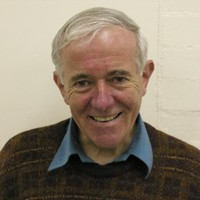 Keith R Godfrey合作论文数School of Engineering, The University of Warwick1
 Martin Raymond Miller合作论文数Institute of Applied Health Sciences, University of Birmingham1




