Objectives/Hypothesis: The American College of Surgeons Thyroid and Parathyroid Ultrasound Skills-Oriented Course (TPUSC) was designed to teach surgeons how to interpret and perform office-based head and neck ultrasound (HNUS). The objective of this study was to survey attendees of the TPUSC to evaluate the usefulness of the course, to track surgeon performed HNUS practice patterns, and to help identify potential roadblocks to incorporation of HNUS into a surgeon's practice.Study Design: Cross-sectional survey.Methods: A Web-based survey was sent to 952 surgeons who completed the TPUSC between 2010 and 2014. Questions included surgeon specialty, practice type, Likert scale rating of the TPUSC, competency with different HNUS procedures, and current HNUS practice patterns.Results: The response rate was 24%. On a scale from 1 (not useful) to 5 (extremely valuable), the mean course usefulness rating was 4.2. Educational goals were met for 194 (92%) surgeons, and 162 (77%) surgeons reported performing HNUS in their practice. Of 48 surgeons not performing HNUS, 24 (50%) attributed insufficient time in their clinic schedule, and 21 (44%) attributed high equipment costs.Conclusions: The TPUSC is a valuable educational experience for surgeons seeking to gain proficiency in HNUS. The majority of TPUSC graduates gain competency with at least one type of HNUS procedure following the course.
Program Description:With the progressive improvements in imaging resolution of ultrasound, concurrent reduction in capital expenditure, and portability, this modality is ideally suited to point‐of‐care use by most otolaryngology practices. Primary and exported training courses through the American College of Surgeons have provided a clinical starting point. An accreditation process for head and neck ultrasound is now available through a joint project between the American Institute for Ultrasound in Medicine and the Academy. This miniseminar presents the practical application of ultrasound to general, pediatric, endocrinologic, and oncologic conditions through a panel of outstanding experts from radiology and otolaryngology.Educational Objectives:(1) Cite the principles of ultrasound and its application to conditions of the head and neck. (2) Recognize the advantages of ultrasound‐guided aspiration of samples for cytology and other chemical tests. (3) Learn ultrasound techniques and eventual accreditation of skills.
Ultrasound is an integral part of the preoperative workup for patients who are being evaluated for thyroid and parathyroid surgery. It helps improve the accuracy of a fine-needle aspiration biopsy and complements other imaging modalities used for planning the extent of surgery. It also allows imaging of vital structures in relation to the thyroid and parathyroid. The compact nature and portability of ultrasound machines in recent years has made it easier for motivated surgeons (head and neck, general, and endocrine surgeons) to incorporate them into their practice. However, successfully setting up such a service needs adequate planning and an understanding of the obstacles that are involved. We aim to discuss these obstacles in detail, with practical suggestions on how to overcome them. This review may serve as a resource when dealing with issues such as purchasing equipment, training, credentialing, billing, documentation, and collaboration. Although these are discussed with respect to surgeons with an interest in endocrine disease, with some modifications they may also apply to any surgeon who uses ultrasound frequently.
Program Description Office‐based ultrasound of the thyroid and neck is now a standard imaging tool for otolaryngologists, endocrinologists, and surgical clinicians. Training courses in the use of ultrasound are now well‐structured and available to clinicians in their related disciplines. Yet, as with all standard technology, new applications and refinements have expanded both the resolution of image quality and uses of ultrasound beyond what was initially envisioned. Through the use of both short didactic presentations and directed questions with clinical case examples from the moderator, this panel of experts will discuss several new frontiers of ultrasound. Techniques that optimize cytologic aspiration, indications and use of core biopsy, alcohol ablation of thyroid cysts, and selection of cases of lymph node ablation with papillary carcinoma, elastography, and the evolving use of radiofrequency ablation and contrast enhanced ultrasound are some of the topics that will be covered. An emphasis on how best to improve the standard use of head and neck ultrasound will make this a practical seminar for the otolaryngologist as well as an important glimpse into the near future. Educational Objectives 1) The seminar attendee should learn new advances in obtaining cytologic samples with ultrasound. 2) The seminar attendee will understand new advances in destroying malignant lymph nodes and cysts of the thyroid gland including advantages and disadvantages of the technology compared with conventional surgery. 3) The seminar attendee will learn new adjunctive technologies to ultrasound such as elastography and contrast imaging.
ObjectiveInvestigate the findings in repeat samples of patients with initial ultrasound‐guided non‐diagnostic fine needle aspirates (FNA) of thyroid tissue performed and interpreted over the most recent 10‐year period at the University of Vermont.MethodRetrospective review of medical records for patients with nondiagnostic thyroid FNAs. Patients were identified using a pathology laboratory information system according to specific search parameters. Patients were excluded if ultrasound guidance was not used and if documentation in the medical records was limited.ResultsA total of 239 patients met inclusion criteria. Of the 239, 145 have undergone follow‐up FNA, surgical intervention, or both. There were 19 documented neoplasms after initial non‐diagnostic FNA, including 2 incidental papillary microcarcinomas, 3 incidental follicular variant of papillary microcarcinomas, 1 follicular carcinoma, 7 papillary carcinomas, 2 follicular variant of papillary carcinomas, 1 non‐Hodgkin DLBCL, 2 Hurthle cell neoplasms, and 1 follicular neoplasm. Excluding the 3 microcarcinomas, there were 13 significant neoplasms in the ipsilateral lobe (5%). For the 61 patients (25%) who were lost to follow‐up before a definitive diagnosis was made, we cannot draw final conclusions regarding malignancy.ConclusionOur study reveals significant potential for malignancy in patients with nondiagnostic FNAs. The IRB has granted permission to contact the 61 patients lost to follow‐up before a definitive diagnosis was made. Delayed samples will be included as a separate analysis to profile this series of patients with nondiagnostic cytology.
This content is designed to acquaint the clinician with some of the more common ultrasonographic manifestations of clinical conditions that the otolaryngologist is likely to encounter in a general practice. The clinician requires a thorough knowledge of head and neck anatomy to best interpret the variations from normal structures demonstrated on ultrasound. A knowledge of sonographic artifacts may assist the examiner in properly identifying the process under review. Ultrasonography may be the best imaging study for certain organs. In many instances it is the first clinical study that directs further imaging. By providing the clinician with clues as to the underlying pathology, it allows a more efficient direction in determining which aspiration techniques to use.
 Lisa A. Orloff合作论文数Otolaryngology (Head and Neck Surgery) Department, Stanford University6
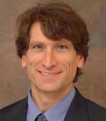 David L. Steward合作论文数Department of Otolaryngology, Head & Neck Surgery, College of Medicine, University of Cincinnati4
 Clayman Gary L合作论文数3
 Gregory W. Randolph合作论文数Department of Otolaryngology Head and Neck Surgery, Harvard Medical School, Harvard University;Division of Thyroid and Parathyroid Endocrine Surgical Service, Massachusetts Eye and Ear Infirmary;Massachusetts General Hospital3Maisie L Shindo合作论文数2Righi P D合作论文数2
 Merry E Sebelik合作论文数2Todd Dray合作论文数2Hh Schmidek合作论文数1A. Cmelak合作论文数1William B. Armstrong合作论文数1DA PLANTE合作论文数1H. Jack Baskin合作论文数1G RANDOLPH合作论文数1Stephen Leech合作论文数1D A Smith合作论文数1Harris Jonathan合作论文数1
 Gregory K Hartig合作论文数Division of Otolaryngology-Head & Neck Surgery, Department of Surgery, University of Wisconsin-Madison1
 Victoria Aviado Flores合作论文数1N Weidner合作论文数1T. Mcculloch合作论文数1GL Clayman合作论文数1K FU合作论文数1James I. Cohen合作论文数1
 R Michael Tuttle合作论文数David H. Koch Center for Cancer Care at Memorial Sloan Kettering Cancer Center1Jay Smith合作论文数1
 Kevin Thomas Robbins合作论文数1Njoku Dolores B合作论文数1Darr E Ashlie合作论文数1Brennan Fred H合作论文数1Sharma Giriraj K合作论文数Department of Neurological Surgery, George Washington University1Ihm P S合作论文数1
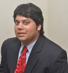 Sushruta Nagarkatti合作论文数Harvard Medical School, Massachusetts General Hospital1
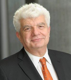 James A. Fagin合作论文数Division of Subspecialty Medicine, Memorial Sloan Kettering Cancer Center1
 Anthony M Reginato合作论文数1Mautner Kenneth R合作论文数Emory Sports Medicine Center1Sritharan Niranjan合作论文数Otolaryngology Head and Neck Surgery Department, The Canberra Hospital1Hammers Lynwood合作论文数1Mekel Michal合作论文数1
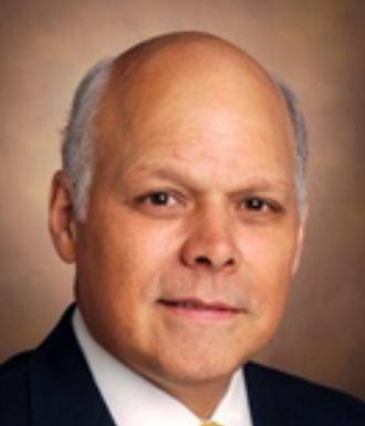 James Netterville合作论文数Vanderbilt Institute for Global Health, Vanderbilt University Medical Center;Department of Otolaryncology, Vanderbilt University Medical Center1Vargas H合作论文数1
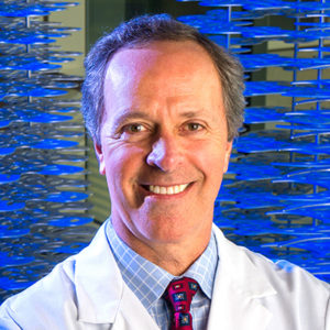 Paul Harari合作论文数Department of Human Oncology, School of Medicine and Public Health, University of Wisconsin-Madison1Jeremy L. Freeman合作论文数Department of Otolaryngology-Head and Neck Surgery;Mount Sinai Hospital1
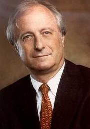 Jack L. Gluckman合作论文数University of Cincinnati1
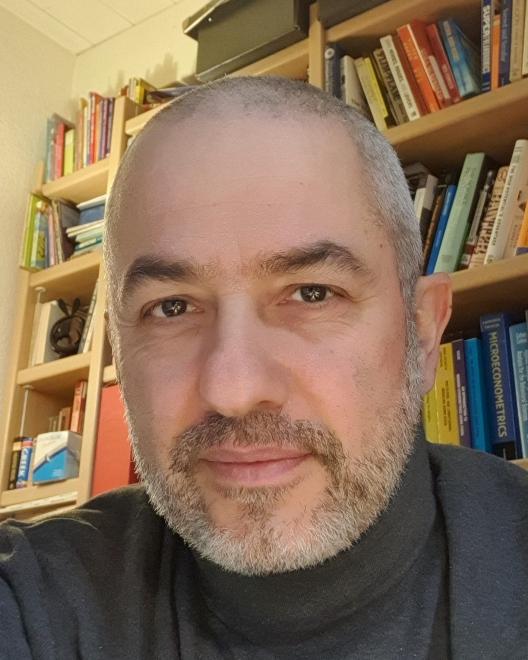 John Antonakis合作论文数Department of Organizational Behavior, Faculty of Business & Economics, University of Lausanne1
 Paul A. Levine合作论文数Department of Otolaryngology, University of Virginia1David N. Krag合作论文数1
 Pradeep Kumar合作论文数Department of Computer Science and Engineering, JSS Academy of Technical Education1
 Jay F. Piccirillo合作论文数Department of Otolaryngology-Head and Neck Surgery, School of Medicine, Washington University in St Louis1




