Recent studies have highlighted the crucial role of microglia (MG) and their interactions with the gut microbiome in post-stroke neuroinflammation. The activation of immunoregulatory pathways, including the aryl hydrocarbon receptor (AHR) pathway, is influenced by a dynamic balance of ligands derived from both the host and microbiota. This study aimed to investigate the association between stroke-induced dysbiosis and the resultant imbalance in AHR ligand sources (loss of microbiota-derived [indole-based] and increase of host-derived [kynurenine-based]) after stroke. Microbiota-derived AHR ligands decreased in human plasma and remained low for days following an ischemic stroke highlighting the translational significance. Transient-middle-cerebral-artery-occlusion was performed in aged wild-type and germ-free male mice. MG-AHR expression and activity increased in both in vivo and ex vivo stroke models. Germ-free mice showed altered neuroinflammation and antigen presentation while aged mice showed reduced infarct volume and neurological deficits following treatment with microbiota-derived AHR ligands after stroke. Restoring a balanced pool of host- and microbiota-derived AHR ligands may be beneficial after stroke and may represent a therapeutic target.
Stroke is the most common cause of long-term disability and places a high economic burden on the global healthcare system. Functional outcomes from stroke are largely determined by the extent of ischemic injury, however, there is growing recognition that systemic inflammatory responses also contribute to outcomes. Mast cells (MCs) rapidly respond to injury and release histamine (HA), a pro-inflammatory neurotransmitter that enhances inflammation. The gut serves as a major reservoir of HA. We hypothesized that cromolyn, a mast cell stabilizer that prevents the release of inflammatory mediators, would decrease peripheral and central inflammation, reduce MC trafficking to the brain, and improve stroke outcomes. We used the transient middle cerebral artery occlusion (MCAO) model of ischemic stroke in aged (18 mo) male mice to investigate the role of MC in neuroinflammation post-stroke. After MCAO we treated mice with 25 mg/kg body weight of cromolyn (MC stabilizer) by oral gavage. Cromolyn was administered at 3 h, 10 h, 24 h and every 24 h for 3 days post-stroke. Three control groups were used. One group underwent a sham surgery and was treated with cromolyn, one received sham surgery with PBS vehicle and the third underwent MCAO with PBS vehicle. Mice were euthanized at 24 h and 3 days post-stroke. Cromolyn administration significantly reduced MC numbers in the brain at both 24 h and 3 days post-stroke. Infarct volume was not significantly different between groups, however improved functional outcomes were seen at 3 days post-stroke in mice that received cromolyn. Treatment with cromolyn reduced plasma histamine and IL-6 levels in both the 24-h and 3-day cohorts. Gut MCs numbers were significantly reduced after cromolyn treatment at 24 h and 3 days after stroke. To determine if MC trafficking from the gut to the brain occurred after injury, GFP + MCs were adoptively transferred to c-kit −/− MC knock-out animals prior to MCAO. 24 h after stroke, elevated MC recruitment was seen in the ischemic brain. Preventing MC histamine release by cromolyn improved gut barrier integrity and an improvement in stroke-induced dysbiosis was seen with treatment. Our results show that preventing MC histamine release possesses prevents post-stroke neuroinflammation and improves neurological and functional outcomes. Graphical abstract
Background:Stroke is a major cause of morbidity and mortality, and its incidence increases with age. While acute therapies for stroke are currently limited to intravenous thrombolytics and endovascular thrombectomy, recent studies have implicated an important role for the gut microbiome in post-stroke neuroinflammation. After stroke, several immuno-regulatory pathways, including the aryl hydrocarbon receptor (AHR) pathway, become activated. AHR is a master regulatory pathway that mediates neuroinflammation. Among various cell types, microglia (MG), as the resident immune cells of the brain, play a vital role in regulating post-stroke neuroinflammation and antigen presentation. Activation of AHR is dependent on a dynamic balance between host-derived and microbiota-derived ligands. While previous studies have shown that activation of MG AHR by host-derived ligands, such as kynurenine, is detrimental after stroke, the effects of post-stroke changes in microbiota-derived ligands of AHR, such as indoles, is unknown. Our study builds on the concept that differential activation of MG AHR by host-derived versus microbiome-derived metabolites affects outcomes after ischemic stroke. We examined the link between stroke-induced dysbiosis and loss of essential microbiota-derived AHR ligands. We hypothesize that restoring the balance between host-derived (kynurenine) and microbiota-derived (indoles) ligands of AHR is beneficial after stroke, offering a new potential avenue for therapeutic intervention in post-stroke neuroinflammation. Method:We performed immunohistochemical analysis of brain samples from stroke patients to assess MG AHR expression after stroke. We used metabolomics analysis of plasma samples from stroke and non-stroke control patients with matched comorbidities to determine the levels of indole-based AHR ligands after stroke. We performed transient middle cerebral artery occlusion (MCAO) in aged (18 months) wild-type (WT) and germ-free (GF) mice to investigate the effects of post-stroke treatment with microbiota-derived indoles on outcome. To generate our results, we employed a range of methodologies, including flow cytometry, metabolomics, and 16S microbiome sequencing. Results:We found that MG AHR expression is increased in human brain after stroke and after ex vivo oxygen-glucose deprivation and reperfusion (OGD/R). Microbiota-derived ligands of AHR are decreased in the human plasma at 24 hours after ischemic stroke. Kynurenine and indoles exhibited differential effects on aged WT MG survival after ex vivoOGD/R. We found that specific indole-based ligands of AHR (indole-3-propionic acid and indole-3-aldehyde) were absent in GF mice, thus their production depends on the presence of a functional gut microbiota. Additionally, a time-dependent decrease in the concentration of these indole-based AHR ligands occurred in the brain within the first 24 hours after stroke in aged WT mice. Post-stroke treatment of GF mice with a cocktail of microbiota-derived indole-based ligands of AHR regulated MG-mediated neuroinflammation and molecules involved in antigen presentation (increased CD80, MHC-II, and CD11b). Post-stroke treatment of aged WT mice with microbiota-derived indole-based ligands of AHR reduced both infarct volume and neurological deficits at 24 hours. Conclusion:Our novel findings provide compelling evidence that the restoration of a well-balanced pool of host-derived kynurenine-based and microbiota-derived indole-based ligands of AHR holds considerable therapeutic potential for the treatment of ischemic stroke.
Background Obstructive sleep apnea (OSA) is an independent risk factor for the development of hypertension. We have demonstrated that OSA induces gut dysbiosis, and this dysbiotic microbiota contributes to hypertension. However, the mechanisms linking gut dysbiosis to blood pressure regulation remain unclear. Recent studies demonstrate that gut dysbiosis can induce a proinflammatory response of the host resulting in peripheral and neuroinflammation, key factors in the development of hypertension. We hypothesized that OSA induces inflammation in the gut that contributes to neuroinflammation and hypertension. Methods and Results OSA was induced in 8‐week‐old male rats. After 2 weeks of apneas, lymphocytes were isolated from aorta, brain, cecum, ileum, mesenteric lymph node, and spleen for flow cytometry. To examine the role of interleukin‐17a, a monoclonal antibody was administered to neutralize interleukin‐17a. Lymphocytes originating from the gut were tracked by labeling with carboxyfluorescein succinimidyl ester dye. OSA led to a significant decrease in T regulatory cells along with an increase in T helper (TH) 17 cells in the ileum, cecum, and brain. Interleukin‐17a neutralization significantly reduced blood pressure, increased T regulatory cells, and decreased TH1 cells in the ileum, cecum, and brain of OSA rats. TH1, TH2, and TH17 cells from the gut were found to migrate to the mesenteric lymph node, spleen, and brain with increased frequency in rats with OSA. Conclusions OSA induces a proinflammatory response in the gut and brain that involves interleukin‐17a signaling. Gut dysbiosis may serve as the trigger for gut and neuroinflammation, and treatments to prevent or reverse gut dysbiosis may prove useful in reducing neuroinflammation and hypertension.
The gut is a major source of bacteria and antigens that contribute to neuroinflammation after brain injury. Colonic epithelial cells (ECs) are responsible for secreting major cellular components of the innate defense system, including antimicrobial proteins (AMP) and mucins. These cells serve as a critical regulator of gut barrier function and maintain host-microbe homeostasis. In this study, we determined post-stroke host defense responses at the colonic epithelial surface in mice. We then tested if the enhancement of these epithelial protective mechanisms is beneficial in young and aged mice after stroke. AMPs were significantly increased in the colonic ECs of young males, but not in young females after experimental stroke. In contrast, mucin-related genes were enhanced in young females and contributed to mucus formation that maintains the distance between the host and gut bacteria. Bacterial community profiling was done using universal amplification of 16S rRNA gene sequences. The sex-specific colonic epithelial defense responses after stroke in young females were reversed with ovariectomy and led to a shift from a predominately mucin response to the enhanced AMP expression seen in males after stroke. Estradiol (E2) replacement prior to stroke in aged females increased mucin gene expression in the colonic ECs. Interestingly, we found that E2 treatment reduced stroke-associated neuronal hyperactivity in the insular cortex, a brain region that interacts with visceral organs such as the gut, in parallel to an increase in the composition of Lactobacillus and Bifidobacterium in the gut microbiota. This is the first study demonstrating sex differences in host defense mechanisms in the gut after brain injury.
Gut dysbiosis, a pathological imbalance of bacteria, has been shown to contribute to the development of hypertension (HT), systemic- and neuro-inflammation, and blood–brain barrier (BBB) disruption in spontaneously hypertensive stroke prone rats (SHRSP). However, to date individual species that contribute to HT in the SHRSP model have not been identified. One potential reason, is that nearly all studies of the SHRSP gut microbiota have analyzed samples from rats with established HT. The goal of this study was to examine the SHRSP gut microbiota before, during, and after the onset of hypertension, and in normotensive WKY control rats over the same age range. We hypothesized that we could identify key microbes involved in the development of HT by comparing WKY and SHRSP microbiota during the pre-hypertensive state and longitudinally. Systolic blood pressure (SBP) was measured by tail-cuff plethysmography and fecal microbiota analyzed by16S rRNA gene sequencing. SHRSP showed significant elevations in SBP, as compared to WKY, beginning at 8 weeks of age ( p < 0.05 at each time point). Bacterial community structure was significantly different between WKY and SHRSP as early as 4 weeks of age, and remained different throughout the study ( p = 0.001–0.01). At the phylum level we observed significantly reduced Firmicutes and Deferribacterota, and elevated Bacteroidota, Verrucomicrobiota, and Proteobacteria, in pre-hypertensive SHRSP, as compared to WKY. At the genus level we identified 18 bacteria whose relative abundance was significantly different in SHRSP versus WKY at the pre-hypertensive ages of 4 or 6 weeks. In an attempt to further refine bacterial candidates that might contribute to the SHRSP phenotype, we compared the functional capacity of WKY versus SHRSP microbial communities. We identified significant differences in amino acid metabolism. Using untargeted metabolomics we found significant reductions in metabolites of the tryptophan-kynurenine pathway and increased indole metabolites in SHRSP versus WKY plasma. Overall, we provide further evidence that gut dysbiosis contributes to hypertension in the SHRSP model, and suggest for the first time the potential involvement of tryptophan metabolizing microbes.
Objective: Obstructive sleep apnea (OSA) is an independent risk factor for the development of hypertension (HT). OSA induces gut dysbiosis, and a dysbiotic microbiota is causal in the development of HT. The mechanisms linking gut dysbiosis to blood pressure (BP) regulation remain unclear. Gut dysbiosis can induce a pro-inflammatory response of the host resulting in peripheral- and neuro-inflammation, key factors in the development of HT. We hypothesized that OSA induced gut dysbiosis elicits a pro-inflammatory response that promotes neuroinflammation and HT. Methods: OSA was induced in 8-week-old male rats (60 apneas/hr for 8 hrs during sleep) by inflating a tracheal balloon. Sham rats underwent balloon implantation without inflations. After 2 weeks of apneas, lymphocytes were isolated from brain, cecum, ileum, mesenteric lymph node (MLN), and spleen for flow cytometry. The role of IL-17 was tested by treating rats with a monoclonal antibody to IL-17 to neutralize circulating IL-17. To track the distribution of lymphocytes originating from the gut, cells in the Peyer’s patches of the small intestine were labelled by injection of carboxyfluorescein succinimidyl ester (CFSE) dye. Results: Following 2 weeks of OSA we found a significant decrease in anti-inflammatory regulatory T (Treg) cells along with an increase in T H 17 (IL-17+) in the brain, cecum, and ileum (n=7, p<0.05 for each). To examine the role of T H 17, we injected an IL-17 neutralizing antibody or control IgG during the 2 weeks of OSA. Compared to OSA rats receiving IgG control, neutralization of IL-17 significantly reduced BP of OSA rats (n=6, p<0.05). IL-17 neutralization also resulted in a significantly increased Tregs and decreased Th1 cells in brain, cecum and ileum of OSA rats (n=6, p<0.01 for each). To examine the distribution of lymphocytes originating from the gut, cells in the Peyer’s patches were labelled with CFSE. We observed significant increases in CFSE+ Th1, Th2, and Th17 cells in the brain, MLN and spleen of OSA as compared to sham rats (n=6, p<0.05). Conclusion: OSA induced gut dysbiosis is associated with a pro-inflammatory response in the gut and brain that involves IL-17 signaling. Treatment strategies to prevent gut dysbiosis may prove useful in reducing neuroinflammation and HT.
The microbiota-gut-brain-axis (MGBA) is a bidirectional communication network between gut microbes and their host. Many environmental and host-related factors affect the gut microbiota. Dysbiosis is defined as compositional and functional alterations of the gut microbiota that contribute to the pathogenesis, progression and treatment responses to disease. Dysbiosis occurs when perturbations of microbiota composition and function exceed the ability of microbiota and its host to restore a symbiotic state. Dysbiosis leads to dysfunctional signaling of the MGBA, which regulates the development and the function of the host’s immune, metabolic, and nervous systems. Dysbiosis-induced dysfunction of the MGBA is seen with aging and stroke, and is linked to the development of common stroke risk factors such as obesity, diabetes, and atherosclerosis. Changes in the gut microbiota are also seen in response to stroke, and may impair recovery after injury. This review will begin with an overview of the tools used to study the MGBA with a discussion on limitations and potential experimental confounders. Relevant MGBA components are introduced and summarized for a better understanding of age-related changes in MGBA signaling and its dysfunction after stroke. We will then focus on the relationship between the MGBA and aging, highlighting that all components of the MGBA undergo age-related alterations that can be influenced by or even driven by the gut microbiota. In the final section, the current clinical and preclinical evidence for the role of MGBA signaling in the development of stroke risk factors such as obesity, diabetes, hypertension, and frailty are summarized, as well as microbiota changes with stroke in experimental and clinical populations. We conclude by describing the current understanding of microbiota-based therapies for stroke including the use of pre-/pro-biotics and supplementations with bacterial metabolites. Ongoing progress in this new frontier of biomedical sciences will lead to an improved understanding of the MGBA’s impact on human health and disease.
Microbiome-derived ligands of the aryl hydrocarbon receptor (AHR) including tryptophan-derived indole acetic acid (IAA) have anti-inflammatory effects in some tissues. However, their effect on neuroinflammation after stroke is unknown. Brain-derived ligands of AHR (e.g. kynurenine) increase post-ischemia and are detrimental. Consistently, pharmacological inhibition of AHR after stroke reduces deleterious effects of kynurenine-mediated activation of AHR and improves outcome. However, whether IAA-mediated activation of AHR is detrimental or beneficial after stroke is unknown. We hypothesized that post-stroke treatment with IAA will reduce neuroinflammation and improve outcomes via beneficial activation of microglial (MG) AHR. We used a reversible middle cerebral artery occlusion (MCAO) model in aged (18mo) WT male mice to investigate temporal changes in biome-derived (IAA) versus host-derived (kynurenine) AHR ligands. Using metabolomics analysis, we determined that plasma levels of IAA can be restored in naïve aged mice by oral probiotics administration of AHR ligand producers. We found that brain kynurenine increases but plasma IAA decreases as early as 3 hours after MCAO in aged mice (n=4/gp, p=0.0029 ) while brain IAA levels remain unchanged. Our 16S rRNA-sequencing shows that aging leads to reduction in AHR ligand-producers (e.g. Bifidobacterium [B] and Lactobacillus [L] ). Oral gavage with AHR ligand-producing BBL-probiotic cocktail restored the age-related decline in plasma IAA both acutely (24 hours post-treatment) and chronically (weekly for 6 weeks, n=8/gp, p=0.0086 and p=0.0073 , respectively). Further, the increase in plasma levels of IAA after probiotic bacteriotherapy with AHR ligand producers was associated with modulation of AHR activity in the brain (decreased AHR expression in MG, n=8/gp, p=0.0119 ) and reduced MG activation ( p=0.0069 ). Our results show that IAA modulates MG-mediated neuroinflammation after stroke. We plan to utilize post-stroke treatment with IAA in aged WT mice and in inducible knock-out mice with microglial Ahr deletion to further validate our hypothesis. Future studies are needed to focus on the regulatory function of other biome-derived AHR ligands in post-stroke neuroinflammation.
Extracellular vesicles (EVs) are membrane-enclosed structures that aid in intercellular communication and other biological processes. Recent studies implicate EVs in various acute and chronic disease states, including hypertension. We have previously demonstrated that the gut microbiota plays a causal role in the pathogenesis of hypertension. However, the mechanisms by which the microbiota influences host blood pressure are largely unknown. We hypothesized that EVs act as mediators between the gut microbiota and host, exerting bidirectional effects to regulate blood pressure. For our experiments, we used the spontaneously hypertensive stroke prone rat (SHRSP), a well-known animal model of essential hypertension, and its corresponding normotensive control, the Wistar-Kyoto rat (WKY). To demonstrate the impact of EVs on host blood pressure, we transplanted plasma EVs from SHRSP and WKY donors into SHRSP and WKY recipients via intravenous injection. Treatment duration was 4 weeks, during which systolic blood pressure (SBP) measurements were taken. In SHRSP, WKY-EVs had a statistically significant SBP lowering effect over time compared with SHRSP-EVs (p=0.03), with a reduction of SBP of 23 mmHg at 4 weeks (166 ± 1 vs. 189 ± 3 mmHg; p=0.05). In WKYs, EV type had a significant main effect on SBP (p=0.05), with those receiving SHRSP-EVs exhibiting elevated SBP. We then performed flow cytometry analysis on various tissues from the treatment groups to investigate if immune and inflammatory processes could underlie EV action. We found that SHRSP that received SHRSP-EVs (SHRSP control) had increased Th17 cells (p<0.0001) and decreased regulatory T (Treg) cells (p<0.05) in ileum compared to WKY that received WKY-EVs (WKY control). These results are consistent with previous reports citing Th17/Treg imbalance toward Th17 dominance as a critical factor in hypertension. Interestingly, WKY-EV treatment in SHR restored Th17/Treg balance, decreasing Th17 numbers in brain (p<0.0001), ileum (p<0.0001) and cecum (p<0.01) and increasing Treg numbers in ileum (p<0.001) compared to SHRSP control. Conversely, WKY that received SHR-EVs had fewer Tregs in ileum (p<0.05) compared to WKY control, demonstrating that SHRSP-EV treatment in WKY resulted in deleterious inflammatory effects on gut. Finally, to examine if the effects of EVs on SBP may be mediated through alteration of the gut microbiota, we administered SHRSP-EVs or WKY-EVs to germ free rats lacking a gut microbiota. Germ free rats receiving SHRSP-EVs exhibited significantly elevated SBP as compared to those receiving WKY-EVs (150 ± 5 vs. 129 ± 4 mmHg; p=0.01). This finding further demonstrates the SBP elevating effect of SHRSP-EVs, and suggests EV alteration of the gut microbiota is not required to influence host blood pressure. Future studies will seek to establish how microbiota may alter circulating EV profile and function to promote the pathogenesis of hypertension. Overall, our data suggest that EVs play an important role in regulation of host blood pressure and gut inflammation, in part by alteration of the Th17/Treg balance.
In recent years, it has become apparent that the gut microbiome can influence the functioning and pathological states of organs and systems throughout the body. In this study, we tested the hypothesis that the gut microbiome has a major role in the disruption of the blood-brain barrier (BBB) in the spontaneously hypertensive stroke prone rats (SHRSP), an animal model for hypertensive cerebral small vessel disease (CSVD). Loss of BBB is thought to be an early and initiating component to the full expression of CSVD in animal models and humans. To test this hypothesis, newly born SHRSP pups were placed with foster dams of the SHRSP strain or dams of the WKY strain, the control strain that does not demonstrate BBB dysfunction or develop hypertensive CSVD. Similarly, WKY pups were placed with foster dams of the same or opposite strain. The rationale for cross fostering is that the gut microbiomes are shaped by environmental bacteria of the foster dam and the nesting surroundings. Analysis of the bacterial genera in feces, using 16S rRNA analysis, demonstrated that the gut microbiome in the rat pups was influenced by the foster dam. SHRSP offspring fostered on WKY dams had systolic blood pressures (SBPs) that were significantly decreased by 26 mmHg (P < .001) from 16-20 weeks, compared to SHRSP offspring fostered on SHRSP dams. Similarly WKY offspring fostered on SHRSP dams had significantly increased SBP compared to WKY offspring fostered on WKY dams, although the magnitude of SBP change was not as robust. At ~20 weeks of age, rats fostered on SHRSP dams showed enhanced inflammation in distal ileum regardless of the strain of the offspring. Disruption of BBB integrity, an early marker of CSVD onset, was improved in SHRSPs that were fostered on WKY dams when compared to the SHRSP rats fostered on SHRSP dams. Although SHRSP is a genetic model for CSVD, environmental factors such as the gut microbiota of the foster dam have a major influence in the loss of BBB integrity.
Studies have demonstrated that disruption of the gut microbiota, termed gut dysbiosis, plays a causal role in the development of hypertension (HT) in animal models and patients. Prevention of this dysbiosis can attenuate or abolish HT. Translational mechanisms to prevent gut dysbiosis as well as an understanding of the mechanisms linking gut dysbiosis to HT are lacking. Recent studies revealed that intermittent fasting alters the gut microbiota and the production of microbial metabolites. Thus, we hypothesized that every‐other‐day‐fasting (EODF) would prevent elevations of blood pressure (BP) in the spontaneously hypertensive stroke prone rat (SHRSP) by maintaining a healthy gut microbiota. Five‐week old SHRSP rats and normotensive Wistar Kyoto (WKY) rats were randomized to be fed ad lib or on EODF for 10 weeks. BP was measured weekly, and cecal content and plasma were collected at the end of the study. To examine the roles of gut microbiota and microbial metabolites in HT, we performed whole‐genome shotgun sequencing on cecal samples and non‐targeted metabolomics on cecal contents and plasma. We found that ten‐weeks EODF was able to prevent elevations of systolic BP (SBP) in SHRSP compared to ad lib fed SHRSP (~220 vs. ~170 mmHg; n=6‐8, p<0.05). Principle coordinate analysis showed that EODF significantly altered the overall composition of both WKY and SHRSP microbiota (WKY p<0.01, SHRSP p<0.009). Multi‐omics analysis indicates distinct microbiome and metabolome in SHRSP compared to WKY, and significant alterations to each induced by EODF. To examine the direct effects of the EODF altered microbiota on BP regulation and eliminate the confounding variable of fasting, pooled cecal contents of SHRSP and WKY animals fed ad lib or EODF were given to germ‐free (GF) rats by oral gavage. GF rats transplanted with SHRSP ad lib microbiota had a significantly higher SBP as compared to those transplanted with SHRSP EODF microbiota (~152 vs. ~140 mmHg; n=6‐7, p<0.01), indicating that microbiota and their metabolites are accountable for the effects of EODF. These findings suggest that EODF is able to prevent HT in SHRSP, and this involves altering the gut microbiota and metabolome.
Exciting recent findings have identified that gut microbiota plays an essential role in blood pressure (BP) regulation. Studies from our lab and others have shown a causal role of gut dysbiosis in hypertension development in the spontaneously hypertensive stroke-prone (SHRSP) rat and several other animal models. One key mechanism by which the microbiota influences the host is through the generation/modification of metabolites. Among these metabolites, bile acids (BAs), are derived from the liver and modified by gut bacteria. Many BA species cross the gut epithelial barrier into the host circulation and influence inflammation, metabolism, and vascular function through the binding and activation of BA receptors, including TGR5. We hypothesized that disrupted BA signaling, as a result of gut dysbiosis, contributes to the development of hypertension. We observed a significant reduction of several BAs in systemic plasma of SHRSP as compared to WKY, including the TGR5 agonists, cholic acid (CA), hyocholic acid (HCA), hyodeoxycholic acid (HDCA), and tauro-lithocholic acid (TLCA) (p<.05). We next supplemented WKY and SHRSP rats with 0.5% CA in the diet and measured systolic blood pressure (SBP) bi-weekly. After 15 weeks of treatment, systemic plasma was collected for BA analysis and endothelium-dependent dilation was assessed in the isolated aorta. We observed that CA supplementation significantly increased CA, HCA, HDCA, and TLCA in systemic plasma of SHRSP (p<.0001). CA supplementation also significantly reduced SBP of SHRSPs, with an average decrease of 23 mmHg over the final 6 weeks of treatment (p<.05) compared to SHRSP controls. Chronic CA treatment also significantly improved vasodilatory response to increasing doses of acetylcholine (ACh) in the isolated aorta of SHRSPs (p<.05). Next, we determined if BAs had their beneficial effects through activation of the BA receptor TGR5. Beginning at 6 weeks of age, SHRSPs were randomized into vehicle control or oleanolic acid (OA, 10 mg/kg IP daily), a selective TGR5 agonist, treatment groups. OA significantly decreased SBP by an average of 15 mmHg over the final 6 weeks of the treatment, compared to controls (p<.05). Furthermore, 15 weeks of OA treatment significantly improved vasodilatory response to increasing doses of ACh in isolated mesenteric arteries of SHRSPs (p<.05). Because systemic and neuronal inflammation also contributes to the development of hypertension, we examined the effects of TGR5 activation by OA treatment on inflammation in the gut and brain of SHRSPs. Using flow cytometry, we determined that OA treatment increased the percentage of anti-inflammatory Tregs and decreased pro-inflammatory T helper 17 cells in both cecum and brain of SHRSP (p<.05). OA treatment also significantly reduced interferon-γ+ T cells in the cecum of SHRSP. These data suggest an anti-inflammatory role of TGR5 signaling in the gut and brain. We conclude that reduced BA-mediated TGR5 signaling contributes to the development of hypertension in SHRSPs, and that activating TGR5 may be a potential therapeutic approach to attenuate vascular endothelial dysfunction and associated hypertension.
Studies have demonstrated that disruption of the gut microbiota, termed gut dysbiosis, plays a causal role in the development of hypertension (HT) in animal models and patients. Recent studies revealed that intermittent fasting alters the gut microbiota and the production of microbial metabolites. Thus, we hypothesized that every-other-day-fasting (EODF) would prevent elevations of blood pressure (BP) in spontaneously hypertensive stroke prone rat (SHRSP) by maintaining a healthy gut microbiota. Five-week old SHRSP rats and normotensive Wistar Kyoto (WKY) rats were randomized to be fed ad lib or on EODF for 10 weeks. BP was measured weekly, and cecal content and plasma were collected at the end of the study. To examine the roles of gut microbiota and microbial metabolites in hypertension, we performed whole-genome shotgun sequencing on cecal samples and non-targeted metabolomics on cecal contents and plasma. To examine the direct effects of the EODF altered microbiota on BP regulation and eliminate the confounding variable of fasting, pooled cecal contents of SHRSP and WKY animals fed ad lib or EODF were given to germ free (GF) rats by oral gavage. We found that ten-weeks EODF was able to prevent elevations of systolic BP (SBP) in SHRSP compared to ad lib fed SHRSP (~220 vs. ~170mmHg; n=6-8, p<0.05), and that germ free rats transplanted with SHRSP ad lib microbiota had a significantly higher SBP as compared to those transplanted with SHRSP EODF microbiota (~152 vs. ~140 mmHg; n=6-7, p<0.01), indicating that microbiota and their metabolites are accountable for the effects of EODF. Principle coordinate analysis showed that EODF significantly altered the overall composition of both WKY and SHRSP microbiota (WKY p<0.01, SHRSP p<0.009). Multi-omics analysis indicates distinct microbiome and metabolome in SHRSP compared to WKY, and significant alterations to each induced by EODF. These findings suggest that EODF is able to prevent hypertension in SHRSP, and this involves altering the gut microbiota and metabolome.
Aging is a non-modifiable risk factor for stroke. Aging is accompanied by chronic low-grade inflammation and gut dysbiosis (a pathological imbalance of microbial organisms in the gut). Age-related gut dysbiosis exacerbates stroke outcomes and can be reversed by manipulation of the gut microbiota (GM) via fecal microbiota transplants (FMTs) from young animals, or “rejuvenation.” But, the mechanisms that mediate these effects are poorly understood. Dendritic cells (DCs) are potent antigen presenting cells and uniquely equipped to mediate the effects of dysbiosis. DCs constantly sample their environment to regulate the inflammatory response to antigens and tissue injury. In this study we investigated the role of intestinal DCs in mediating the detrimental effects of dysbiosis on stroke outcomes. We hypothesize that age-related dysbiosis exacerbates stroke outcomes by inducing an inflammatory and migratory phenotype in intestinal DCs. We studied four cohorts of C57Bl6 mice consisting of (1) naïve young (4mo), (2) naïve aged (22-26mo), (3) middle-aged (14mo) with FMT from aged donors, and (4) naïve young with 60-min middle cerebral artery occlusion (MCAO). Phenotyping of DCs by flow cytometry was performed. Results: In our MCAO cohort, we found a significant increase in activated DCs in the gut (1.4% vs. 7.6%, p = 0.051) but a decrease in frequency of activated DCs in the brain (8.4% vs. 3.9%, p = 0.042). In our FMT cohort, frequency of intestinal DCs was altered in a subset-specific manner after FMT from aged donors. Specifically, our data showed that the MHC-II expression by DC subsets with a migratory phenotype (CD11b + DCs) and resident DCs (CD103 + DCs) were significantly increased when middle-aged mice received FMT from aged donors (p < 0.05). In our naïve cohorts, we found a significant decrease of MHC-II surface expression in brain DCs (p = 0.044) and a significant increase in splenic DCs (p = 0.049) with aging. Conclusion: Our findings show that frequency and maturity state of DCs significantly differ with aging in a tissue- specific manner and can be influenced by manipulation of the gut microbiota. Our data also support the notion that intestinal DCs are involved in mediating the detrimental effects of age-related gut dysbiosis on stroke outcomes.
Aging is associated with dysfunction of the gut microbiota-immune-brain axis, a major regulatory axis in both brain health and in central nervous system (CNS) diseases. Antigen presenting cells (APCs) play a major role in sensing changes in the gut microbiota and regulation of innate and adaptive immune responses. APCs have also been implicated in various chronic inflammatory conditions, including age-related neurodegenerative diseases. The increase in chronic low-level inflammation seen with aging has also been linked to behavioral decline. Despite their acknowledged importance along the gut microbiota-immune-brain axis, there is limited evidence on how APCs change with aging. In this study, we examined age-related changes in myeloid APCs in the gut, spleen, and brain as well as changes in the gut microbiota and behavioral phenotype in mice ranging in age from 2 months up to 32 months of both sexes. Our data show that the number of peripherally-sourced myeloid APCs significantly increases with advanced aging in the brain. In addition, our data showed that age-related changes in APCs are subset-specific in the gut and sexually dimorphic in the spleen. Our work highlights the importance of studying myeloid APCs in an age-, tissue-, and sex-specific manner.
Background Risk of stroke-related morbidity and mortality increases significantly with age. Aging is associated with chronic, low-grade inflammation, which is thought to contribute to the poorer outcomes after stroke seen in the elderly. Histamine (HA) is a major molecular mediator of inflammation and mast cells residing in the gut are a primary source of histamine. Methods Stroke was induced in male C57BL/6J mice at 3 months (young) and 20 months (aged) of age. Role of histamine after stroke was examined using young (Yg) and aged (Ag) mice, mice underwent MCAO surgery and were euthanized at 6h, 24h and 7 days post-ischemia; sham mice received the same surgery but no MCAO. In this work, we evaluated whether worsened outcomes after experimental stroke in aged mice was associated with age-related changes in mast cells, histamine levels, and histamine receptor expression in the gut, brain, and plasma. Results We found increased numbers of mast cells in the gut and the brain with aging. Using the middle cerebral artery occlusion (MCAO) model of ischemic stroke, we demonstrate that stroke leads to increased numbers of mast cells and histamine receptors in the gut. These gut-centric changes are associated with elevated levels of HA and other pro-inflammatory cytokines including IL-6, G-CSF, TNF-α, and IFN-γ in the peripheral circulation. Our data also shows that post-stroke gut inflammation led to a significant reduction of mucin-producing goblet cells and a loss of gut barrier integrity. Lastly, gut inflammation after stroke is associated with changes in the composition of the gut microbiota as early as 24 hours post-stroke. Conclusion An important theme emerging from our results is that acute inflammatory events following ischemic insults in the brain persist longer in the aged mice when compared to younger animals. Taken together, our findings implicate mast cell activation and histamine signaling as a part of peripheral inflammatory response after ischemic stroke, which are profound in aged animals. Interfering with histamine signaling orally might provide translational value to improve stroke outcome.
RATIONALE: The elderly experience profound systemic responses after stroke, which contribute to higher mortality and more severe long-term disability. Recent studies have revealed that stroke outcomes can be influenced by the composition of gut microbiome. However, the potential benefits of manipulating the gut microbiome after injury is unknown. OBJECTIVE: To determine if restoring youthful gut microbiota after stroke aids in recovery in aged subjects, we altered the gut microbiome through young fecal transplant gavage in aged mice after experimental stroke. Further, the effect of direct enrichment of selective bacteria producing short-chain fatty acids (SCFAs) was tested as a more targeted and refined microbiome therapy. METHODS AND RESULTS: Aged male mice (18-20 months) were subjected to ischemic stroke by middle cerebral artery occlusion. We performed fecal transplant gavage 3 days after middle cerebral artery occlusion using young donor biome (2-3 months) or aged biome (18-20 months). At day 14 after stroke, aged stroke mice receiving young fecal transplant gavage had less behavioral impairment, and reduced brain and gut inflammation. Based on data from microbial sequencing and metabolomics analysis demonstrating that young fecal transplants contained much higher SCFA levels and related bacterial strains, we selected 4 SCFA-producers (Bifidobacterium longum, Clostridium symbiosum, Faecalibacterium prausnitzii, and Lactobacillus fermentum) for transplantation. These SCFA-producers alleviated poststroke neurological deficits and inflammation, and elevated gut, brain and plasma SCFA concentrations in aged stroke mice. CONCLUSIONS: This is the first study suggesting that the poor stroke recovery in aged mice can be reversed via poststroke bacteriotherapy following the replenishment of youthful gut microbiome via modulation of immunologic, microbial, and metabolomic profiles in the host.
Background: Cerebral amyloid angiopathy (CAA) is associated with both ischemic and hemorrhagic stroke, is a known cause of vascular cognitive impairment (VCI), and predicts worsened outcome after stroke. A growing body of literature has highlighted the importance of the gut microbiome in stroke outcome and neurodegenerative diseases such as Alzheimerz’s. However, little is known about how changes in the microbiome can affect CAA progression. The gut-brain axis is highly involved in the systemic inflammatory response following stroke. Furthermore, the gut is a primary source of bacterial translocation resulting in cerebral inflammation which further may contribute to vascular pathology. Therefore, the cross talk between CAA, stroke, and gut function could be key in our understanding and treatment of stroke in patients with CAA. Methods: Symptomatic male Tg-SwDI (4 mths old) and C57BL/6 wild type (WT) mice underwent a 60 minute transient MCAO. Stroke was confirmed with cresyl violet staining and presence of amyloid β (Aβ) plaques were demonstrated with thioflavin. Post-stroke motor function was assessed at day 4 with open field-testing and results were compared to pre-stroke baseline and WT values and cognitive assessment was performed with the Y-maze test. Furthermore, PCR was used to identify the presence of Firmicutes and Bacteroidetes ratio (F:B) in both brain tissue and gut content. Results: We demonstrate a significant decrease in the total distance traveled in both open field and cognitive Y-maze (p<0.05 and p<0.01 respectively) at baseline of Tg-SwDI mice. This was associated with the presence of Aβ plaque in the brain. PCR did not reveal any conclusive evidence for bacterial translocation in the brain at day 4. However, there was a pathogenic shift in the gut F:B ratio following stroke in Tg-SwDI mice compared to sham Tg-SwDI or sham WT controls and this will be confirmed by 16S rRNA gene sequencing. Conclusion: Symptomatic CAA mice exhibit decreased motor and cognitive function compared to WT controls. Furthermore, 16S sequencing was performed to look at the bacterial translocation in detail from the gut to the brain. This is the first study to link CAA, stroke, and the gut brain axis, that may be crucial in understanding the complexity of stroke pathology.
Cerebral Amyloid Angiopathy (CAA) is an emerging cause of vascular cognitive impairment in the elderly. CAA is characterized by amyloid-β (Aβ) deposition in the central nervous system (CNS) vasculature and is associated with increased neuroinflammation. Aging is risk factor for CAA and is accompanied by low-grade inflammation and gut dysbiosis (a pathological imbalance of microbial organisms in the gut). Activation of peripheral immunity by a dysfunctional gut-immune axis may contribute to CAA progression. We hypothesize that CAA-induced gut dysbiosis leads to activation of a peripheral immune response which can accelerate CAA progression. We used the Tg-SwDI mouse (harboring Swedish, Dutch, and Iowa mutations of human amyloid precursor protein (APP), “CAA mice”) model that develops cerebral Aβ deposits and cognitive deficits around 3-4 months. We used 2mo mice as pre-onset and 10mo mice as post-onset groups. Results: Our preliminary fecal 16S rRNA sequencing data showed higher microbiome alpha- (or “within-sample”) diversity in CAA mice (n=207, Inverse-Simpson diversity score, p=0.036). Upon visualization of beta- (or “between samples”) diversity in CAA and WT animals, with weighted-UniFrac-distances by principal coordinate analysis (PCoA), we found a notable clustering by strain (34.6% and 26.4% PCoA axes, p=0.001). Immunophenotyping of liver showed significant decrease in the B:T cell ratios when comparing the pre- to post-onset CAA mice (0.53 vs. 0.19, p = 0.005). Additionally, we observed a significant increase in the relative frequency of MHC-II (high) non-myeloid cells, when comparing the pre- to post-onset CAA (11.5% vs. 41.8%, p = 0.027). Conclusion: Our findings suggest that gut dysbiosis occur early in CAA pathogenesis and may be responsible for ongoing increased peripheral and CNS inflammation. This work is significant if follow-up studies confirm that manipulation of the gut microbiota can modulate the peripheral immune response to reduce neuroinflammation in CAA and improve CAA-associated cognitive phenotype.
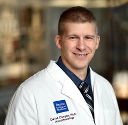 David J Durgan合作论文数46
 Lloyd Eric合作论文数27Bhanu P Ganesh合作论文数22Louise D McCullough合作论文数18
 Sean P Marrelli合作论文数18
 Pedram Honarpisheh合作论文数Univ Texas Hlth Sci Ctr Houston, McGovern Med Sch, Dept Neurol, 6431 Fannin St, Houston, TX 77030 USA12James Nelson合作论文数11Maria P Blasco Conesa合作论文数10Crossland Randy F合作论文数10Pandit Lavannya M合作论文数10Andresen Jon合作论文数9
 Joseph Petrosino合作论文数Center for Metagenomics and Microbiome Research, Baylor College of Medicine;Dan L Duncan Comprehensive Cancer Center, Baylor College of Medicine9Sharon Phillips合作论文数9Bojun Zhang合作论文数7
 Nadim Ajami合作论文数The University of Texas MD Anderson Cancer Center7
 Frank Blixt合作论文数Department of Clinical Sciences, Lund University7
 Sriram Ayyaswamy合作论文数Texas Children's Hospital, Baylor College of Medicine7Nadeem I. Shafi合作论文数6
 John d'Aigle合作论文数6
 Huanan Shi合作论文数Baylor Coll Med, Dept Anesthesiol, Houston, TX 77030 USA6Chauhan Anjali合作论文数Neuropharmacology Laboratory, All India Institute of Medical Sciences6Maniskas Michael合作论文数6Elke M. Golding合作论文数6Venugopal Reddy Venna合作论文数5
 Julia O Reynolds合作论文数5
 Juneyoung Lee合作论文数Department of Biostatistics, College of Medicine, Korea University5Louise McCullough合作论文数4G. Taffet合作论文数4Darius F. Miranda-Sohrabji合作论文数4Pedram Peesh合作论文数4Janelle M Korf合作论文数4Melanie G Kitagawa合作论文数4William Won合作论文数4Anik Banerjee合作论文数4Reddy Anilkumar K合作论文数4
 Claudia Sue Robertson合作论文数Department of Neurosurgery, Baylor College of Medicine4
 Nagireddy Putluri合作论文数Department of Molecular and Cell Biology, Baylor College of Medicine;Dan L Duncan Comprehensive Cancer Center, Baylor College of Medicine4Tan Chunfeng合作论文数4
 Craig J. Hartley合作论文数Baylor College of Medicine4Khodadad Namiranian合作论文数School of Medicine, Tehran University of Medical Sciences4
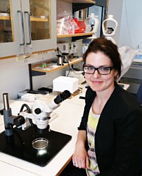 Hilda Ahnstedt合作论文数Department of Clinical Sciences, Lund University4m keith wilkerson合作论文数3Kyung Ae Ko合作论文数3
 Paul A Rogers合作论文数3Romeesa Khan合作论文数3Parisa Honarpisheh合作论文数3
 Rhonda Prisby合作论文数Kinesiology, University of Texas at Arlington3Emily Wilson合作论文数3




