Synthetic Computed Tomography (sCT) is required to provide electron density information for MR-only radiotherapy. Deep-learning (DL) methods for sCT generation show improved dose congruence over other sCT generation methods (e.g. bulk density). Using 30 female pelvis datasets to train a cycleGAN-inspired DL model, this study found mean dose differences between a deformed planning CT (dCT) and sCT were 0.2 % (D98 %). Three Dimensional Gamma analysis showed a mean of 90.4 % at 1 %/1mm. This study showed accurate sCTs (dose) can be generated from routinely available T2 spin echo sequences without the need for additional specialist sequences.
AIMS:Variability in the target and organs at risk (OARs) in cervical cancer treatment presents challenges for precise radiotherapy. Adaptive radiotherapy (ART) offers the potential to enhance treatment precision and outcomes. However, the increased workload and a lack of consensus on the most suitable ART approach hinder its clinical adoption. This systematic review aims to assess the current use of adaptive strategies for cervical cancer and define the optimal approach. MATERIALS AND METHODS:A systematic review of current literature published between January 2012 and May 2023 was conducted. Searches used PubMed/Medline, Cochrane Library, and Web of Science databases, supplemented with the University of Manchester, Google Scholar, and papers retrieved from reference lists. The review assessed workflows, compared dosimetric benefits, and examined resources for each identified strategy. Excluded were abstracts, conference abstracts, reviews, articles unrelated to ART management, proton therapy, brachytherapy, or qualitative studies. A narrative synthesis involved data tabulation, summarizing selected studies detailing workflow for cervical cancer and dosimetric outcomes for targets and OARs. RESULTS:Sixteen articles met the inclusion criteria; these were mostly retrospective simulation planning studies, except four studies that had been clinically implemented. We identified five approaches for ART radiotherapy for cervical cancer: reactive and scheduled adaptation, internal target volume (ITV)-based approach using library of plans (LOP), fixed-margin approach using LOP, and real-time adaptation, with each approach reducing irradiated volumes without compromising target coverage compared to the non-ART approach. The LOP-based ITV approach is the most used and clinically assessed. CONCLUSION:Identifying the optimal strategy is challenging due to dosimetric assessment limitations. Implementing cervical cancer ART necessitates strategic optimization of clinical benefits and resources through research, including studies to identify the optimal frequency, and prospective evaluations of toxicity.
Introduction This work reports on a systematic approach to select MRI sequences, quantify inter-observer image registration variation and determine patient positioning for the clinical implementation of MR-guided adaptive radiotherapy (MRgRT) in patients with oropharyngeal (H&N) and lung cancer. Methods A total of 30 participants (N=10 H&N and N=10 lung cancer patients and N=10 healthy participants) were scanned on the Elekta Unity Magnetic Resonance Linear Accelerator (MRL). Participant experience questionnaires were used to determine the most appropriate positioning device for lung treatments and tolerability of H&N immobilization devices within the confined MR Linac environment. Visual guided assessments (VGAs) completed by three observers (one oncologist and two radiographers) were used to determine the most suitable tissue weighting (using vendor-provided 3D T1w and T2w sequences) for online image registration. Offline MRI to CT and MRI to MRI rigid registrations were undertaken by nine radiographers using bony and soft tissue matching. Single-factor ANOVA and paired t-tests were utilized to determine the interobserver variation. Results Based on oncologist and patient feedback, lung cancer patients would be treated in a vac-bag with their arms by their sides, while H&N cancer patients would be immobilized using a 5-point fixation device and 5-point personalized thermoplastic shell. There was no clear preference for T1w or T2w images in the H&N cohort. However, observers preferred T2w sequences for tumour and organ at risk (OAR) visualization in the lung images. When a bony match was conducted, single-factor ANOVA tests showed no statistically significant differences between all H&N image registration types (p=0.09). For the soft-tissue registrations, T1w-CT and T1w-T1w registrations showed a statistically significant (p=0.01) reduction in inter-observer variability over T2w-CT registrations. Paired t-tests showed no statistically significant differences for bony or soft tissue matches using T1w or T2w sequences to the planning CT in the lung cohorts (p=0.63 and p=0.52, respectively). Conclusion We describe the systematic approach to the selection of strategies for imaging, immobilization, and online image registration we used for H&N and lung cancer treatments on the MRL. This has facilitated the selection of the most appropriate adaptive MRgRT strategies for treating these sites at our institution.
Purpose/ObjectiveTo determine the impact of a MR-based contouring atlas for male pelvis radiotherapy delineation on inter-observer variation to support radiographer led real-time magnetic resonance image guided adaptive radiotherapy (MRgART).Material/MethodsEight RTTs contoured 25 MR images in the Monaco treatment planning system (Monaco 5.40.01), from 5 patients. The prostate, seminal vesicles, bladder, and rectum were delineated before and after the introduction of an atlas developed through multi-disciplinary consensus.Inter-observer contour variations (volume), time to contour and observer contouring confidence were determined at both time-points using a 5-point Likert scale. Descriptive statistics were used to analyse both continuous and categorical variables. Dice similarity coefficient (DSC), Dice-Jaccard coefficient (DJC) and Hausdorff distance were used to calculate similarity between observers.ResultsAlthough variation in volume definition decreased for all structures among all observers post intervention, the change was not statistically significant. DSC and DJC measurements remained consistent following the introduction of the atlas for all observers. The highest similarity was found in the bladder and prostate whilst the lowest was the seminal vesicles. The mean contouring time for all observers was reduced by 50% following the introduction of the atlas (53 to 27 minutes, p=0.01). For all structures across all observers, the mean contouring confidence increased significantly from 2.3 to 3.5 out of 5 (p=0.02).ConclusionAlthough no significant improvements were observed in contour variation amongst observers, the introduction of the consensus-based contouring atlas improved contouring confidence and speed; key factors for a real-time RTT-led MRgART.
A feasibility study has been performed to investigate the viability of applying auto-segmentation methods to the delineation of regions of interest (ROIs) in the treatment of cervical cancer using Image Guided Brachytherapy (IGBT). The introduction of auto-segmentation in IGBT aims to improve outlining consistency while improving patient experience by reducing the time taken to plan treatments. An anonymised database of MRI images and corresponding clinical ROI outlines was curated, categorised by brachytherapy treatment applicator type. This database was then used to train and test an auto-segmentation model to contour the Bladder using three established algorithms, U-Net, SegNet and PSPNet. Quantitatively the U-Net model was found to produce contours geometrically closest to the original manual contours with a mean Dice Similarity Coefficient (DSC) of 0.942 compared to 0.919 and 0.879 for SegNet and PSPNet respectively and a mean Mean Distance to Agreement (mDTA) value of 0.46mm compared to 0.66mm and 0.89mm for SegNet and PSPNet. Visual assessment of the resulting contours demonstrated good agreement for the U-Net and SegNet produced outlines, particularly in the region of clinical significance, with greater variations seen at the extremities of the contour. In conclusion this feasibility study has shown that auto-segmentation methods can be applied to MRI IGBT contour delineation with a method established to facilitate further investigations in the application to all clinical ROIs and brachytherapy applicator types.
Background and purpose: Treatment planning for MR-guided stereotactic body radiotherapy (SBRT) for pancreatic tumors can be challenging, leading to a wide variation of protocols and practices. This study aimed to harmonize treatment planning by developing a consensus planning protocol for MR-guided pancreas SBRT on a 1.5 T MRLinac. Materials and methods: A consortium was founded of thirteen centers that treat pancreatic tumors on a 1.5 T MRLinac. A phased planning exercise was conducted in which centers iteratively created treatment plans for two cases of pancreatic cancer. Each phase was followed by a meeting where the instructions for the next phase were determined. After three phases, a consensus protocol was reached. Results: In the benchmarking phase (phase I), substantial variation between the SBRT protocols became apparent (for example, the gross tumor volume (GTV) D99% ranged between 36.8 - 53.7 Gy for case 1, 22.6 - 35.5 Gy for case 2). The next phase involved planning according to the same basic dosimetric objectives, constraints, and planning margins (phase II), which led to a large degree of harmonization (GTV D99% range: 47.9-53.6 Gy for case 1, 33.9-36.6 Gy for case 2). In phase III, the final consensus protocol was formulated in a treatment planning system template and again used for treatment planning. This not only resulted in further dosimetric harmonization (GTV D99% range: 48.2-50.9 Gy for case 1, 33.5-36.0 Gy for case 2) but also in less variation of estimated treatment delivery times. Conclusion: A global consensus protocol has been developed for treatment planning for MR-guided pancreatic SBRT on a 1.5 T MR-Linac. Aside from harmonizing the large variation in the current clinical practice, this
Introduction This work reports on an unusual finding observed during image quality assessment in the preparation for the clinical implementation of breast magnetic resonance image-guided radiotherapy (MRIgRT) on a 1.5 Tesla (T) magnetic resonance linear accelerator (MR-Linac) (Elekta AB, Stockholm, Sweden). Case and outcomes A patient with T2 N0 M0 right breast invasive ductal carcinoma, receiving adjuvant radiotherapy, underwent two imaging sessions on the MR-Linac. The imaging protocol included T1- and T2-weighted (W) turbo spin echo (TSE) sequences, a T1W mDixon, and a T2W TSE navigated sequence acquired on end-expiration. All images were reconstructed in the axial plane.Images were assessed for image quality and appropriateness for use within the treatment pathway using visual grading analysis (VGA). An artefact in the right breast was noted independently by all observers. The patient's skin and medical notes were reviewed for possible explanation. The findings were discussed with the patient's responsible clinician, and subsequent referral to the local multi-disciplinary team (MDT) for radiologist review was made.On further investigation, the patient's images demonstrated a signal void in the subareolar region of the right breast coinciding with the surgical site. This was distal from the tumour bed and deemed unlikely to be related to a Magseed marker or intraoperative clips. The patient reported no history of nipple tattoo or piercing. There was nothing on clothing that this could be attributed to. Discussion Following MDT review, where all potential sources of signal void were considered, it was concluded that the cause was Magtrace, a superparamagnetic iron oxide tracer, recommended for sentinel lymph node localisation in patients with breast cancer in the United Kingdom. The artefact was characteristic of a magnetic susceptibility artefact. These can arise from local magnetic field inhomogeneities caused by the presence of the metal compounds in MagTrace. For breast MRIgRT on the MR-Linac, treatment verification and the possibility of real-time replanning is a critical aspect. The magnetic susceptibility artefact significantly inhibited plan adaption and confidence in the online image registration process making the patient ineligible for treatment on the MR-Linac. Conclusion As part of ongoing work-up for breast MRIgRT, the screening of patients for Magtrace is now included. Optimisation of MR imaging sequences for radiotherapy planning and image review to minimise distortion are being developed.
Background and purpose: Tumours in the liver often develop on a background of liver cirrhosis and impaired liver function. As a result, radiotherapy treatments are limited by radiation-induced liver disease, parameterised by the liver mean dose (LMD). Liver function is highly heterogeneous, especially in liver cancer, but the use of LMD does not take this into account. One possible way to improve liver treatments is to use quantitative imaging techniques to assess liver health and prioritise the sparing of healthy liver tissue. Materials and methods: Anatomical T2 and quantitative iron-corrected T1 (cT1) images were made available for 10 patients with liver metastases. Functional liver volumes were automatically segmented on the quantitative images using a threshold. Liver stereotactic ablative body radiotherapy (SABR) plans were made using a departmental protocol. Liver-sparing plans were then made by reducing the dose to the functional sub-volume. Results: The sparing plans achieved a statistically significant (p=0.002) reduction in the functional liver mean dose, with a mean reduction of 1.4 Gy. The LMD was also significantly different (p=0.002) but had a smaller magnitude with a mean reduction of 0.7 Gy. There were some differences in the planning target volume D99% (p=0.04) but the sparing plans remained within the optimal tolerance and the D95% was not significantly different (p=0.2). Conclusions: This study has, for the first time, demonstrated the use of cT1 maps in radiotherapy showing significant reductions in dose to the healthy liver. Further work is needed to validate this in liver cancer patients, who would likely benefit most.
The impact of fat on abdominal compression effectiveness in abdominal cancers was determined using magnetic resonance imaging (MRI). Visceral and subcutaneous fat were delineated on T2W 3D MRI, and motion change with compression was measured on 2D cine MRI. Results from 16 participants showed no correlation between fat percentage, body mass index (BMI), and motion change. Median BMI was 28.7 (SD, 4.9). Mean motion reduction was 7.8 mm (IQR, 5.0; p = 0.001) with compression. While no direct link was found between fat, BMI, and compression effectiveness, abdominal compression remains crucial for motion management in radiotherapy planning, providing dosimetric benefits.
Purpose: To investigate whether a novel signal derived from tumor motion allows more precise sorting of 4D-magnetic resonance (4D-MR) image data than do signals based on normal anatomy, reducing levels of stitching artifacts within sorted lung tumor volumes.Methods: (4D-MRI) scans were collected for 10 lung cancer patients using a 2D T2-weighted single-shot turbo spin echo sequence, obtaining 25 repeat frames per image slice. For each slice, a tumor-motion signal was generated using the first principal component of movement in the tumor neighborhood (TumorPC1). Signals were also generated from displacements of the diaphragm (DIA) and upper and lower chest wall (UCW/LCW) and from slice body area changes (BA). Pearson r coefficients of correlations between observed tumor movement and respiratory signals were determined. TumorPC1, DIA, and UCW signals were used to compile image stacks showing each patient's tumor volume in a respiratory phase. Unsorted image stacks were also built for comparison. For each image stack, the presence of stitching artifacts was assessed by measuring the roughness of the compiled tumor surface according to a roughness metric (Rg). Statistical differences in weighted means of Rg between any two signals were determined using an exact permutation test.Results: The TumorPC1 signal was most strongly correlated with superior inferior tumor motion, and had significantly higher Pearson r values (median 0.86) than those determined for correlations of UCW, LCW, and BA with superior-inferior tumor motion (p < 0.05).Weighted means of ratios of Rg values in TumorPC1 image stacks to those in unsorted, UCW, and DIA stacks were 0.67, 0.69, and 0.71, all significantly favoring TumorPC1 (p = 0.02-0.05). For other pairs of signals, weighted mean ratios did not differ significantly from one.Conclusion: Tumor volumes were smoother in 3D image stacks compiled using the first principal component of tumor motion than in stacks compiled with signals based on normal anatomy.
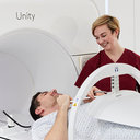
Using a 4-stage non-comparative prospective feasibility study has facilitated clinical implementation MRIgRT of multiple treatment sites at our institution.
 McWilliam Alan合作论文数35
 Marcel Van Herk合作论文数Division of Cancer Sciences, School of Medical Sciences, The University of Manchester28
 Cynthia L Eccles合作论文数Faculty of Biology, Medicine and Health, The University of Manchester23
 Ananya Choudhury合作论文数Cancer Research UK Clinical Centre, Leeds Institute of Molecular Medicine23
 Peter Hoskin合作论文数Mount Vernon Cancer Centre;Cancer Research UK Manchester Centre, University of Manchester;Christie Hospital;University College Hospital14
 Emma Jane Bradshaw合作论文数13
 Omar Almaini合作论文数School of Physics and Astronomy, The University of Nottingham;Faculty of Science, The University of Nottingham13
 William G. Hartley合作论文数ETH;Université de Genève;University College London13C. Nelder合作论文数11P. Whitehurst合作论文数11
 Ranald I Mackay合作论文数11
 Christopher J. Conselice合作论文数School of Physics and Astronomy, University of Manchester;Alan Turing Institute;University of Padova11
 Michele Cirasuolo合作论文数European Southern Observatory11
 Ross McLure合作论文数School of Physics and Astronomy, The University of Edinburgh11Eliana Vasquez Osorio合作论文数Division of Cancer Sciences, School of Medical Sciences, Faculty of Biology, Medicine and Health, The University of Manchester10
 Corinne Faivre-Finn合作论文数Institute of Cancer Sciences, University of Manchester10
 James Dunlop合作论文数Institute for Astronomy, School of Physics and Astronomy, University of Edinburgh10
 Jane Shortall合作论文数Division of Cancer Sciences, School of Medical Sciences, Faculty of Biology, Medicine and Health, The University of Manchester9
 Chris Simpson合作论文数Gemini Observatory9
 Sebastien Foucaud合作论文数9
 D.C.P. Cobben合作论文数the Groningen University Hospital, University Hospital9L. McDaid合作论文数8A. Clough合作论文数8Andrew Green合作论文数Division of Cancer Sciences, School of Medical Sciences, Faculty of Biology, Medicine and Health, The University of Manchester8
 Marianne Aznar合作论文数Division of Cancer Sciences, University of Manchester8
 Katie Lichter合作论文数Department of Radiation Oncology, University of California San Francisco7Alice Mortlock合作论文数7Ganesh Radhakrishna合作论文数7L. Freear合作论文数6h j pearce合作论文数6
 M. Daly合作论文数Div Canc Sci, Univ Manchester6R. Benson合作论文数6Michael Dubec合作论文数6Speight Richard J合作论文数6Gareth Price合作论文数6
 Fernando Buitrago合作论文数School of Physics;University of Nottingham;School of Physics, University of Nottingham5J. Berresford合作论文数5Claire Nelder合作论文数4Abigael Clough合作论文数4
 A. Cree合作论文数Clatterbridge Canc Ctr4
 Rosie Beech Hales合作论文数4Rebecca Benson合作论文数4Lisa McDaid合作论文数4
 Andrew Mcpartlin合作论文数The Christie NHS Foundation Trust4Philip Whitehurst合作论文数4
 Ann Henry合作论文数School of Medicine, University of Leeds;St James's Institute of Oncology, University of Leeds4Ruth Grutzbauch合作论文数4
 Asa Bluck合作论文数4




