Purpose: To report a rare complication of pneumatic retinopexy involving intraocular gas entrapment in the canal of Petit and its successful noninvasive management. Methods: A single case was reviewed. Results: A 49-year-old woman underwent pneumatic retinopexy for a macula-on rhegmatogenous retinal detachment and subsequently developed a distinctive incomplete “donut-shaped” peripheral gas collection consistent with entrapment in the canal of Petit. With strict prone face-down positioning, the gas migrated posteriorly into the vitreous cavity within 50 min, as documented on sequential ultra-widefield imaging. The retina remained reattached without the need for any surgical intervention. Conclusions: This case highlights that gas entrapment in the canal of Petit after pneumatic retinopexy may resolve with positioning alone and underscores the utility of serial ultra-widefield imaging for diagnosis and monitoring, potentially preventing unnecessary surgical intervention.
PURPOSE:To review current and emerging biomaterials in retinal surgery - vitreous tamponade agents, retinal prostheses, retinal pigment epithelium (RPE) transplant scaffolds, intraocular drug delivery platforms, and gene therapy vectors - with emphasis on the authors' oxime-crosslinked hyaluronic acid vitreous substitute. DESIGN:Narrative expert review. Systematic review methodology was not used; no protocol registration, pre-specified eligibility criteria, dual independent screening, or PRISMA flow diagram was undertaken. SUBJECTS, PARTICIPANTS, AND/OR CONTROLS:Published preclinical animal studies, Phase I-III clinical trials, US Food and Drug Administration approvals, and real-world evidence on biomaterials in posterior segment surgery. No individual patient data were accessed. METHODS, INTERVENTION, OR TESTING:A structured literature search of PubMed/MEDLINE, supplemented by Google Scholar, was performed from January 2018 to March 2026 across five domains: vitreous tamponade agents; retinal prostheses; scaffold-based RPE transplantation; nanoparticle and sustained-release drug delivery; and gene therapy vectors. Peer-reviewed investigations, trial reports, and regulatory data were prioritized. The authors' preclinical hydrogel data are presented as original data within a review context. MAIN OUTCOME MEASURES:Best-corrected visual acuity, intraocular pressure, electroretinography, retinal microstructure on optical coherence tomography, prosthetic visual acuity, ellipsoid zone area loss, injection frequency, and in vivo biocompatibility. RESULTS:The PRIMA photovoltaic subretinal implant produced a clinically meaningful improvement in visual acuity in 26 of 32 patients (81%) with geographic atrophy at 12 months. A 44-channel suprachoroidal prosthesis showed no device-related serious adverse events at 2.7 years. The authors' oxime-crosslinked hyaluronic acid hydrogel demonstrated density 1.01 g/mL and refractive index 1.356, with preserved electroretinography and intraocular pressure of 13.5-25.9 mmHg in rabbits. Scaffold-based RPE transplantation has entered Phase I/IIa trials. 2025 approvals included a refillable ranibizumab port delivery system for diabetic retinopathy and the first encapsulated cell therapy implant for macular telangiectasia type 2. Three Phase 3 anti-VEGF gene therapy trials are ongoing. CONCLUSIONS:Biomaterial engineering is transforming retinal disease management from stabilization toward tissue replacement and vision restoration. Principal limitations are the preclinical status of the hydrogel data and early-phase status of RPE scaffold trials. Outstanding challenges include long-term in vivo stability, immunological management of allogeneic constructs, scalable manufacturing, and definitive efficacy trials.
INTRODUCTION:Vitreous substitutes are crucial adjuncts during vitreoretinal surgery for retinal detachment, macular holes, and vitreous hemorrhage. However, current vitreous substitutes pose limitations, including but not limited to their postoperative decrease in visual sharpness, nonfavorable refractive indices, and complications such as glaucoma, cataract, and retinal toxicity. METHODS:We conducted a systematic review to evaluate the biological, physical, and clinical properties of novel experimental vitreous substitutes in animal and human models. We performed a systematic search on Ovid Medline, Embase, and Scopus from January 1, 1990, to October 10, 2025. Studies were included if they reported on any of the following three aspects: optical properties, biological interactions, or clinical outcomes. RESULTS:Out of 11,045 search results, 37 studies investigating novel hydrogels (n = 15), polymers (n = 5), liquids (n = 5), and devices (n = 12) were included. Among these, 16/37 substitutes had > 80% success in tamponade of retinal detachments, 12/37 showed high tissue biocompatibility, 15/37 demonstrated low ocular inflammation, 13/37 exhibited optical properties similar to native vitreous, and 14/37 were associated with no significant changes in intraocular pressure. CONCLUSIONS:Novel experimental vitreous substitutes, such as UV-CHA, Vitargus ABV-1701, PanaceaGel SPG-178, E10KDC18, or FCVB, show the most promising biological, optical, and physical properties for additional investigation as vitreous substitutes.
Introduction: Anecdotal reports and limited reports suggest a possible link between activities involving rapid acceleration and retinal detachment. We present two novel such cases and review existing literature to investigate the plausibility of this association and delineate in what populations such an association may be more likely. Case Presentation: We report 2 cases of retinal detachment following roller coaster riding. The first, a 24-year-old woman with a family history of retinal detachment, presented with floaters after consecutive rides and was found to have an inferior temporal macula-sparing retinal detachment with associated retinal breaks. The second case, a 25-year-old female with a history of high myopia, presented with visual field defect and was found to have a macula-on retinal detachment with an accompanying tear at the edge of an area of lattice degeneration. Both were successfully treated with pneumatic retinopexy followed by laser retinopexy. Conclusion: Rapid acceleration/deceleration forces, such as those experienced on roller coasters, could potentially lead to retinal detachment. Structural predisposition is likely necessary for acceleration/deceleration injury to lead to retinal detachment, with all known cases having risk factors, including high myopia and positive family history. These same forces in eyes without structural predisposition have resulted in hemorrhage, but not detachment.
Purpose: Morvan syndrome (MoS) is a neurologic disorder belonging to a spectrum of autoimmune encephalitis, Contactin-associated protein-like 2 (Caspr2) antibody syndrome. We report a case of bilateral panuveitis associated with MoS. Methods: Case report and review of the literature. Results: A 57-year-old male with Morvan syndrome presented with painless vision loss and floaters. Initial visual acuities were 20/50 and 20/60. Hallmarks of this uveitis included persistently active vitritis, and nonhemorrhagic retinitis with nonperfusion and neovascularization. Uveitis consistently flared with attempted immunosuppressive tapers. Vision deteriorated to count fingers (2 ft) OU over 2.5 years despite corticosteroids, mycophenolate mofetil, intravenous immunoglobulin, adalimumab, and rituximab. Explanations for reduced final visual acuity included macular atrophy, disruption of retinal architecture, epiretinal membrane, vitritis, and cataract. Conclusions: This case constitutes the first report of uveitis associated with MoS and Caspr2 antibody syndrome, raising the question of autoimmunity targeting the retinal inner and/or outer plexiform layers.
This study reports the repeatability of 20 s-duration fixation stability measurements recorded with the Macular Integrity Assessment (MAIA) microperimeter in patients with central vision loss, in contrast to the Nidek MP1 microperimeter. Fixation stability was recorded in 39 eyes of 25 patients with macular disease using MAIA and the MP1 for 20 s intervals, twice for each eye, with each instrument. Twenty eyes were identified as the better eye (BE) and 19 eyes as the worse eye (WE). Fixation stability was quantified with the 95
Background: The purposes of this pilot retrospective observational cohort study are to determine: 1) the safety and efficacy of corneal photovitrification (CPV), a corneal laser procedure, for vision improvement in patients with neovascular age-related macular degeneration (nAMD) and other retinal disorders involving central vision loss, and 2) the correlation between post-treatment (post-treatment (Tx)) best-corrected distance visual acuity (BCDVA) and pre-Tx potential visual acuity (PVA). Methods: BCDVA measurements using ETDRS charts and PVA measurements using Gonzalez-Markowitz PVA charts were analyzed for 72 eyes that received a single CPV treatment. Results: Safety - No clinically significant complications or serious adverse events occurred. Efficacy – Mean BCDVA improved significantly from 20/303 (1.18 log of the minimum angle of resolution (logMAR), 26 letters) at pre-Tx to 20/208 (1.02 logMAR, 34 letters) at 1m post-Tx (p = 0.000001) for a cohort of 57 eyes, improving to 20/198 (1.00 logMAR, 35 letters; 11.6 mean letters gained) at 12m (p = 0.0009) for a cohort of 29 eyes. Mean pre-Tx PVA gains correlated well with mean BCDVA improvements at 1m, 3m, 6m and 12m post-Tx. Conclusions: Subject to a small sample size, the CPV corneal laser procedure is safe and efficacious for vision improvement in patients with nAMD and other retinal disorders involving central vision loss. The pre-Tx PVA correlates well with post-Tx BCDVA; the PVA test may be useful for screening of candidate eyes for CPV treatment and for managing expectations.
PURPOSE:To compare the efficacy of topical nepafenac 0.1% versus intravitreal triamcinolone acetonide (IVTA) at the conclusion of vitrectomy surgery versus no adjuvant therapy (NAT) in improving macular morphology post-operatively in patients undergoing vitrectomy for epiretinal membrane (ERM), as measured by optical coherence tomography (OCT) imaging and best-corrected visual acuity (BCVA).METHODS:Design: Prospective randomized clinical trial Setting: Multi-centre 80 patients scheduled to undergo vitrectomy surgery for idiopathic ERM were randomized to receive either IVTA (4 mg/0.1 cc) at the end of surgery, topical nepafenac sodium 0.1% TID for 1 month post-operation or no adjuvant treatment (NAT). Optical coherence tomography (OCT) imaging, best-corrected visual acuity and intraocular pressure (IOP) were measured before surgery, and 1 and 2 months post-operation.RESULTS:Although all three groups showed reduction in macular thickness post-operation, the NAT group showed the most improvement, with a reduction of 136.18 ± 29.84 μm at two months. There was no statistically significant difference in macular thickness between the groups at each time point, p = 0.158. The NAT group also had the best recovery in BCVA with an improvement of 0.207 logMAR (10.35 letters) at two months post-operation. There was no statistically significant difference in BCVA between the groups, p = 0.606. There was statistically significant difference in the IOP between the three groups, p = 0.04 only at 1-month visit. The IVTA group had the highest rise in average IOP at both 1 and 2 months post-operation (2.72 and 1.58 mmHg, respectively).CONCLUSION:Our study data suggest there was no advantage in the use of topical nepafenac or IVTA for post-vitrectomy ERM surgery.
Vitreous substitutes are clinically used to maintain retinal apposition and preserve retinal function; yet the most used substitutes are gases and oils which have disadvantages including strict face-down positioning post-surgery and the need for subsequent surgical removal, respectively. We have engineered a vitreous substitute comprised of a novel hyaluronan-oxime crosslinked hydrogel. Hyaluronan, which is naturally abundant in the vitreous of the eye, is chemically modified to crosslink with poly(ethylene glycol)-tetraoxyamine via oxime chemistry to produce a vitreous substitute that has similar physical properties to the native vitreous including refractive index, density and transparency. The oxime hydrogel is cytocompatible in vitro with photoreceptors from mouse retinal explants and biocompatible in rabbit eyes as determined by histology of the inner nuclear layer and photoreceptors in the outer nuclear layer. The ocular pressure in the rabbit eyes was consistent over 56 d, demonstrating limited to no swelling. Our vitreous substitute was stable in vivo over 28 d after which it began to degrade, with approximately 50% loss by day 56. We confirmed that the implanted hydrogel did not impact retina function using electroretinography over 90 days versus eyes injected with balanced saline solution. This new oxime hydrogel provides a significant improvement over the status quo as a vitreous substitute.
PURPOSE:There are conflicting reports as to whether there is a binocular advantage or disadvantage when reading with central vision loss. This study examined binocular reading summation in patients with macular degeneration.METHODS:Seventy-one patients with bilateral central vision loss [mean age: 63 (S.D. = 21) years] participated. Reading performances during binocular and monocular viewing with the better eye (i.e., the eye with the best monocular visual acuity) were evaluated using different versions of the Italian MNREAD reading chart (www.precision-vision.com). Fixation stability and preferred retinal loci (PRLs) were recorded monocularly for each eye. The overall sample was split into inhibition, equality, and summation groups based on the binocular ratio (i.e., binocular/monocular) of the maximum reading speed.RESULTS:41% of patients experienced binocular inhibition, 42% summation, and 17% equality. Binocular reading speed of the inhibition group was approximately 30 words per minute slower than those of the equality and summation groups, although the inhibition group had the best visual acuity. These patients generally had monocular PRLs in non-corresponding locations temporal or nasal to the scotoma, had the largest interocular acuity difference and lacked residual stereopsis. The three groups did not differ in fixational control, contrast sensitivity or critical print size.CONCLUSIONS:Equal proportions of patients with central vision loss show binocular reading summation and inhibition. Patients with binocular reading inhibition have poorer reading performance and different clinical characteristics than those with binocular reading summation and equality.
Purpose: To analyze and provide an overview of the incidence, management, and prevention of conjunctival erosion in Argus II clinical trial subjects and postapproval patients. Methods: This retrospective analysis followed the results of 274 patients treated with the Argus II Retinal Prosthesis System between June 2007 and November 2017, including 30 subjects from the US and European clinical trials, and 244 patients in the postapproval phase. Results were gathered for incidence of a serious adverse event, incidence of conjunctival erosion, occurrence sites, rates of erosion, and erosion timing. Results: Overall, 60% of subjects in the clinical trial subjects versus 83% of patients in the postapproval phase did not experience device- or surgery-related serious adverse events. In the postapproval phase, conjunctival erosion had an incidence rate of 6.2% over 5 years and 11 months. In 55% of conjunctival erosion cases, erosion occurred in the inferotemporal quadrant, 25% in the superotemporal quadrant, and 20% in both. Sixty percent of the erosion events occurred in the first 15 months after implantation, and 85% within the first 2.5 years. Conclusion: Reducing occurrence of conjunctival erosion in patients with the Argus II Retinal Prosthesis requires identification and minimization of risk factors before and during implantation. Implementing inverted sutures at the implant tabs, use of graft material at these locations as well as Mersilene rather than nylon sutures, and accurate Tenon's and conjunctiva closure are recommended for consideration in all patients.
PURPOSE:The LIGHTSITE I study investigated the efficacy and safety of photobiomodulation (PBM) treatment in subjects with dry age-related macular degeneration. METHODS:Thirty subjects (46 eyes) were treated with the Valeda Light Delivery System, wherein subjects underwent two series of treatments (3× per week for 3-4 weeks) over 1 year. Outcome measures included best-corrected visual acuity, contrast sensitivity, microperimetry, central drusen volume and drusen thickness, and quality of life assessments. RESULTS:Photobiomodulation-treated subjects showed a best-corrected visual acuity mean letter score gain of 4 letters immediately after each treatment series at Month 1 (M1) and Month 7 (M7). Approximately 50% of PBM-treated subjects showed improvement of ≥5 letters versus 13.6% in sham-treated subjects at M1. High responding subjects (≥5-letter improvement) in the PBM-treated group showed a gain of 8 letters after initial treatment (P < 0.01) and exhibited earlier stages of age-related macular degeneration disease. Statistically significant improvements in contrast sensitivity, central drusen volume, central drusen thickness, and quality of life were observed (P < 0.05). No device-related adverse events were reported. CONCLUSION:Photobiomodulation treatment statistically improved clinical and anatomical outcomes with more robust benefits observed in subjects with earlier stages of dry age-related macular degeneration. Repeated PBM treatments are necessary to maintain benefits. These pilot findings support previous reports and suggest the utility of PBM as a safe and effective therapy in subjects with dry age-related macular degeneration.
The Argus II retinal prosthesis (Second Sight Medical Products, Sylmar, CA, USA) is an electronic epiretinal implant designed for use in patients with end-stage retinitis pigmentosa, as well as for other outer retinal degenerative conditions, such as choroideremia, Leber’s congenital amaurosis, Bardet-Biedl syndrome, Usher syndrome and rod–cone disease.1 This device was tested in a clinical trial of 30 subjects in the USA and in Europe to measure the rate of serious adverse events (SAEs) and to assess its effects on functional vision and visual acuity.2–4 Subjects performed better on visual tasks with the device turned on and retained these improvements at 5 years postsurgery.4 Additionally, an acceptable safety profile enabled approval of the device for consumer use by the European regulatory authorities in 2011 (Conformite Europeenne (CE) mark) and by the US Food and Drug Administration (FDA) in 2013 (FDA approval). The implant is composed of an extraocular component, namely, an electronics case and implant coil, as well as an intraocular component involving a 60-electrode array that is tacked over the macula (figure 1). The extraocular and intraocular components are connected by a trans-scleral silicone-coated polyamide electrode cable, and the implantation procedure involves a 5.2 mm pars plana sclerotomy incision to insert the electrode array and the electrode cable into the eye. The sclerotomy is then sutured around the cable and covered by an allograft or autograft material.5 Figure 1 The Argus II retinal prosthesis. Labelled image of the Argus II implant (adapted from Humayun et al 2). As with any system having both intraocular and extraocular components, the trans-scleral electrode cable that traverses the sclerotomy permanently is a high-risk feature of the device, as leakage through the pars plana incision can result in chronic hypotony. The percentages of clinical trial subjects who experienced hypotony were 10.0% at …
PURPOSE:To examine the relationship between retinal detachment and retrobulbar cysts in patients with optic nerve coloboma (ONC) and Morning Glory syndrome (MGS).METHODS:Patients diagnosed with either ONC or MGS were identified through a search of the Sick Kids database. Seventy-one patients either agreed to come in for a B-scan or had an incidental orbital B-scan or magnetic resonance imaging or both. Eyes with orbital B-scan ultrasound and/or magnetic resonance imaging images were assessed independently by two ophthalmologists and a radiologist for the presence of retrobulbar cysts. Retinal detachment was identified clinically with either indirect ophthalmoscopy or from fundus photographs.RESULTS:Forty-five of 71 (63%) and 26/71 (37%) patients had ONC and MGS, respectively. Retinal detachment occurred significantly more often in eyes with MGS than with ONC (9/17 [53%] vs. 5/45 [11%], P = 0.03, respectively). Retrobulbar cysts were not detected more often in MGS than in ONC (11/45 [24%] vs. 7/26 [27%]; P = 1.0). Eyes with retrobulbar cysts were significantly more likely to be associated with retinal detachment than those without (7/18 [39%] vs. 7/53 [13%]; P = 0.04).CONCLUSION:Retinal detachment occurs more frequently in MGS than in ONC in a cohort of patients referred to a specialist children's retinal service. Eyes with retrobulbar cysts are more likely to be associated with retinal detachment.
The Argus II retinal prosthesis is the first commercially available device for restoration of vision in patients with Retinitis Pigmentosa or with similar retinal pathology who still have minimal residual native vision. The technology is able to restore vision with production of artificial visual percepts which usually are given adequate useful interpretation by the visual system in most implanted patients. The technology usually produces visual perception at the level of shape identification or better in some cases enabling in many less dependence on vision substitution devices and skills. There is no consensus among vision rehabilitation practitioners on single methods for assessments, outcome measures and training, yet there is constant progress in these areas of concern. Hence the current vision rehabilitation practice related to the implantation of the Argus II retinal prosthesis is a work in progress with many learning opportunities for all involved. All agree that implementation of this technology in clinical practice requires the combined work of a multi-disciplinary team which includes a specialized surgical team as well as a specialized rehabilitation team in order to obtain optimal results. Our own experience is presented in this paper and indicates so far that the Argus II technology is beneficial to patients and that it could be successfully managed within the Canadian heath care system.
 Chris Hudson合作论文数School of Optometry and Vision Science, University of Waterloo;Department of Ophthalmology and Vision Science, University of Toronto18
 John G. Flanagan合作论文数Herbert Wertheim School of Optometry & Vision Science, University of California, Berkeley15
 WC LAM合作论文数12
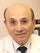 Samuel N. Markowitz合作论文数University of Toronto12
 Tien Yin Wong (黄天荫)合作论文数School of Clinical Medicine, Tsinghua University12Wai Ching Lam合作论文数10
 Michael H Brent合作论文数Department of Ophthalmology and Vision Sciences, Temerty Faculty of Medicine, University of Toronto10
 Peter Kertes合作论文数Department of Ophthalmology & Vision Sciences, University of Toronto10Mark Mandelcorn合作论文数10Kit Guan合作论文数8r k nrusimhadevara合作论文数7m kisilevsky合作论文数6Mandelcorn Mark S合作论文数6Berger Alan合作论文数6
 Weiqing Lin (林伟青)合作论文数Li Ka Shing Faculty of Medicine, The University of Hong Kong5
 Jean-Francois Korobelnik合作论文数4Michael Walker合作论文数4
 Peng Yan合作论文数Department of Ophthalmology, University of Toronto4Puzyeyeva Olena合作论文数4Chaudhary Varun合作论文数4Stanislao Rizzo合作论文数3Ravi K. Nrusimhadevara合作论文数3Patricia T. Harvey合作论文数3Graham Merry合作论文数3
 Robert Dotson合作论文数3
 Suber Huang合作论文数3
 Mickael Dang合作论文数3
 David N. Zacks合作论文数Department of Ophthalmology and Visual Sciences, Kellogg Eye Center, University of Michigan;Retina & Uveitis Clinic, Kellogg Eye Center, University of Michigan3
 K. Thiran Jayasundera合作论文数Department of Ophthalmology and Visual Sciences, Kellogg Eye Center, Medical School, University of Michigan-Ann Arbor3
 Jennifer I. Lim合作论文数Department of Ophthalmology, College of Medicine, University of Illinois3Valerie A. Wallace合作论文数3
 Allen Chiang合作论文数3
 Robert J. Greenberg合作论文数3
 Molly Shoichet合作论文数Donnelly Centre for Cellular & Biomolecular Research, University of Toronto;PRiME3Edward D. Gilmore合作论文数3
 Paul Hahn合作论文数Ophthalmol, Duke Univ3Ninel Z. Gregori合作论文数Bascom Palmer Eye Institute, University of Miami3
 Luminita Tarita-Nistor合作论文数Vision Science Research Program, Toronto Western Hospital3
 Raymond Iezzi合作论文数3Alexander E. G. Baker合作论文数2Ramin Tadayoni合作论文数2Mark Rankin合作论文数2Sally Justus合作论文数2Beatriz E Patino合作论文数2
 Harneel Kaur合作论文数2Thomas Wright合作论文数2e palka合作论文数2Michael D Mills合作论文数2




