Understanding the biological mechanisms behind fracture nonunions and their treatments remains incomplete. Since these mechanisms are linked to phenotypic expression, studying the longitudinal metabolic profile of bone regeneration has emerged as a potential way to better understand healing progress and treatment effectiveness. In this study, we tracked the serum metabolome of rats with critically-sized femoral defects treated with either a control, a minimum effective dose, or a supraphysiologic dose of rhBMP-2 over time. We observed distinct changes in the metabolome across control, impaired healing, and rhBMP-2-treated animals. Early in the process, we saw increased levels of metabolites that support angiogenesis in the rhBMP-2 groups. Supraphysiologic doses of rhBMP-2 significantly affected metabolite expression, especially in pathways related to angiogenesis, fatty acid metabolism, and ATP production. These longitudinal trends and individual metabolite changes provide a deeper understanding of rhBMP-2's mechanisms, improve our knowledge of its biological effects—guiding appropriate dosing—and offer a metabolic profile that may indicate healing impairment.
Revascularization remains a challenge for regenerative medicine strategies. Extensive research has been done to identify key moments of the dynamic wound healing cascade where targeted therapies can elicit a proregenerative response. However, the influence of oxygenation, temperature, and their temporal variation during healing are often challenging to promote tissue regeneration. This study investigated the effects of temporally varied oxygenation and temperature conditions on angiogenesis using an in vitro model of rat-derived, intact microvascular fragments in a collagen type-I hydrogel. By generating culture conditions that are similar to the accepted wound healing time course, the angiogenic response depended critically on both the timing of stimulus initiation and the magnitude of deviation from model conditions. Dynamic stimuli activated distinct biological pathways, as evidenced by qPCR analysis, revealing mechanistic links between environmental perturbations and the angiogenic response. This work emphasizes the need for regenerative medicine strategies to consider varying environmental stimuli to improve revascularization outcomes.
Abstract Nonunion and delayed union, which occur in approximately 5–10% of major long bone fracture cases, significantly complicate treatment and negatively impact patients' quality of life. This results in the need for novel treatment strategies to enhance healing outcomes. Researchers have explored the role of fluid flow in stimulating bone cells and regulating bone regeneration; however, many of these studies are conducted in silico or in vitro. This study introduces a novel magnetohydrodynamic (MHD) device integrated into a bone fixation plate for investigating the effects of fluid flow on bone regeneration in vivo. An implantable MHD device was designed and fabricated to induce fluid flow in vivo. To validate the device's safety and biocompatibility, it was implanted in female Wistar rats upon the creation of a 2 mm unilateral segmental bone defect for 4 weeks. The results show complete device functionality throughout seven postinjury fluid flow treatments administered over 28 days. The device is designed to induce fluid flow of varying magnitudes directly at the fracture site, enabling investigation of its potential impact on bone healing. In addition, the device did not induce adverse effects such as fibrous encapsulation or tissue necrosis in vivo, supporting its potential for investigating fluid flow stimulation as a treatment approach for improving healing in delayed and nonunion fractures. The potential of a fluid flow treatment holds great significance for researchers, addressing a critical knowledge gap regarding the impact of fluid flow on healing calluses, while offering an alternative regimen for the elderly and those with multiple injuries.
Introduction Bone stress injuries (BSIs) are common overuse injuries in athletes and are associated with prolonged recovery and high rates of reinjury. Despite their clinical importance, there is no consensus on how to define or assess BSI healing. Clinical, functional and imaging outcomes for BSI healing are used inconsistently across studies, limiting comparability and the development of evidence-based return-to-sport guidelines. This study aims to identify and characterise candidate outcomes for assessing BSI healing in runners, including their time course and variability across function, imaging and clinical domains.Methods and analysis This is a 1 year, multi-site prospective cohort study of male and female runners with a recent MRI-confirmed BSI of the tibia, metatarsals, femoral neck or sacrum. We will enrol participants across four clinical and academic research sites in the USA and Canada, within 3 weeks of diagnostic MRI and follow them longitudinally for 1 year through recovery, return to sport and full sport participation. Candidate outcomes for BSI healing include time to pain-free functional milestones (eg, hopping, jogging), completion of a return-to-run protocol, medical clearance for unrestricted activity, and changes in MRI grade and clinical severity scores. Secondary and exploratory measures include training patterns, wearable-derived activity and sleep metrics, biomechanical assessments, and serum proteomic profiles. We will use descriptive statistics to characterise the time course and variability of candidate outcomes. We will explore associations between outcomes and subsequent BSIs within 1 year using regression models.Ethics and dissemination This study has received institutional review board approval at all participating institutions. We will obtain written informed consent from all participants prior to any study procedures (and assent with parental consent for minors). We will disseminate results through peer-reviewed publications and scientific conferences.
Bone morphogenetic proteins (BMPs) are among the most potent clinical therapeutics for promoting bone regeneration, yet their use is limited by potential adverse events such as heterotopic bone formation, osteolysis, and soft tissue inflammation. Sustained release delivery systems may reduce the minimum effective BMP dose needed to induce functional bone healing but have yet to be fully characterized or translated to clinical use. Here, we utilized a rat model of segmental femur defect healing to investigate the dose-dependent inflammatory effects of BMP delivery in a hydrogel-based system by profiling local and systemic immune responses. Defects were treated with the delivery vehicle only (control), a minimum effective dose of BMP-2, or a high dose of BMP-2. The minimum effective BMP-2 dose induced comparable bone formation within the defects but significantly less ossification outside the defect region relative to the high dose. Immune profiling revealed significant dose-dependent effects of BMP-2 delivery on cellular and proteomic signatures both locally and systemically. Notably, however, no significant differences in immune response markers were detected between the vehicle only control and minimum effective dose groups at any timepoint. This study confirms that BMP-2 delivered at high doses incites strong inflammatory responses locally and systemically, but sustained delivery of a minimum effective dose can regenerate bone without provoking bone formation outside the intended delivery location or inflammatory side effects. The results of this study are limited to only two BMP-2 doses and a sustained-release delivery system, and may not remain true in the FDA-approved collagen sponge.
In this study, we demonstrate an automated approach to efficiently and reproducibly manufacture perforated poly(ε-caprolactone) (PCL) solution electrospun tubular meshes designed for critically-sized bone defect repair. The workflow improves reproducibility and reduces fabrication time by 67% (8.7 vs. 2.7 h per 10 meshes). By directly electrospinning PCL onto a rotating cylindrical mandrel, seam-related discontinuities are eliminated, and subsequent use of an automated soldering iron system enables precise 1 mm perforations that promote vascular ingrowth during bone healing. Despite the decrease in mass of the new design compared with the original design (18.24 ± 1.5 mg for old vs. 11.48 ± 1.2 mg for new design), mechanical testing revealed similar resistance to lateral compression compared with semimanually assembled meshes. This is important to prevent collapse during surgical placement and injection of osteoinductive treatments. Further, eliminating surgical glue improves the manufacturing simplicity and scaffold reproducibility. Following implantation with bone morphogenic protein-2 loaded alginate, the new design performed similarly to the original: in vivo microcomputed tomography confirmed bone formation that significantly increased (p ≤ 0.05) over 8-weeks in an established rat femoral defect model. This study provides a novel production method of tubular scaffolds with variable dimensions and flexible perforation patterns and demonstrates improvements in fabrication efficiencies and reproducibility.
Effective regeneration of skeletal muscle with highly aligned fiber architecture remains a significant challenge in tissue engineering. Structural alignment of muscle constructs along with mechanical integrity are crucial for effective engineering of grafts and microphysiological systems. This study introduced a composite microfiber-hydrogel platform using melt electrowriting (MEW) with a reinforcing architecture for aligned fiber scaffolds that maintains directional consistency while providing mechanical cohesion. Three MEW scaffold designs (Isotropic, Aligned T with perpendicular fiber reinforcements, and Aligned X with local cross-bridging fiber reinforcements) were developed and fabricated into composite scaffolds with collagen hydrogels and seeded with myoblasts. Aligned X scaffolds with cross-bridge reinforcements exhibited enhanced mechanical strength and continuous alignment without structural interruption that led to highly aligned and multinucleated cellular organization. The incorporation of collagen hydrogel improved cell seeding efficiency, viability, and metabolic activity compared to scaffolds alone. All scaffold designs provided fiber reinforcement that prevented hydrogel contraction. The Aligned X architecture increased myogenic differentiation, evidenced by increased myosin heavy chain expression and myotube diameter. This effect was attributed to topographical cues of alignment and mechanotransduction signaling measured by YAP nuclear localization. Overall, this composite microfiber-hydrogel approach provides structurally stable and highly aligned platform for enhanced muscle tissue engineering applications, representing an advancement towards clinical challenges associated with muscle injuries. Statement of Significance The study introduces large and highly aligned tissue engineered composite constructs of skeletal muscle using a natural hydrogel and a microfiber scaffold. The approach enables large and scalable tissue constructs with robust mechanical integrity that resists soft tissue contraction, a challenge in tissue engineering. Moreover, a new reinforcing scheme for aligned scaffolds is used to fabricate a scaffold architecture termed Aligned X, that provided structural integrity and geometrical continuity of aligned fiber walls. The composite system enabled formation of highly aligned and multinucleated myotubes while maintaining bulk construct shape by resisting cell-mediated contractions. The Aligned X architecture significantly improved myogenic differentiation and maturation, and induced increased mechanosensitive signaling.
Angiogenesis, the growth of vasculature from existing blood vessels, requires the coordinated secretion of multiple angiogenic growth factors that each stimulate the cellular recruitment, patterning, and morphogenesis inherent to vascular network formation. Among these secreted factors, vascular endothelial growth factor (VEGF), fibroblast growth factor-2 (FGF-2), and platelet derived growth factor (PDGF) amplify key stages of angiogenesis. Disruptions in their secretion have been implicated in poor vascular network formation. Current methods for exploring variations in the phased presentation of multiple different proteins are limited, which has restricted our ability to explore the effect of growth factor timing on angiogenesis. To address this knowledge gap, we developed affibodies, which are alpha-helical binding proteins, to phase the release of VEGF-165, FGF-2, and PDGF-BB from a single delivery vehicle via specific protein-affibody affinity interactions. We used yeast surface display to engineer three VEGF-, three FGF-2-, and two PDGF-specific affibodies with a wide range of affinities for their target proteins spanning dissociation constants of 3.08 ± 0.21 nM to 4550 ± 590 nM. We demonstrated that the cumulative release of VEGF and FGF-2 is inversely correlated with the strength of the protein-affibody affinity interaction and that hydrogels containing multiple protein-specific affibodies can control the release of VEGF, FGF-2, and PDGF, largely in accordance with the strength of the affinity interactions. Using a rat-derived intact microvascular fragment (MVF) model of in vitro angiogenesis, we revealed that sequential delivery of soluble VEGF, followed by FGF-2, and then PDGF enhances vascular network length by 2.8-fold and branching by 4.1-fold compared to untreated MVFs. We then designed an affibody-conjugated polyethylene glycol maleimide (PEG-MAL) hydrogel to mimic this sequence of protein delivery, resulting in a 3.0-fold increase in vascular network length and a 2.3-fold increase in vascular branching compared to all other hydrogel compositions and the sequential delivery of soluble growth factors. Changing temporal growth factor presentation with affibody-conjugated hydrogels altered the expression of key angiogenic genes involved in vessel stabilization and destabilization and matrix remodeling. Perivascular coverage measured by the colocalization of lectin and alpha smooth muscle actin staining was similar between all treatment groups, suggesting pericyte recruitment to stabilize expanded vascular networks created by the soluble and affibody-mediated delivery of the optimal sequence of proteins. Overall, this work establishes a new biomaterial platform for modulating the timing of growth factor delivery, enabling the exploration of how temporal variations in protein secretion impact regeneration and development.
Macrophages play a central role in early immune response after injury that can shape the success or failure of craniomaxillofacial (CMF) bone repair. While mineralized collagen glycosaminoglycan (GAG) scaffolds have been developed to support osteogenesis, here we define how scaffold pore size, pore alignment, and glycosaminoglycan (GAG) composition influence human monocyte-derived macrophage polarization. We establish flow cytometry, secretome, and gene expression benchmarks to assess primary macrophage polarization toward M1 versus M2 phenotypes in response to cytokine cocktails in 2D culture and 3D scaffolds. We then define the kinetics macrophage polarization in response to scaffold pore architecture and composition in the absence of exogenous cytokines. All scaffold variants support an early pro-inflammatory response followed by a shift toward M2-like phenotypes over seven days reflected by increased CD206 expression, secretion of pro-healing factors such as CCL18, and upregulation of M2a- and M2c-associated genes. Anisotropic scaffolds with smaller pores more robustly drove angiogenic and extracellular matrix related gene expression as well as earlier emergence of M2-like phenotypes. Scaffold GAG chemistry provided an additional tuning mechanism, with chondroitin-6-sulfate variants promoting the greatest late-stage M2 surface marker expression, heparin variants accelerating early M2 and pro-angiogenic phenotypes, and chondroitin-4-sulfate variants dampening both M1 and M2 phenotypes at early timepoints. These findings demonstrate that mineralized collagen scaffolds intrinsically guide macrophage polarization toward pro-regenerative states but that scaffold structure and composition can be used to shape the kinetics and intensity of these responses. These insights provide a critical foundation for immuno-instructive biomaterial designs that enhance CMF bone repair.
Post-traumatic osteoarthritis (PTOA) frequently arises following knee injury, such as anterior cruciate ligament (ACL) rupture. Small animal models are critical for studying PTOA pathology and translating therapeutic interventions. Here our objective was to identify key structural PTOA pathologies and evaluate their associations with aberrant pain and function after a clinically relevant ACL injury in a small animal model. This work leveraged a suite of quantitative cartilage and bone analysis techniques using high-resolution contrast-enhanced microcomputed tomography to investigate the associations between pain-related behaviors and structural osteoarthritis pathogenesis throughout the knee joint in rats. We hypothesized that ACL rupture would increase pain sensitization and limb dysfunction and that this would correlate with the development of clinically relevant osteophytes and cartilage lesions at moderate (4 weeks) and severe disease stages (8 weeks). We showed that ACL rupture induced hyperalgesia and prolonged hindlimb weight-bearing dysfunction; these pain-related behaviors were strongly correlated with changes in the tibiofemoral subchondral bone plate, but not with osteophyte formation. Moreover, we show that patella bone pathology correlates strongly with pain sensitivity and limb function. Joint degeneration initially manifested in the posterior aspect of the medial tibiofemoral compartments, as indicated by full-thickness femoral cartilage lesions and tibial osteophytes at 4 weeks and corresponding tibial cartilage hypertrophy and subchondral sclerosis by 8 weeks. The established outcome parameters and association of structural pathogenesis with pain and dysfunction provide a foundation for studying fundamental PTOA disease etiology and testing therapeutic efficacy in a rodent preclinical model.
Bone healing is a complex physiological process modulated by coordinated biological and mechanical factors. Despite bone’s inherent regenerative capabilities, approximately 5-10
Satellite Cells (SCs) and Fibro-Adipogenic Progenitors (FAPs) are muscle-resident cell populations crucial for maintaining skeletal muscle homeostasis and coordinating regeneration after injuries. However, primary human SCs and FAPs are difficult to co-isolate, and their broad use in translational research has been limited by a lack of standardized biobanking protocols. Recently, we published a protocol for efficient co-isolation of SCs and FAPs from human skeletal muscle. Here, we extend those efforts to establish a comprehensive pipeline for the cryopreservation, cold-chain transport, and independent-site utilization of human SCs and FAPs. Cells taken through this pipeline maintained lineage-specific markers, including Pax7, MyoD and CD56 for SCs, and PDGFRα and TE7 for FAPs, indicating retention of their pre-biobanking phenotype. Furthermore, SCs demonstrate robust myogenic differentiation capacity, and FAPs demonstrate both fibrogenic and adipogenic differentiation capacity post-transport. Finally, previously biobanked SCs were incorporated into in vitro 3D muscle constructs, demonstrating their utility for human-based New Approach Methodologies (NAMs). This framework for multi-site collaboration facilitates broader access to human primary muscle cells, which will improve the scalability and translatability of human-based NAMs for skeletal muscle research.
Musculoskeletal trauma is exceedingly common and experiences a high incidence of complications, including bone non-union. Previous studies have implicated a dysregulated systemic immune response after injury as an important factor contributing to complications. We have previously identified a negative correlation between circulating myeloid-derived suppressor cells (MDSCs) and bone regeneration in a rat model of composite bone and muscle trauma. MDSCs are an immature, heterogeneous cell population of myeloid lineage that expands after injury and possesses potent immunosuppressive functions including the inhibition of T cells and expansion of regulatory T cells (Tregs), which could contribute to poor outcomes. As such, they may represent a novel therapeutic target in bone trauma. All-trans retinoic acid (ATRA) is a vitamin A derivative that has previously been shown to reduce MDSCs in cancer models by promoting their differentiation into mature myeloid cell populations. However, ATRA’s effects on MDSCs have not been explored in the context of trauma. Here, we investigated the effects of ATRA on peripheral blood immune cells both in vitro and in vivo using a rodent model of bone and muscle trauma. Treatment with ATRA depleted MDSCs in blood derived from traumatized rats in vitro. Further, in vivo studies revealed that a specific subset of MDSCs were depleted by systemic ATRA administration. Tregs were also depleted and correlated negatively with endpoint bone healing, although healing overall was not altered by ATRA treatment. Additionally, higher doses of ATRA had the unanticipated effect of increasing rather than decreasing circulating MDSC levels. Further studies will be necessary to develop and optimize immunomodulatory interventions targeting systemic immune dysregulation after trauma and to determine whether such interventions possess the potential to improve functional bone regenerative outcomes.
Rehabilitation can help promote functional restoration following surgical reconstruction of severe bone injuries, yet prescribed regimens are often conservative due to limited understanding of their impact on healing. This study examined rodent physical activity parameters, including wheel running duration, distance, bout frequency, bout duration, velocity, and rest time, assessing their combined impact on bone healing in 2 and 3 mm segmental defects. Artifical intelligence (AI)-based genetic programming generated high-accuracy nonlinear models, revealing a “goldilocks” phenomenon: some rehabilitation stimulates bone formation, but excessive activity is detrimental. Subject-specific finite element modeling showed that local defect compressive strains decreased only in injuries that achieved union and that the correlation between strain and healing evolved over time. These findings highlight the dynamic healing process, necessitating a subject-specific approach. While bone healing is often thought to be primarily driven by achieving optimal strain magnitudes, our results suggest a more complex reality. Bone healing depends not only on optimal strain magnitude but also on balancing activity and rest, which shifts with injury severity and healing progression. Overall, effective rehabilitation must consider injury stabilization, severity, and healing status while ensuring adequate rest to promote optimal bone regeneration.
Bone homeostasis depends on spatially orchestrated interactions among osteoclasts, osteoblasts, and osteocytes that are embedded within a unique extracellular matrix that is mineralized on the nanoscale to define the structure and function of bone. Reconstructing these interactions to enable autonomous cell differentiation and tissue remodeling has remained a significant challenge towards mimicking adequate bone physiology in-vitro. Here, we present an engineered model that spatially defines the paracrine communication of heterogeneous cell populations within bone tissue that support the rapid maturation of primary osteoblasts into osteocytes, the differentiation of macrophages into osteoclasts, and calcified tissue resorption within a mineralized cell-laden bone-like tissue. We demonstrate that nanoscale mineralization of cell-laden collagen hydrogels on-a-chip enhances osteoblast to osteocyte differentiation, whereas osteocytes in the matrix accelerate osteoclastogenesis and remodeling in a spatially defined manner without the need for exogenous growth factors. Osteocyte-dependent osteoclastogenesis on-a-chip outperformed conventional stimulation with RANKL and M-CSF, reproduced the clinical response of anti-resorptive drugs, and mimicked established tumor-bone interactions observed in invasive oral cancer. By replicating essential aspects of bone composition and function, this system provides a robust, self-regulated microphysiologic model to investigate bone remodeling, cancer-bone crosstalk, and therapeutic interventions.
Polytrauma with significant bone and volumetric muscle loss presents substantial clinical challenges. Although immune responses significantly influence fracture healing post-polytrauma, the cellular and molecular underpinnings of polytrauma-induced immune dysregulation require further investigation. While previous studies examined either injury site tissue or systemic tissue (peripheral blood), our study uniquely investigated both systemic and local immune cells at the same time to better understand polytrauma-induced immune dysregulation and associated impaired bone healing. Using single-cell RNA sequencing (scRNA-seq) in a rat polytrauma model, we analyzed blood, bone marrow, and the local defect soft tissue to identify potential cellular and molecular targets involved in immune dysregulation. We identified a trauma-associated immunosuppressive myeloid (TIM) cell population that drives systemic immune dysregulation, immunosuppression, and potentially impaired bone healing. We found CD1d as a global marker for TIM cells in polytrauma. In the local defect tissue, we observed Spp1+ monocytes/macrophages mediating inflammatory, fibrotic, and impaired adaptive immune responses. Finally, our findings highlighted increased signaling via Anxa1-Fpr2 and Spp1-Cd44 axes. This comprehensive analysis enhances our understanding of immune dysregulation-mediated nonunion following traumatic injury and provides biomarkers that could function as treatment targets.
Background Tensioning and relaxation of grafts in tendon and ligament reconstruction surgeries significantly influence operation success, yet there are no clinically available technologies that can effectively measure both graft tension intra-operatively and during post-surgical recovery and rehabilitation. Objective To address the lack of an effective technology to measure graft tension intra-operatively and post-operatively, an implantable sensor was developed to provide real-time suture loading biofeedback both during reconstruction for surgeons and during rehabilitation exercises for physical therapists. Methods This paper introduces a passively powered wireless sensor designed to monitor tension of tendon and ligament sutures fixed in orthopedic reconstruction surgeries. The inductive-capacitive-resistive (LCR) based sensor was designed to be used along with commercially available suture buttons to monitor loading in reconstructed tendons and ligaments. Results Sensor loading experiments demonstrated a detection range from 5 N to 180 N with high repeatability (1.8 % change in sensitivity over 10,000 cycles). Additionally, the effects of depth, alignment, and orientation on signal transmission between the implantable sensor and an external detection device were characterized. Finally, the sensor was deployed in an anterior cruciate ligament reconstructed cadaver knee and used to detect graft loading during various knee joint movements. Conclusion This study established the design and basic functionality of a suture button accessory loading sensor to potentially assist orthopedic surgeons with surgical technique and help physical therapists with optimizing rehabilitation protocols for enhanced recovery in an effort to reduce failure rates.
Bone fracture non-unions are common and often lead to costly revision surgeries, long-term patient pain and loss of function. Identifying fractures at-risk for non-union remains challenging due to an incomplete understanding of underlying mechanisms. Preclinical and clinical studies have shown that dysregulated immune responses are linked to impaired healing. These studies have also identified fracture characteristics, biologic factors, and lifestyle habits associated with a higher risk of poor healing. However, the impact of exercise history on the immune response to fracture remains underexplored. Load-bearing aerobic exercise is known to modulate properties of bone and systemic inflammation, suggesting that exercise history could influence post-fracture immune responses and healing outcomes. Using a rat treadmill exercise and femoral segmental defect model, this study sought to determine if regular exercise pre-fracture affects the systemic immune response and healing outcomes. We hypothesized that pre-fracture treadmill running would attenuate immunosuppressive mediators—shown previously to correlate with poor healing—and improve bone regeneration compared to sedentary controls. Subjects that exercised before fracture had decreased post-fracture circulating immunosuppressive myeloid-derived suppressor cells and pain sensitivity, however there was no significant effect of prehabilitation on bone repair volume, defect bridging rate, or biomechanical properties.
Longitudinal estimation of local strain distributions within the regenerative niche of segmental femoral fractures is important for understanding mechanobiology principles for bone healing to design more effective rehabilitation regimens and mitigate nonunion complications. Finite element (FE) modeling is the standard for investigating these biomechanical parameters, yet most existing models lack clinical relevance due to their use of generic data and computational inefficiency. This study developed a subject-specific FE workflow aimed at accurate biomechanical predictions based on subject-specific data while addressing the limitations of previous approaches. For the experimental study, near-critical-sized segmental bone defects were created in the femurs of Wistar rats and stabilized with internal fixators before rehabilitation. Subject-specific geometries of the defect were generated from in vivo micro-CT scans, which were also used to assign material coefficients. Generalized geometries of the cortical and trabecular bone and fixator were integrated to increase computational efficiency. In addition, axial strain data from strain gauges on the fixators were used to define subject-specific boundary conditions, enabling a longitudinal study of the healing process. Sensitivity analyses revealed that incorporating subject-specific boundary conditions significantly enhanced model accuracy, a factor often overlooked in conventional approaches. The workflow was used to build six defect models to approximate compressive strains within the defect and the joint contact force. Strain distributions correlated with experimentally observed mineralization and better predicted functional bone bridging (union) compared to bone volume metrics. This efficient workflow facilitates the assessment of local biomechanics during bone healing and highlights their influence on adaptive regeneration. Further, the findings support the potential application of the subject-specific modeling workflow to guide clinical decision-making and improve therapeutic outcomes for treating bone fractures.
Up to 40% of elite athletes experience bone stress injuries (BSIs), with 20-30% facing reinjury. Early identification of runners at high risk of subsequent BSI could improve prevention strategies. However, the complex etiology and multifactorial risk factors of BSIs makes identifying predictive risk factors challenging. In a study of 30 female recreational athletes with tibial BSIs, 10 experienced additional BSIs over a 1-year period, prompting investigation of systemic biomarkers of subsequent BSIs using aptamer-based proteomic technology. We hypothesized that early proteomic signatures could discriminate runners who experienced subsequent BSIs. 1,500 proteins related to metabolic, immune, and bone healing pathways were examined. Using supervised machine learning and genetic programming methods, we analyzed serum protein signatures over the 1-year monitoring period. Models were also created with clinical metrics, including standard-of-care blood analysis, bone density measures, and health histories. Protein signatures collected within three weeks of BSI diagnosis achieved the greatest separation by sparse partial least squares discriminant analysis (sPLS-DA), clustering single and recurrent BSI individuals with a mean accuracy of 96 ± 0.02%. Genetic programming models independently verified the presence of candidate biomarkers, including fumarylacetoacetase, osteopontin, and trypsin-2, which significantly outperformed clinical metrics. Time-course differential expression analysis highlighted 112 differentially expressed proteins in individuals with additional BSIs. Gene set enrichment analysis mapped these proteins to pathways indicating increased fibrin clot formation and decreased immune signaling in recurrent BSI individuals. These findings provide new insights into biomarkers and dysregulated protein pathways associated with recurrent BSI and may lead to new preventative or therapeutic intervention strategies. One Sentence Summary:Our study identified candidate serum biomarkers to predict subsequent bone stress injuries in female runners, offering new insights for clinical monitoring and interventions.
 Nick Willett合作论文数62
 Hazel Y Stevens合作论文数George W Woodruff Sch Mech Engn, Georgia Inst Technol48
 Angela Lin合作论文数The Knight Campus for Accelerating Scientific Impact, University of Oregon44
 Angela S. P. Lin合作论文数40
 Barbara D. Boyan合作论文数Institute for Engineering and Medicine, College of Engineering, Virginia Commonwealth University;Departments of Pediatrics, Biochemistry, and Orthopaedics, Medical School, Virginia Commonwealth University;Department of Biomedical Engineering; School of Materials Science and Engineering, Georgia Institute of Technology32
 Krishnan Laxminarayanan合作论文数Parker H Petit Inst Bioengn & Biosci, Georgia Inst Technol31Ken Gall合作论文数27
 Zvi Schwartz合作论文数Department of Biomedical Engineering, College of Engineering, Virginia Commonwealth University;Department of Periodontics, Faculty of Dental Medicine, The Hebrew University Hadassah;Departments of Periodontics, The University of Texas Health Science Center at San Antonio26
 Andrés J. García合作论文数George W. Woodruff School of Mechanical Engineering, Georgia Institute of Technology;Parker H. Petit Institute for Bioengineering and Bioscience, Georgia Institute of Technology26Joel D. Boerckel合作论文数Department of Bioengineering, School of Engineering and Applied Science, University of Pennsylvania23
 Keat Ghee Ong合作论文数Dept Biomed Engn, Michigan Technol Univ22
 Dietmar W Hutmacher合作论文数School of Mechanical, Medical and Process Engineering, Faculty of Engineering, Queensland University of Technology20
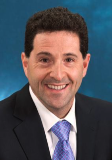 Edward M. Schwarz合作论文数Center for Musculoskeletal Research, University of Rochester Medical Center;Department of Orthopaedics, University of Rochester19
 Safranski David L合作论文数MedShape Solutions17
 Torstrick F Brennan合作论文数Parker H. Petit Institute for Bioengineering and Bioscience, Georgia Institute of Technology15
 Kenneth M. Dupont合作论文数George W. Woodruff School of Mechanical Engineering, Georgia Institute of Technology14
 Yash Kolambkar合作论文数14
 Kelly Leguineche合作论文数Knight Campus Accelerating Sci Impact, Univ Oregon13
 Marian Hettiaratchi合作论文数The Wallace H. Coulter Department of Biomedical Engineering, Georgia Institute of Technology &Emory University;The Wallace H. Coulter Department of Biomedical Engineering, Georgia Institute of Technology & Emory University13
 Robert N. Taylor合作论文数Department of Obstetrics and Gynecology, Jacobs School of Medicine & Biomedical Sciences, University at Buffalo13Uhrig Brent A合作论文数12
 Ruehle Marissa A合作论文数12Thote T合作论文数12
 Gordon L Warren合作论文数Department of Physical Therapy;Georgia State University;Department of Physical Therapy, Georgia State University12
 Craig L. Duvall合作论文数Vanderbilt Institute of Nanoscale Science and Engineering, Vanderbilt University;Department of Biomedical Engineering, Vanderbilt University12
 Johnna S. Temenoff合作论文数Wallace H. Coulter Department of Biomedical Engineering, College of Engineering, Georgia Institute of Technology;School of Materials Science and Engineering, Georgia Institute of Technology12
 Brett S. Klosterhoff合作论文数Parker H Petit Inst Bioengn & Biosci, Georgia Inst Technol11Salil Sidharthan Karipott合作论文数11
 Regis O'Keefe合作论文数Department of Orthopaedic Surgery, School of Medicine, Washington University;Division of Musculoskeletal Oncology, Washington University;Barnes-Jewish Hospital;Shriners Hospital for Children - St. Louis;St. Louis Children's Hospital11
 Jeffrey Weiss合作论文数Department of Biomedical Engineering, University of Utah11
 Hani A. Awad合作论文数Department of Orthopaedics, University of Rochester Medical Center11A. S. Lin合作论文数10
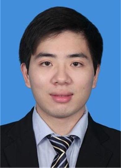 Xinping Zhang 合作论文数10
 Salazar-Noratto Giuliana E合作论文数10
 Allen Ashley B合作论文数1 Wallace H. Coulter Department of Biomedical Engineering, Parker H. Petit Institute for Bioengineering &Bioscience, Georgia Institute of Technology;Parker H. Petit Institute for Bioengineering & Bioscience, Georgia Institute of Technology10
 Krishnendu Roy合作论文数Vanderbilt Institute for Infection, Immunology and Inflammation, Vanderbilt University Medical Center;School of Engineering, Vanderbilt University10
 Scott Hollister合作论文数Wallace H. Coulter Department of Biomedical Engineering, College of Engineering, Georgia Institute of Technology10
 Megan E. Oest合作论文数10
 Marc Levenston合作论文数Department of Mechanical Engineering, Stanford University10
 Nagaraja Srinidhi合作论文数Center for Devices and Radiological Health, U.S. Food and Drug Administration9Cheng Albert合作论文数George W. Woodruff School of Mechanical Engineering, Georgia Institute of Technology;Petit Institute for Bioengineering &Bioscience, Georgia Institute of Technology;Georgia Institute of Technology, Georgia Institute of Technology9
 Susan Y Smith合作论文数9
 Todd McDevitt合作论文数Genentech9
 Alexandra Peister合作论文数Center for Gene Therapy, Tulane University Health Sciences Center9Casey E. Vantucci合作论文数Parker H Petit Inst Bioengn & Biosci, Georgia Inst Technol8
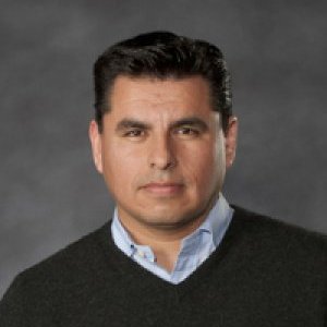 René Olivares-Navarrete合作论文数Department of Biomedical Engineering, College of Engineering, Virginia Commonwealth University8
 Don P. Giddens合作论文数Wallace H. Coulter Department of Biomedical Engineering, College of Engineering, Georgia Institute of Technology;The Parabola Group LLC;Covanos8
 Subbiah Ramesh合作论文数Center for Biomaterials, Korea Institute of Science and Technology;Dept. of Biomedical Engineering, University of Science and Technology;Korea Institute of Science and Technology, University of Science and Technology8
 Bradley Nelson合作论文数Orthopaed Bioengn Res Lab, Colorado State Univ7




