OBJECTIVES:Motor cortex stimulation (MCS) is an effective technique in treating chronic intractable pain for some patients. However, most studies are small case series (n < 20). Heterogeneity in technique and patient selection makes it difficult to draw consistent conclusions. In this study, we present one of the largest case series of subdural MCS. MATERIALS AND METHODS:Medical records of patients who underwent MCS at our institute between 2007 and 2020 were reviewed. Studies with at least 15 patients were summarized for comparison. RESULTS:The study included 46 patients. Mean age was 56.2 ± 12.5 years (SD). Mean follow-up was 57.2 ± 41.9 months. Male-to-female ratio was 13:33. Of the 46 patients, 29 had neuropathic pain in trigeminal nerve territory/anesthesia dolorosa; nine had postsurgical/posttraumatic pain; three had phantom limb pain; two had postherpetic pain, and the rest had pain secondary to stroke, chronic regional pain syndrome, and tumor. The baseline numeric rating pain scale (NRS) was 8.2 ± 1.8 of 10, and the latest follow-up score was 3.5 ± 2.9 (mean improvement of 57.3%). Responders comprised 67% (31/46)(NRS ≥ 40% improvement). Analysis showed no correlation between percentage of improvement and age (p = 0.352) but favored male patients (75.3% vs 48.7%, p = 0.006). Seizures occurred in 47.8% of patients (22/46) at some point but were all self-limiting, with no lasting sequelae. Other complications included subdural/epidural hematoma requiring evacuation (3/46), infection (5/46), and cerebrospinal fluid leak (1/46). These complications resolved with no long-term sequelae after further interventions. CONCLUSION:Our study further supports the use of MCS as an effective treatment modality for several chronic intractable pain conditions and provides a benchmark to the current literature.
Microglia have fundamental roles in health and disease, however effects of age, sex and genetic factors on human microglia have not been fully explored. We applied bulk and single cell approaches to comprehensively characterize human microglia transcriptomes and their associations with age, sex and APOE. We identified a novel microglial signature, characterized its expression in bulk tissue and single cell microglia transcriptomes. We discovered microglial co-expression network modules associated with age, sex and APOE-ε4 that are enriched for lipid and carbohydrate metabolism genes. Integrated analyses of modules with single cell transcriptomes revealed significant overlap between age-associated module genes and both pro-inflammatory and disease-associated microglial clusters. These modules and clusters harbor known neurodegenerative disease genes including APOE, PLCG2 and BIN1. Meta-analyses with published bulk and single cell microglial datasets further supported our findings. Thus, these data represent a well-characterized human microglial transcriptome resource; and highlight age, sex and APOE-related microglial immunometabolism perturbations with potential relevance in neurodegeneration.
Anti-seizure medications are the first-line treatments for those with a tendency toward recurrent seizures, or epilepsy and chronically alter the excitability of many or most cerebral neurons. There are three brain stimulation approaches that are US Food and Drug Administration-approved for the treatment of epilepsy: vagus nerve stimulation (VNS), responsive neurostimulation, and thalamic deep-brain stimulation. This chapter focuses on these invasive approaches. Overall response to VNS was predicted by non-lesional epilepsy. Long-term safety data suggest a 3% risk of permanent vagus nerve damage and 5% risk of lead fracture. VNS reduces the risk of sudden unexpected death in epilepsy from approximately 6–7 per 1000 patient years for patients to approximately 2 per 1000 patient years, an effect that seems to strengthen over time. In many regards, neuromodulation for epilepsy remains in its infancy, with underlying pathophysiology poorly understood despite clear evidence of efficacy via multiple stimulation approaches.
Seizure freedom is the single best predictor of quality of life in epilepsy, as recurrent seizures lead to significant morbidity and potentially increased mortality. While anterior temporal lobectomy (ATL) remains the most established and tested surgical therapy for temporal lobe epilepsy (TLE), new minimally invasive options have catalyzed a resurgence of interest in epilepsy surgery. This chapter highlights the role of radiofrequency thermoablation, stereotactic radiosurgery, and laser interstitial thermal therapy in the treatment of patients with drug-resistant focal seizures. The temporal lobe epilepsies are the most common type of drug-resistant epilepsy in adults, with mesial temporal sclerosis as the most common cause of mesial TLE. Surgical treatment of this disorder includes procedures such as ATL, sublobar ATL and selective amygadalohippocampectomy. Many types of lesions causing drug-resistant epilepsy have been treated with laser interstitial thermal therapy guided by magnetic resonance thermography.
Objective To prospectively evaluate safety and efficacy of brain-responsive neurostimulation in adults with medically intractable focal onset seizures (FOS) over 9 years. Methods Adults treated with brain-responsive neurostimulation in 2-year feasibility or randomized controlled trials were enrolled in a long-term prospective open label trial (LTT) to assess safety, efficacy, and quality of life (QOL) over an additional 7 years. Safety was assessed as adverse events (AEs), efficacy as median percent change in seizure frequency and responder rate, and QOL with the Quality of Life in Epilepsy (QOLIE-89) inventory. Results Of 256 patients treated in the initial trials, 230 participated in the LTT. At 9 years, the median percent reduction in seizure frequency was 75% (p < 0.0001, Wilcoxon signed rank), responder rate was 73%, and 35% had a ≥90% reduction in seizure frequency. We found that 18.4% (47 of 256) experienced ≥1 year of seizure freedom, with 62% (29 of 47) seizure-free at the last follow-up and an average seizure-free period of 3.2 years (range 1.04–9.6 years). Overall QOL and epilepsy-targeted and cognitive domains of QOLIE-89 remained significantly improved (p < 0.05). There were no serious AEs related to stimulation, and the sudden unexplained death in epilepsy (SUDEP) rate was significantly lower than predefined comparators (p < 0.05, 1-tailed χ2). Conclusions Adjunctive brain-responsive neurostimulation provides significant and sustained reductions in the frequency of FOS with improved QOL. Stimulation was well tolerated; implantation-related AEs were typical of other neurostimulation devices; and SUDEP rates were low. ClinicalTrials.gov identifier NCT00572195. Classification of evidence This study provides Class IV evidence that brain-responsive neurostimulation significantly reduces focal seizures with acceptable safety over 9 years.
Despite advancements in the neurophysiology of language and presurgical evaluation for epilepsy surgery, there is a paucity of information in the literature regarding presurgical evaluation of multilingual patients. We present a case of a 52-year-old right-handed woman with refractory epilepsy who was fluent in six languages and underwent subsequent trilingual presurgical evaluation which included neuropsychological testing, Wada testing, functional magnetic resonance imaging (fMRI), and electrocortical stimulation. These studies suggested a seizure focus in the left temporal lobe and language localization that was predominantly right-hemispheric; she subsequently underwent left temporal laser interstitial thermal therapy without clinical disturbance in language function while remaining seizure-free. A multidisciplinary effort was integral in providing an optimal outcome for this patient.
Functional imaging studies have implicated an area in the left lateral fusiform gyrus, known as the visual word form area (VWFA), in pre-lexical orthographic processing. There are very few studies that have examined the functional specificity of this area in patients with discrete lesions limited to this region. Here we describe a rare opportunity to examine the functional specificity of the VWFA in a patient with stereo EEG (sEEG) electrodes implanted for localization of seizures prior to epilepsy surgery. sEEG offers the opportunity to create a transient and highly localized electrophysiological lesion to examine brain behavior correlates during functional mapping. In this case, word reading and writing as well as a variety of non-orthographic language functions (e.g., picture and face naming, auditory naming, and non-word repetition), were tested during electrical stimulation at a series of different electrode contact sites in the ventral temporal region. Pure alexia resulted from stimulation of the lateral fusiform gyrus at coordinates nearly identical to those published for the VWFA in the functional imaging literature.
OBJECTIVESurgical site infection (SSI) is a rare but significant complication after vagus nerve stimulator (VNS) placement. Treatment options range from antibiotic therapy alone to hardware removal. The optimal therapeutic strategy remains open to debate. Therefore, the authors conducted this retrospective multicenter analysis to provide insight into the optimal management of VNS-related SSI (VNS-SSI).METHODSUnder institutional review board approval and utilizing an institutional database with 641 patients who had undergone 808 VNS-related placement surgeries and 31 patients who had undergone VNS-related hardware removal surgeries, the authors retrospectively analyzed VNS-SSI.RESULTSSixteen cases of VNS-SSI were identified; 12 of them had undergone the original VNS placement procedure at the authors' institutions. Thus, the incidence of VNS-SSI was calculated as 1.5%. The mean (± standard deviation) time from the most recent VNS-related surgeries to infection was 42 (± 27) days. Methicillin-sensitive staphylococcus was the usual causative bacteria (58%). Initial treatments included antibiotics with or without nonsurgical procedures (n = 6), nonremoval open surgeries for irrigation (n = 3), generator removal (n = 3), and total or near-total removal of hardware (n = 4). Although 2 patients were successfully treated with antibiotics alone or combined with generator removal, removal of both the generator and leads was eventually required in 14 patients. Mild swallowing difficulties and hoarseness occurred in 2 patients with eventual resolution.CONCLUSIONSRemoval of the VNS including electrode leads combined with antibiotic administration is the definitive treatment but has a risk of causing dysphagia. If the surgeon finds dense scarring around the vagus nerve, the prudent approach is to snip the electrode close to the nerve as opposed to attempting to unwind the lead completely.
BACKGROUND: Preoperative thalamic targeting methods have historically relied on indirect targeting techniques that do not fully account for variances in anatomy or for thalamic atrophy in epilepsy. We aimed to address variability noted between traditional indirect targeting and direct targeting methods for the anterior nucleus of the thalamus (ANT). METHODS: Fifteen consecutive patients undergoing ANT deep brain stimulator placement were evaluated (30 thalamic nuclei). Direct ANT targeting was performed using a fast gray matter acquisition T1 inversion recovery sequence and compared with standard stereotactic coordinates. Thalamic volumes were calculated for each patient, and degree of thalamic volume loss was assessed compared with matched control subjects. Vertex analysis was performed to assess shape changes in the thalamus compared with age- and sex-matched subjects. RESULTS: There was significant variation between direct and indirect targets in the y-axis and z-axis on both sides. On the left, the direct target was located at y = 2 +/- 1.3 mm and z = 9.3 +/- 1.8 mm (both P = 0.02). On the right, the direct target was located at y = 2.9 +/- 1.8 mm and z = 9.2 +/- 2 mm (both P <= 0.0003). There was no significant difference in the x-coordinate on either side (P > 0.5). Additionally, there was a correlation between thalamic volume and difference between direct and indirect targets in the y-axis and the z-axis. CONCLUSIONS: We showed a significant difference in direct and indirect targeting in the y-axis and z-axis when targeting the ANT for deep brain stimulation for epilepsy. This difference is correlated to thalamic volume, with a larger difference noted in patients with thalamic atrophy.
BACKGROUND: Deep brain stimulation (DBS) of the nuc.eus of the thalamus (ANT) is a recently approved therapy for patients with drug-resistant epilepsy. To date, there is a poor understanding of the mechanism of action and lack of in vivo biomarkers. We propose a method for investigating the in vivo stimulation effects using blood-oxygen-level-dependent (BOLD) magnetic resonance imaging (MRI) and present the brain activation pattern associated with ANT DBS. METHODS: Two patients undergoing ANT DBS for epilepsy underwent BOLD MRI using a block design after the DBS was programmed to alternate ON/OFF in 30-second blocks. The scanner was triggered using surface electrophysiologic recordings to detect the DBS cycle. Nine total runs were obtained and were analyzed using a general linear model. RESULTS: Active ANT stimulation produced activation within several areas of the brain, including the thalamus, bilateral anterior cingulate and posterior cingulate cortex, precuneus, medial prefrontal cortex, amygdala, ventral tegmental area, hippocampus, striatum, and right angular gyrus. CONCLUSIONS: Using block-design BOLD MRI, we were able to show widespread activation resulting from ANT DBS. Overlap with multiple areas of both the default mode and limbic networks was shown, suggesting that these nodes may modulate the effect of seizure control with ANT DBS.
Background. The management of normal pressure hydrocephalus (NPH) can be difficult, partly because there are frequent treatment complications such as overdrainage which, when serious, may require surgical intervention. We previously reported a correlation between the difference of lumbar puncture opening pressure minus the valve opening pressure setting (LPOP - VOP) (which we refer to as the delta) and increased rates of overdrainage. This led to a modification in our practice, whereby we now set the VOP equal to, or close to, the LPOP, resulting in lower deltas. Objective. In this new study, our aim was to compare the rate of overdrainage in our patients with higher and lower deltas and assess the significance of setting the VOP equal, or close, to the patient's LPOP. Methods. 1. We reproduced the association between delta and overdrainage. 2. We compared the incidence of overdrainage in those whose VOP was set close to LPOP (low delta) versus those with VOP setting distant from the LPOP (higher delta). 3. We compared symptom improvement in those with a low versus higher delta. Results. We confirmed the relation between high delta and an increased rate of overdrainage, lower rates of overdrainage in those whose VOP was set close to the LPOP (Delta Adjusted Practice), and better improvement of symptoms when the VOP was set closer to the LPOP. Conclusion. We propose that the initial VOP should be set as close as possible to the patient's LPOP to decrease overdrainage without compromising symptom improvement.
Background: Deep Brain Stimulation (DBS) was approved by the FDA in the 1990s and is used to treat a variety of movement disorders. Patients are increasingly turning to the internet for information regarding their ailments. In this study, we aim to evaluate the accuracy and reliability of information presented in DBS-related YouTube videos. Methods: Using the "Relevance-Based Ranking" strategy for analysis we assessed the first 3 pages of YouTube for each of the following keywords: "Deep Brain Stimulation", "DBS", "DBS for Parkinson's disease", "DBS for essential tremor", and "DBS for movement disorders". Four independent healthcare personnel evaluated the videos' education quality and informational material using the validated DISCERN tool. Results: Our study found that only 24% of the 42 published videos analyzed scored above a 3 on the DISCERN scoring scale (considered a "good" video). The search term "Deep Brain Stimulation" had the highest percentage of good videos (DISCERN > 3) (32%). We also found that the duration of videos was longer for the "good" videos (Good = 25.6 min vs Unhelpful = 3.0 min, P = 0.01). Conclusion: YouTube is one of the largest video platforms; the uploaded videos lack reliability and institutional oversight by the experts. We believe that medical institutions should explore this way of communicating to patients by publishing evidence-based and informative videos on diseases and their management. As it is imperative that the medical field advance to combat medical misinformation. (C) 2020 Elsevier Ltd. All rights reserved.
This study examines whether surgical resection of carotid body tumors (CBTs) is acceptable in light of potential significant neurologic complications. This IRB-approved retrospective study analyzed data from 24 patients undergoing surgical treatment for CBTs between April 1998 and April 2017 at Mayo Clinic (Florida campus only). For patients who underwent multiple CBT resections, only data from the first surgery was used in this analysis. CBT resection occurred in 24 patients with the following demographics: fourteen patients (58.3%) were female, median age was 56.5 years, median BMI was 29. A prior history of neoplasm was found in ten patients (41.7%). A known family history of paraganglioma was present in five patients (20.8%). Two patients were positive for succinate dehydrogenase mutation (8.3%). Multiple paragangliomas were present in seven patients (29.2%). There was nerve sacrifice in three patients (12.5%) during resection. Carotid artery reconstruction and patch angioplasty occurred in one patient (4.2%). Complete resection occurred in 24 patients (100.0%). Postoperatively, one patient (4.2%) suffered stroke. No mortalities occurred within or beyond 30 days of surgery. Persistent cranial nerve injury occurred in two patients (8.3%) with vocal cord paralysis. There was no recurrence of CBT through last follow-up. Five patients (20.8%) were diagnosed with other neoplasms after resection, including basal cell carcinoma, contralateral carotid body tumor, glomus vagale, and glomus jugulare. There was 100% survival at 1 year in patients followed for that time ( n = 17). Surgical treatment remains the first-line curative treatment to relieve symptoms and ensure non-recurrence. While acceptable, neurologic complications are significant and therefore detailed preoperative informed consent is mandatory.
SUMMARY: Deep brain stimulation is an established therapy for multiple brain disorders, with rapidly expanding potential indications. Neuroimaging has advanced the field of deep brain stimulation through improvements in delineation of anatomy, and, more recently, application of brain connectomics. Older lesion-derived, localizationist theories of these conditions have evolved to newer, network-based “circuitopathies,” aided by the ability to directly assess these brain circuits in vivo through the use of advanced neuroimaging techniques, such as diffusion tractography and fMRI. In this review, we use a combination of ultra-high-field MR imaging and diffusion tractography to highlight relevant anatomy for the currently approved indications for deep brain stimulation in the United States: essential tremor, Parkinson disease, drug-resistant epilepsy, dystonia, and obsessive-compulsive disorder. We also review the literature regarding the use of fMRI and diffusion tractography in understanding the role of deep brain stimulation in these disorders, as well as their potential use in both surgical targeting and device programming.
Stereo-EEG (sEEG) is an invasive recording technique used to localize the seizure-onset zone for epilepsy surgery in people with drug-resistant focal seizures. Pathological crying reflects disordered emotional expression and the anterior insula is known to play a role in empathy and socio-emotional processing. We describe a patient where electrical stimulation mapping (ESM) of the anterior insula during sEEG generated pathological crying and profound sadness that was time-locked to the electrical stimulus.We evaluated a 35-year-old left-handed female for repeat epilepsy surgery. The patient had drug resistant focal impaired awareness seizures despite a previous left temporal neocortical resection informed by an invasive study using subdural grid and strip electrodes seven years earlier. She was studied invasively with 10 sEEG electrodes sampling temporal, occipital, and insular targets. In the process of functional mapping, stimulation of the anterior insular cortex provoked tearful crying with sad affect, reproducible upon repeat stimulation.Our case is unique in demonstrating transitory pathological crying with profound sadness provoked by ESM of the left anterior insula. Furthermore we demonstrate repeated time-synched crying from electrical stimulation, which supports the hypothesis that the anterior insula in the brain plays an important role in the biology of emotion, as implicated by previous studies.
OBJECTIVE: The optimal surgical strategy for management of adult patients with craniopharyngioma remains controversial. To analyze the functional outcomes of adult patients with gross total resection (GTR) and subtotal resection (STR) of craniopharyngioma. METHODS: MEDLINE, EMBASE, Scopus, and Cochrane databases were searched from inception to July 19, 2018, for articles comparing postoperative endocrine function, vision, complications, and recurrence rates for adult patients with GTR and STR of craniopharyngioma. The articles were analyzed by meta-analysis of proportions using a random-effects model to calculate summary odds ratios (ORs). RESULTS: The initial search resulted in 2468 studies and 540 studies selected for full text review. Seventeen studies were included in the final analyses with 748 patients in the GTR cohort and 559 patients in the STR cohort. GTR resulted in a significantly lower likelihood of recurrence when compared with STR (OR, 0.106; 95% confidence interval [CI], 0.067-0.168; P < 0.001), hut a significantly greater likelihood of panhypopituitarism (OR, 2.063; 95% CI, 1.058-4.024; P = 0.034) and permanent diabetes insipidus (OR, 2.776; 95% CI, 1.321-5.832; P = 0.007). There was no significant difference between the groups for postoperative worsened vision (P = 0.868), improved vision (P = 0.876), pathologic weight gain (P = 0.724), cerebrospinal fluid leak (P = 0.788), complications (P = 0.656), or death (P = 0.261). CONCLUSIONS: This is the first systematic review of functional outcomes of adult patients with craniopharyngioma. GTR results in decreased likelihood of recurrence, but increased likelihood of postoperative panhypopituitarsm and permanent diabetes insipidus. Surgeons should be aware of these associations when determining the optimal operative strategy for adult patients with craniopharyngioma.
Bulk tissue transcriptome studies can identify expression changes associated with pathology, however bulk tissue represents a heterogeneous mix of cells and the proportion can vary between samples and by diagnosis. It is imperative to account for cell proportion changes in such studies, which typically rely on the expression of cell type marker genes in tissue samples. Isolation of sorted populations of cells can characterize the expression profiles of CNS cell types and improve knowledge of cell type marker genes. We aim to isolate CNS cell types from fresh surgical tissue in greater numbers of older samples to identify cell type marker genes and assess expression profile in the context of sex and APOE genotype, particularly relevant for Alzheimer's disease bulk tissue studies. Mouse brain tissue was obtained from FVB males. Human brain tissue was obtained from neurosurgical procedures for epilepsy and for tumor resection. White matter and excess vascular tissue were removed prior to dissociation. We initially used magnetic-activated cell sorting (MACS) to isolate CD11b+ cells (microglia/macrophage) from mouse and human brain tissue and subsequently characterised these populations using 10X single cell RNAseq. Seurat was used to cluster cells and identify differentially expressed genes. Initial experiments in mouse identified 4 clusters within the CD11b+ population, whereas 5 clusters were observed in the human sample. CD11b+ cells exhibited greater expression of microglial markers, which were absent from CD11b- cells, indicating the presence of microglia. In the human sample, one cluster showed high expression of oligodendrocyte markers, suggestive of potential oligodendrocyte contamination, illustrating the need for multi-marker panels to isolate pure cell populations. Current RNAseq methods calculate expression levels from millions of cells by averaging the signal of individual cells, possibly overlooking rare cell types and other differences within a heterogeneous cell population that may associate with disease progression. Single cell-type and single cell RNAseq provides a higher resolution of cellular differences and gene expression, with the characterization of the CNS cell type-specific expression profiles expected to provide novel insights into brain cell specific changes that are associated with AD risk factors.
 Grewal Sanjeet S合作论文数Department of Surgery, University of Cincinnati Medical Center34
 William Tatum合作论文数Mayo Clinic22
 Ryan J. Uitti合作论文数Department of Neurology, Mayo Clinic21Ronald Reimer合作论文数20
 Gregory A. Worrell合作论文数Department of Neurology, Mayo Clinic;Department of Physiology & Biomedical Engineering, Mayo Clinic15
 H. Gordon Deen合作论文数Department of Neurosurgery, Mayo Clinic Florida12
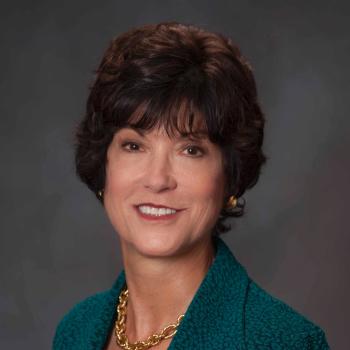 Martha Morrell合作论文数Department of Neurology, School of Medicine, Stanford University;NeuroPace11
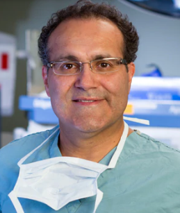 Alfredo Quinones-Hinojosa合作论文数Department of Neurosurgery, Mayo Clinic11
 Robert E. Gross合作论文数Department of Neurosurgery, School of Medicine, Emory University10
 Ryder Gwinn合作论文数Swedish Neuroscience Institute10
 Gregory K. Bergey合作论文数Department of Neurology;Johns Hopkins Epilepsy Center;School of Medicine, Johns Hopkins University9Jerzy Słowiński合作论文数8
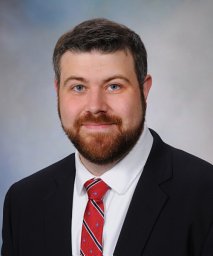 Erik Middlebrooks合作论文数8
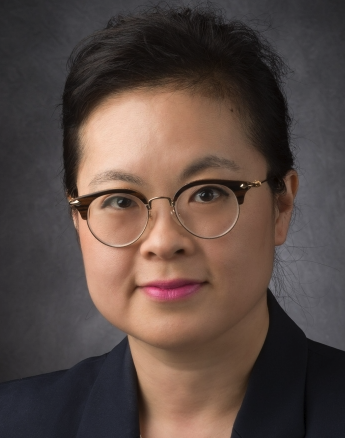 Betty Kim合作论文数Department of Neurosurgery, Division of Surgery, University of Texas MD Anderson Cancer Center7Eric Geller合作论文数7
 Vivek Gupta合作论文数7
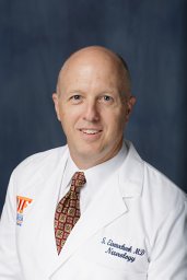 Stephan Eisenschenk合作论文数College of Medicine, University of Florida7
 Karim ReFaey合作论文数Dept Neurosurg, Mayo Clin7David Spencer合作论文数Department of Neurology, Oregon Health & Science University7Salanova Vicenta合作论文数7
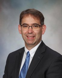 Pirris Stephen M合作论文数Department of Neurological Surgery, University of Pittsburgh Medical Center7
 Dileep Nair合作论文数Epilepsy Center, Cleveland Clinic Foundation7Cole Andrew J合作论文数7Heck Christianne N合作论文数7
 Alica Goldman合作论文数Baylor College of Medicine, Baylor College of Medicine7
 Eric Nottmeier合作论文数Department of Neurological Surgery, Mayo Clinic Jacksonville7Nathan B. Fountain合作论文数7Barbara C. Jobst合作论文数Department of Medicine, Dartmouth Medical School7Gregory L. Barkley合作论文数7
 William D. Freeman合作论文数Mayo Clinic7Jonathan C. Edwards合作论文数6
 Anthony Murro合作论文数6Felice T Sun合作论文数6
 Paul C. Van Ness合作论文数6David King-Stephens合作论文数6Putzke D John合作论文数6James F. Meschia合作论文数6Lawrence J. Hirsch合作论文数6
 Jerry J. Shih合作论文数6Paul Rutecki合作论文数6
 Robert Duckrow合作论文数6Christopher Skidmore合作论文数6
 John A Lucas合作论文数5
 Oluwaseun Akinduro合作论文数Dept Neurosurg, Mayo Clin5Srinivasan Shraddha合作论文数5
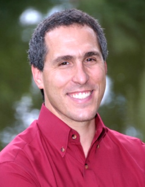 Sydney Cash合作论文数Division of Epilepsy, Department of Neurology, Mass General Brigham;Department of Neurology, Massachusetts General Hospital;Harvard Medical School, Harvard University5
 Anteneh Feyissa合作论文数Department of Pharmacology and Therapeutics, NUI Galway5Brown Benjamin L合作论文数5
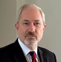 William P Cheshire合作论文数Department of Neurology, Mayo Clinic in Florida5




