SIGNIFICANCE:Veridical depictions of scene appearance with scotomas allow better understanding of the impact of field loss and may improve the development and implementation of rehabilitation. Explanation and depiction of the invisibility of scotoma may lead to patients' understanding and thus better compliance with related treatments. PURPOSE:Simulations of perception with scotomas guide training, patient education, and rehabilitation research. Most simulations incorrectly depict scotomas as black patches, although the scotomas and the missing contents are usually invisible to patients. We present a novel approach to capture the reported appearance of scenes with scotomas. METHODS:We applied a content-aware image resizing algorithm to carve out the content elided under the scotomas. With video sequences, we show how and why eye movements fail to increase the visibility of the carved scotomas. RESULTS:Numerous effects, reported by patients, emerge naturally from the scotoma carving. Carving-eliminated scotomas over natural images are barely visible, despite causing substantial distortions. Low resolution and contrast sensitivity at farther eccentricities and saccadic blur reduce the visibility of the distortions. In a walking scenario, static objects moving smoothly to the periphery disappear into and then reemerge out of peripheral scotomas, invisibly. CONCLUSIONS:Scotoma carving provides a viable hypothetical simulation of vision with scotomas due to loss of neurons at the retinal ganglion cell level and higher. As a hypothesis, it generates predictions that lend themselves to future clinical testing. The different effects of scotomas due to loss of photoreceptors are left for follow-up work.
OBJECTIVE Medial thalamotomy has been shown to benefit patients with neuropathic pain, but widespread adoption of this procedure has been limited by reporting of clinical outcomes in studies without a control group. This study aimed to minimize confounders associated with medial thalamotomy for treating chronic pain by using modern MRI-guided stereotactic lesioning and a rigorous clinical design. METHODS This prospective, double-blinded, randomized controlled trial in 10 patients with trigeminal neuropathic pain used sham procedures as controls. Participants underwent assessments by a pain psychologist and pain management clinician, including use of the following measures: the Numeric Pain Rating Scale (NPRS); patient-reported outcome measures; and patient’s impression of improvement at baseline, 1 day, 1 week, 1 month, and 3 months postprocedure. Patients in the treated group underwent bilateral focused ultrasound (FUS) medial thalamotomy targeting the central lateral nucleus. Patients in the control group underwent sham procedures with energy output disabled. The primary efficacy outcome measure was between-group differences in pain intensity (using the NPRS) at baseline and at 3 months postprocedure. Adverse events were measured for safety and included MRI analysis. Exploratory measures of connectivity and metabolism were analyzed using diffusion tensor imaging, functional MRI, and PET, respectively. RESULTS There were no serious complications from the FUS procedures. MRI confirmed bilateral medial thalamic ablations. There was no significant improvement in pain intensity from baseline to 3 months, either for patients undergoing FUS medial thalamotomy or for sham controls; and the between-group change in NPRS score as the primary efficacy outcome measure was not significantly different. Patient-reported outcome assessments demonstrated improvement (i.e., a decrease) only in pain interference with enjoyment of life at 3 months. There was a perception of benefit at 1 week, but only for patients treated with FUS and not for the sham cohort. Advanced neuroimaging showed that these medial thalamic lesions altered structural connectivity with the postcentral gyrus and demonstrated a trend toward hypometabolism in the insula and amygdala. CONCLUSIONS This randomized controlled trial of bilateral FUS medial thalamotomy did not reduce the intensity of trigeminal neuropathic pain, although it should be noted that the ability to estimate the magnitude of treatment effects is limited by the small cohort.
Eye and head movements are used to scan the environment when driving. In particular, when approaching an intersection, large gaze scans to the left and right, comprising head and multiple eye movements, are made. We detail an algorithm called the gaze scan algorithm that automatically quantifies the magnitude, duration, and composition of such large lateral gaze scans. The algorithm works by first detecting lateral saccades, then merging these lateral saccades into gaze scans, with the start and end points of each gaze scan marked in time and eccentricity. We evaluated the algorithm by comparing gaze scans generated by the algorithm to manually marked “consensus ground truth” gaze scans taken from gaze data collected in a high-fidelity driving simulator. We found that the gaze scan algorithm successfully marked 96% of gaze scans and produced magnitudes and durations close to ground truth. Furthermore, the differences between the algorithm and ground truth were similar to the differences found between expert coders. Therefore, the algorithm may be used in lieu of manual marking of gaze data, significantly accelerating the time-consuming marking of gaze movement data in driving simulator studies. The algorithm also complements existing eye tracking and mobility research by quantifying the number, direction, magnitude, and timing of gaze scans and can be used to better understand how individuals scan their environment.
Purpose Using a driving simulator, we quantified the effects of age and central field loss (CFL) on head scanning when approaching an intersection and investigated the role of inadequate head scanning in detection failures. Methods Participants with CFL (n = 20) and with normal vision (NV; n = 29), middle-aged (36–60 years) or older (67–87 years), drove along city routes with multiple intersections while head movements were recorded. The effects of age and CFL on scanning were analyzed at 32 intersections with stop/yield signs. The relationships between age, CFL, scanning, and detection were examined at four additional intersections with a pedestrian appearing on the far left. Results Older NV participants made fewer total scans than middle-aged NV participants and had smaller maximum scan magnitudes. Head scanning of older CFL and NV participants did not differ, but middle-aged CFL participants made fewer head scans, had higher rates of failing to scan, and made smaller head scans than middle-aged NV participants. For the older NV and both CFL groups, detection failures were high (≥58%); head scan magnitudes were 15° smaller when the pedestrian was not detected than when it was detected. Conclusions Both older NV and CFL participants exhibited head scanning deficits relative to middle-aged NV participants. Unexpectedly, however, it was the middle-aged CFL group that performed least well when scanning, a finding that warrants further investigation. Translational Relevance Failing to head scan sufficiently far at intersections may place older drivers and drivers with vision impairment at a higher risk for causing collisions.
PURPOSE:Peripheral field loss (PFL) due to retinitis pigmentosa, choroideremia, or glaucoma often results in a highly constricted residual central field, which makes it difficult for patients to avoid collision with approaching pedestrians. We developed a virtual environment to evaluate the ability of patients to detect pedestrians and judge potential collisions. We validated the system with both PFL patients and normally sighted subjects with simulated PFL. We also tested whether properly placed high-power prisms may improve pedestrian detection.METHODS:A virtual park-like open space was rendered using a driving simulator (configured for walking speeds), and pedestrians in testing scenarios appeared within and outside the residual central field. Nine normally sighted subjects and eight PFL patients performed the pedestrian detection and collision judgment tasks. The performance of the subjects with simulated PFL was further evaluated with field of view expanding prisms.RESULTS:The virtual system for testing pedestrian detection and collision judgment was validated. The performance of PFL patients and normally sighted subjects with simulated PFL were similar. The prisms for simulated PFL improved detection rates, reduced detection response times, and supported reasonable collision judgments in the prism-expanded field; detections and collision judgments in the residual central field were not influenced negatively by the prisms.CONCLUSIONS:The scenarios in a virtual environment are suitable for evaluating PFL and the impact of field of view expanding devices.TRANSLATIONAL RELEVANCE:This study validated an objective means to evaluate field expansion devices in reproducible near-real-life settings.
Purpose: Oncoming headlight glare (HLG) reduces the visibility of objects on the road and may affect the safety of nighttime driving. With cataracts, the impact of oncoming HLG is expected to be more severe. We used our custom HLG simulator in a driving simulator to measure the impact of HLG on pedestrian detection by normal vision subjects with simulated mild cataracts and by patients with real cataracts. Methods: Five normal vision subjects drove nighttime scenarios under two HLG conditions (with and without HLG: HLGY and HLGN, respectively), and three vision conditions (with plano lens, simulated mild cataract, and optically blurred clip-on). Mild cataract was simulated by applying a 0.8 Bangerter diffusion foil to clip-on plano lenses. The visual acuity with the optically blurred lenses was individually chosen to match the visual acuity with the simulated cataract clip-ons under HLGN. Each nighttime driving scenario contains 24 pedestrian encounters, encompassing four pedestrian types; walking along the left side of the road, walking along the right side of the road, crossing the road from left to right, and crossing the road from right to left. Pedestrian detection performances of five patients with mild real cataracts were measured using the same setup. The cataract patients were tested only in HLGY and HLGN conditions. Participants' visual acuity and contrast sensitivity were also measured in the simulator with and without stationary HLG. Results: For normal vision subjects, both the presence of oncoming HLG and wearing the simulated cataract clip-on reduced pedestrian detection performance. The subjects performed worst in events where the pedestrian crossed from the left, followed by events where the pedestrian crossed from the right. Significant interactions between HLG condition and other factors were also found: (1) the impact of oncoming HLG with the simulated cataract clip-on was larger than with the plano lens clip-on, (2) the impact of oncoming HLG was larger with the optically blurred clip-on than with the plano lens clip-on, but smaller than with the simulated cataract clip-on, and (3) the impact was larger for the pedestrians that crossed from the left than those that crossed from the right, and for the pedestrians walking along the left side of the road than walking along the right side of the road, suggesting that the pedestrian proximity to the glare source contributed to the performance reduction. Under HLGN, almost no pedestrians were missed with the plano lens or the simulated cataract clip-on (0 and 0.5%, respectively), but under HLGY, the rate of pedestrian misses increased to 0.5 and 6%, respectively. With the optically blurred clip-on, the percent of missed pedestrians under HLGN and HLGY did not change much (5% and 6%, respectively). Untimely response rate increased under HLGY with the plano lens and simulated cataract clip-ons, but the increase with the simulated cataract clip-on was significantly larger than with the plano lens clip-on. The contrast sensitivity with the simulated cataract clip-on was significantly degraded under HLGY. The visual acuity with the plano lens clip-on was significantly improved under HLGY, possibly due to pupil myosis. The impact of HLG measured for real cataract patients was similar to the impact on performance of normal vision subjects with simulated cataract clip-ons. Conclusion: Even with mild (simulated or real) cataracts, a substantial negative effect of oncoming HLG was measurable in the detection of crossing and walking-along pedestrians. The lowered pedestrian detection rates and longer response times with HLGY demonstrate a possible risk that oncoming HLG poses to patients driving with cataracts.
PURPOSE:Efficacy of current visual prostheses in object recognition is limited. Among various limitations to be addressed, such as low resolution and low dynamic range, here we focus on reducing the impact of background clutter on object recognition. We have proposed the use of motion parallax via head-mounted camera lateral scanning and computationally stabilizing the object of interest (OI) to support neural background decluttering. Simulations in head-mounted displays (HMD), mimicking the proposed effect, were used to test object recognition in normally sighted subjects. METHODS:Images (24° field of view) were captured from multiple viewpoints and presented at a low resolution (20 × 20). All viewpoints were centered on the OI. Experimental conditions (2 × 3) included clutter (with or without) × head scanning (single viewpoint, 9 coherent viewpoints corresponding to subjects' head positions, and 9 randomly associated viewpoints). Subjects used lateral head movements to view OIs in the HMD. Each object was displayed only once for each subject. RESULTS:The median recognition rate without clutter was 40% for all head scanning conditions. Performance with synthetic background clutter dropped to 10% in the static condition, but it was improved to 20% with the coherent and random head scanning (corrected P = 0.005 and P = 0.049, respectively). CONCLUSIONS:Background decluttering using motion parallax cues but not the coherent multiple views of the OI improved object recognition in low-resolution images. The improvement did not fully eliminate the impact of background. TRANSLATIONAL RELEVANCE:Motion parallax is an effective but incomplete decluttering solution for object recognition with visual prostheses.
Purpose: Drivers with homonymous hemianopia (HH) were previously found to have impaired detection of blind-side hazards, yet in many jurisdictions they may obtain a license. We evaluated whether oblique 57Δ peripheral prisms (p-prisms) and perceptual-motor training improved blind-side detection rates. Methods: Patients with HH (n = 11) wore p-prisms for 2 weeks and then received perceptual-motor training (six visits) detecting and touching stimuli in the prism-expanded vision. In a driving simulator, patients drove and pressed the horn upon detection of pedestrians who ran toward the roadway (26 from each side): (1) without p-prisms at baseline; (2) with p-prisms after 2 weeks acclimation but before training; (3) with p-prisms after training; and (4) 3 months later. Results: P-prisms improved blind-side detection from 42% to 56%, which further improved after training to 72% (all P < 0.001). Blind-side timely responses (adequate time to have stopped) improved from 31% without to 44% with p-prisms (P < 0.001) and further improved with training to 55% (P = 0.02). At the 3-month follow-up, improvements from training were maintained for detection (65%; P = 0.02) but not timely responses (P = 0.725). There was wide between-subject variability in baseline detection performance and response to p-prisms. There were no negative effects of p-prisms on vehicle control or seeing-side performance. Conclusions: P-prisms improved detection with no negative effects, and training may provide additional benefit. Translational Relevance: In jurisdictions where people with HH are legally driving, these data aid in clinical decision making by providing evidence that p-prisms improve performance without negative effects.
PURPOSE:We investigated whether people with homonymous hemianopia (HH) were able to spontaneously (without training or instructions) adapt their blind-side scan magnitudes in response to differing scanning requirements for detection of pedestrians in a driving simulator when differing cues about pedestrian eccentricities and movement behaviors were available in the seeing hemifield. METHODS:Twelve HH participants completed two sessions in a driving simulator pressing the horn when they detected a pedestrian. Stationary pedestrians outside the driving lane were presented in one session and approaching pedestrians on a collision course in the other. Gaze data were analyzed for pedestrians initially appearing at approximately 14° in the blind hemifield. No instructions were given regarding scanning. RESULTS:After appearing, the stationary pedestrians' eccentricity increased rapidly to a median of 31° after 2.5 seconds, requiring increasingly larger blind-side gaze scans for detection, while the approaching pedestrians' eccentricity remained constant at approximately 14°, requiring a more moderate scan (∼14°) for detection. Although median scan magnitudes did not differ between the two conditions (approaching: 14° [IQR 9°-15°]; stationary: 13° [IQR 9°-20°]; P = 0.43), three participants showed evidence of adapting (increasing) their blind-side scan magnitudes in the stationary condition. CONCLUSIONS:Three participants (25%) appeared to be able to apply voluntary cognitive control to modify their blind-side gaze scanning in response to the differing scanning requirements of the two conditions without explicit training. TRANSLATIONAL RELEVANCE:Our results suggest that only a minority of people with hemianopia are likely to be able to spontaneously adapt their blind-side scanning in response to rapidly changing and unpredictable situations in on-road driving.
Purpose: Peripheral prisms (p-prisms) shift peripheral portions of the visual field of one eye, providing visual field expansion for patients with hemianopia. However, patients rarely show adaption to the shift, incorrectly localizing objects viewed within the p-prisms. A pilot evaluation of a novel computerized perceptual-motor training program aiming to promote p-prism adaption was conducted.Methods: Thirteen patients with hemianopia fitted with 57D oblique p-prisms completed the training protocol. They attended six 1-hour visits reaching and touching peripheral checkerboard stimuli presented over videos of driving scenes while fixating a central target. Performance was measured at each visit and after 3 months.Results: There was a significant reduction in touch error (P = 0.01) for p-prism zone stimuli from pretraining median of 16.6 degrees (IQR 12.1 degrees-19.6 degrees) to 2.7 degrees (IQR 1.0 degrees-8.5 degrees) at the end of training. P-prism zone reaction times did not change significantly with training (P > 0.05). P-prism zone detection improved significantly (P = 0.01) from a pretraining median 70% (IQR 50%-88%) to 95% at the end of training (IQR 73%-98%). Three months after training improvements had regressed but performance was still better than pretraining.Conclusions: Improved pointing accuracy for stimuli detected in prism-expanded vision of patients with hemianopia wearing 57D oblique p-prisms is possible and training appears to further improve detection.Translational Relevance: This is the first use of this novel software to train adaptation of visual direction in patients with hemianopia wearing peripheral prisms.
BACKGROUND:Visual impairment associated with central field loss may make vehicle control more difficult due to the degraded view of the road. We evaluated how central field loss affects vehicle control in a driving simulator.METHODS:Nineteen participants with binocular central field loss (acuity 6/9 to 6/60) and 15 controls with normal vision drove 10 scenarios, each about eight to 12 minutes. Speed, lane offset and steering wheel reversal rate were measured on straights, left and right curves, along city (approximately 50 km/h) and rural highway (approximately 100 km/h) routes. Following distance was measured on two city straight segments.RESULTS:Subjects with central field loss had higher steering wheel reversal rates (0.55 versus 0.45 reversals per second, p = 0.015), suggesting that the steering task was more demanding for them, requiring more steering corrections; however, they did not differ in other performance measures. Nearly all maintained a safe following distance, although they were more likely than controls with normal vision to lose sight of the lead car in scenarios that required following a car.CONCLUSIONS:Most measures of vehicle control did not significantly differ between participants with central field loss and those with normal vision; however, the higher steering wheel reversal rates suggest that, in compensating for their vision impairment, drivers with central field loss had to allocate extra steering effort to maintain their lane position, which in turn could reduce attentional resources for other driving tasks.
Patients with peripheral field loss complain of colliding with other pedestrians in open-space environments such as shopping malls. Field expansion devices (e.g., prisms) can create artificial peripheral islands of vision. We investigated the visual angle at which these islands can be most effective for avoiding pedestrian collisions, by modeling the collision risk density as a function of bearing angle of pedestrians relative to the patient. Pedestrians at all possible locations were assumed to be moving in all directions with equal probability within a reasonable range of walking speeds. The risk density was found to be highly anisotropic. It peaked at ≈45° eccentricity. Increasing pedestrian speed range shifted the risk to higher eccentricities. The risk density is independent of time to collision. The model results were compared to the binocular residual peripheral island locations of 42 patients with forms of retinitis pigmentosa. The natural residual island prevalence also peaked nasally at about 45° but temporally at about 75°. This asymmetry resulted in a complementary coverage of the binocular field of view. Natural residual binocular island eccentricities seem well matched to the collision-risk density function, optimizing detection of other walking pedestrians (nasally) and of faster hazards (temporally). Field expansion prism devices will be most effective if they can create artificial peripheral islands at about 45° eccentricities. The collision risk and residual island findings raise interesting questions about normal visual development.
In a prior study, intersection detection failures of individuals with hemianopia were strongly associated with inadequate head scanning; however, eye position was not tracked. In this pilot study, the authors tracked eye and head movements, and examined the relationship between gaze scanning and detection of pedestrians at intersections in a driving simulator. Gaze scan deficits, in particular not scanning sufficiently far into the blind hemifield, were the main reason for detection failures at the extreme edge of the clear-sight triangle in the blind hemifield. In addition, the gaze data revealed detection failures due to looked-but-failed-to-see events. The results suggest that homonymous hemianopia (HH) drivers may be at increased risk for collisions at intersections.
PURPOSE Although the impact of homonymous visual field defects (HFDs) on mobility has been investigated previously, the emphasis has been on obstacle detection. Relatively little is known about HFD patients' ability to judge collisions once an obstacle is detected. We investigated this using a walking simulator. METHODS Patients with HFDs (n = 29) and subjects with normal vision (NV; n = 21) were seated in front of a large screen on which a visual simulation of walking was displayed. They made collision judgments for a human figure that appeared for 1 second at lateral offsets from the virtual walking path. A perceived-collision threshold was calculated for right and left sides. RESULTS Symmetrical collision thresholds (same on left and right sides) were measured for participants with NV (n = 21), and right (n = 9) and left (n = 7) HFD without hemispatial neglect. Participants with left neglect (n = 10) showed significant asymmetry with thresholds smaller (compared to the NV group and other HFD groups) on the blind (P < 0.001) and larger on the seeing (P = 0.05) sides. Despite the asymmetry, the overall width of the zone of perceived collision risk was not different, suggesting a relatively uniform rightward deviation in judgments of the left neglect group. CONCLUSIONS Left neglect was associated with rightward asymmetry in collision judgments, which may cause collisions on the left side even when an obstacle is detected. These behaviors may represent the spatial misperceptions in body midline described previously in patients with left neglect.
We determined whether binocular central scotomas above or below the preferred retinal locus affect detection of hazards (pedestrians) approaching from the side. Seven participants with central field loss (CFL), and seven age-and sex-matched controls with normal vision (NV), each completed two sessions of 5 test drives (each approximately 10 minutes long) in a driving simulator. Participants pressed the horn when detecting pedestrians that appeared at one of four eccentricities (-14°, -4°, left, 4°, or 14°, right, relative to car heading). Pedestrians walked or ran towards the travel lane on a collision course with the participant’s vehicle, thus remaining in the same area of the visual field, assuming participant's steady forward gaze down the travel lane. Detection rates were nearly 100% for all participants. CFL participant reaction times were longer (median 2.27s, 95% CI 2.13 to 2.47) than NVs (median 1.17s, 95%CI 1.10 to 2.13; difference p<0.01), and CFL participants would have been unable to stop for 21% of pedestrians, compared with 3% for NV, p<0.001. Although the scotomas were not expected to obscure pedestrian hazards, gaze tracking revealed that scotomas did sometimes interfere with detection; late reactions usually occurred when pedestrians were entirely or partially obscured by the scotoma (time obscured correlated with reaction times, r = 0.57, p<0.001). We previously showed that scotomas lateral to the preferred retinal locus delay reaction times to a greater extent; however, taken together, the results of our studies suggest that any binocular CFL might negatively impact timely hazard detection while driving and should be a consideration when evaluating vision for driving.
 Eliezer Peli合作论文数Tufts Medical Center, Tufts University School of Medicine;Schepens Eye Research Institute, Harvard Medical School;Dalian Maritime University50
 Alexandra A. Bowers合作论文数Harvard Medical School, Harvard University26
 Russell L. Woods合作论文数Department of Ophthalmology, Harvard Medical School, Harvard University;Boston Children's Hospital;Schepens Eye Research Institute, Massachusetts Eye and Ear9
 P. Matthew Bronstad合作论文数Department of Psychology, The University of Texas9
 Albu Amanda合作论文数8
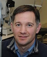 Kevin E. Houston合作论文数Harvard Medical School, Harvard University
8
Kevin E. Houston合作论文数Harvard Medical School, Harvard University
8 Jae-Hyun Jung合作论文数NVIDIA Corporation6Aaron J Mandel合作论文数6
 Gang Luo合作论文数Vision Research and Technology Lab, Schepens Eye Research Institute, Harvard Medical School, Harvard University;Massachusetts Eye and Ear5Qiu Cheng合作论文数Department of Psychology, University of Minnesota4
 Lauren Spano合作论文数Department of Psychology, University of Massachusetts Amherst4Concetta F. Alberti合作论文数Department of General Psychology, University of Padova4Henry L. Apfelbaum合作论文数4
 Merve Tuccar-Burak合作论文数3jeffrey churchill合作论文数3jean paul wiegand合作论文数2
 Daejoon Alex Hwang合作论文数Harvard Medical School, Harvard University
2
Daejoon Alex Hwang合作论文数Harvard Medical School, Harvard University
2 Eliot L. Berson合作论文数Department of Ophthalmology, Harvard Medical School, Harvard University2
 Rui Liu (刘睿)合作论文数2
 Elizabeth Dugan合作论文数From the Section on Social Sciences and Health Policy, Department of Public Health Sciences, Bowman Gray School of Medicine, Winston-Salem, North Carolina2Robert G. Giorgi合作论文数2Egor Ananev合作论文数1Zoraiz Qureshi合作论文数1
 Long To合作论文数1
 Kassandra R. Lee合作论文数Integrat Neurosci, Univ Nevada1
 Steven Savage合作论文数Dept Ophthalmol, Schepens Eye Res Inst Massachusetts Eye & Ear1Lily Zhang合作论文数1Joseph Donahue合作论文数1Kundu Bijoy K合作论文数Department of Radiology and Medical Imaging, University of Virginia1
 Sheldon Sarah合作论文数1Qu Tang合作论文数1
 Moosa Shayan合作论文数Department of Neurological Surgery, University of Virginia Health System1Patel Sohil H合作论文数1
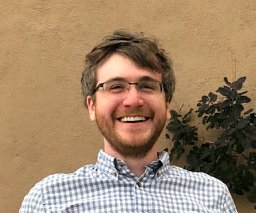 Swan Garrett合作论文数State College, Penn State University1P. Thomas Fletcher合作论文数University of Virginia;Scientific Computing and Imaging Institute, School of Computing, University of Utah1
 Ishaque Mariam合作论文数1
 Tanya K. Murphy合作论文数Department of Psychiatry, University of Florida1
 Xianping Fu (付先平)合作论文数Information Science and Technology College, Dalian Maritime University1Gary R. Geffken合作论文数1
 Eric Alan Storch合作论文数Department of Psychiatry & Behavioral Sciences, Baylor College of Medicine1Lawrence E. Arend合作论文数1Schepens Eye合作论文数1Christina Gambacorta合作论文数1
 Patrick H Finan合作论文数Department of Psychology, Arizona State University1Tamara Bond合作论文数1
 Wayne Goodman合作论文数Lee and Joe Jamail Specialty Care Center, Menninger Department of Psychiatry and Behavioral Sciences, Baylor College of Medicine1Randy D. Easton合作论文数1James Barabas合作论文数MIT1K Wooledge合作论文数1




