INTRODUCTION:Total body irradiation (TBI) practice varies globally. The optimal technique for TBI has not yet been determined, although guidelines and recommendations are available. Five years after the initial Australia and New Zealand (ANZ) practice survey in 2019, we report updated 2024 TBI practice patterns from 19 ANZ centres who are members of the ANZ Transplant and Cellular Therapies (ANZTCT) TBI Special Interest Group (TBISIG). METHOD:The 2024 survey was distributed in November 2024. Survey questions covered patient cohorts and caseloads, TBI regimens and dose-fractionations, TBI techniques, planning considerations, quality assurance, and treatment delivery. For all centres, one staff member from each discipline (Radiation Oncology, Medical Physics, and Radiation Therapy) was invited to respond. RESULTS:Responses were received from all 19 centres. Seven centres use modulated techniques in 2024, whereas only one centre delivered modulated TBI in 2019. Twelve centres use large open fields in 2024, six of which are considering changing to volumetric modulated arc therapy (VMAT) TBI. Compared to large open fields, modulated techniques (1) require dedicated patient immobilisation equipment and treatment planning software, (2) reduce dose to organs at risk (such as lungs and kidneys) below the prescription dose, (3) have higher dose rates, and (4) require more time for planning and treatment delivery. CONCLUSION:The 2024 survey showed diverse TBI practice patterns in ANZ. Compared to 2019, more centres are using or actively considering the implementation of modulated techniques, such as VMAT TBI, which adds to the complexity of treatment planning and delivery.
Purpose Cutaneous T-cell lymphomas (CTCL) are rare with increasing incidence. Most patients have skin-confined mycosis fungoides: a typically indolent, highly morbid, incurable disease requiring multi-lined treatment. For these patients, radiation therapy (RT) is efficacious, well-tolerated, and cost-effective; total skin electron beam therapy (TSEBT) is a specialized technique with proven quality-of-life benefits. Consensus on optimal treatment sequencing is lacking, creating heterogeneity in care. International data suggest declining RT-utilization. Using a state-wide, population-based, Australian cancer registry with linked data sets, we investigated RT-utilization for CTCL, examining for geographically based disparities. Correlations between dermatologist-availability and linac-density on incidence and RT-utilization rates were also explored. Methods and Materials Retrospective analysis of New South Wales Cancer Registry data set of all patients diagnosed with CTCL from 2009 to 2018, with data linkage to the Outpatient Radiation Oncology Database. Actual RT-utilization rates included all treatment lines, calculated per person. Results A total of 553 patients were diagnosed with CTCL; age-standardized incidence was 7.19/million/year. Geographically, 35% resided in regional/rural local health districts (LHDs); 13% in underaccessible locations; 15% >50 km road distance from the nearest RT center. RT-utilization and TSEBT-utilization rates were 29.0% and 1.8%, respectively. Variation existed between LHDs, with greater RT-utilization in regional/rural over metropolitan LHDs (odds ratio: 1.66; P = .01). Patients residing in major cities had lower RT-utilization than those in inner regional areas (odds ratio: 2.12; P = .001). Shortest road distance to the nearest RT center did not predict for greater RT-utilization. Disparities in access to specialist services existed between LHDs. Dermatologist-availability significantly predicted for diagnosis rates (R2 = 0.34; F(1,11) = 5.58; P = .04). The absence of RT facilities was associated with poor RT-utilization rates. Conclusions Over 10 years, only 29% of patients with CTCL received RT across all treatment lines, with significant geographic-based heterogeneity observed. Contrary to expectation, residence in major cities and proximity to RT facilities were associated with lower RT-utilization rates. Further advocacy is encouraged to overcome diagnostic and treatment disparities for patients with CTCL.
OBJECTIVES:The presence of nodal disease at presentation of a head and neck mucosal-based squamous cell carcinoma has a significant impact upon outcomes. METHODS:This is a retrospective, ethics-approved study in which patients with squamous cell carcinoma of the larynx, oropharynx, hypopharynx and oral cavity were reviewed and compared with respect to nodal disease (N0 vs N1-N2 vs N3). Patient, disease and treatment parameters were evaluated with ultimate local control, regional control, cancer-specific survival and overall survival investigated. RESULTS:In the cohort of 1265 patients, 764 presented with nodal disease (N3 = 60). The majority of the N3 group had oropharynx squamous cell carcinoma (52%) and experienced worse ultimate local control (63%; p < 0.001), regional control (67%; p < 0.001) and both squamous cell carcinoma and overall survival (log rank p < 0.001). CONCLUSION:Patients presenting with N3 nodal disease had poor regional control, a lower cancer-specific survival and a worse overall survival compared to patients with lesser to no nodal disease.
Primary laryngeal synovial sarcoma is a rare head and neck cancer. We describe a case of synovial sarcoma of the larynx in a previously well 9-year-old boy with a one-month history of a progressively enlarging neck lump. He was referred to our institution after incomplete surgical excision of the then undifferentiated neck mass. A partial laryngectomy including wide local excision of the residual mass was performed. An ipsilateral level I-III neck dissection was also performed concurrently. Clear re-excision margins were achieved. The neck nodes were all negative for metastatic disease. Adjuvant local radiotherapy treatment was administered to reduce the probability of local recurrence. Four years following treatment completion, the patient remains in remission with no signs of treatment-related morbidity. A review of the published literature on laryngeal synovial sarcoma was undertaken. This case represents the youngest patient to be diagnosed with the condition. Surgical excision represents the mainstay of treatment of laryngeal synovial sarcoma. At more common sites of disease, adjuvant radiotherapy has been associated with lower rates of recurrence. However, there is the potential for significant morbidity from irradiating the neck of a paediatric patient. This case report explores the challenges in treating young patients with aggressive head and neck cancers when faced with little available evidence to guide decision-making.
Searchable abstracts of presentations at key conferences in endocrinology ISSN 1470-3947 (print) | ISSN 1479-6848 (online)
Linear accelerator (LINAC) stereotactic radiosurgery (SRS) is a specialised treatment used for various brain conditions, including arteriovenous malformations (AVMs). This paper investigates the obliteration rate of AVMs treated by the LINAC-based facility at Prince of Wales Hospital, defines factors influencing obliteration rate, time to obliteration and complications post-SRS. A retrospective audit review of patient notes sourced from electronic medical records was conducted. During the study period 219 patients received treatment, of which the final status of 136 AVMs was known. Overall obliteration rate was 75.7 %, with obliteration rates of 5 %, 30 % and 46 % at one, three and four years, respectively. Post analysis, a radiosurgery dose of ≥ 18 Gray (Gy) was predictive of achieving obliteration (Odds Ratio (OR) 4.2, 95 % Confidence Interval (CI) 1.61–10.83, p = 0.003) whilst a nidus size of 3–6 cm was less likely of achieving obliteration (OR 0.2, CI 0.10–0.57, p = 0.001). Multivariate analysis showed a radiosurgery dose of ≥ 18 Gy remained predictive (OR 4.7, CI 1.69–13.25, p = 0.003) and a nidus size of 3–6 cm remained less likely to achieve obliteration (OR 0.2, CI 0.10–0.57, p = 0.001). Females were predictive of developing temporary complications post-SRS in multivariate analysis (OR 2.8, CI 1.24–6.13, p = 0.013), and having > 1 SRS session was predictive of developing permanent complications post-SRS (OR 7.1, CI 2.44–20.53, p < 0.001). The obliteration rate achieved from our study and the predictive nature to achieve obliteration with a radiosurgery dose of ≥ 18 Gy is comparable to existing literature including that using the Gamma-Knife system.
The impact of computed tomography-defined sarcopenia on outcomes in head and neck cancer has been well described. Sarcopenic obesity (SO) (depleted muscle mass combined with obesity) may pose a more serious risk than either condition alone. We investigated SO and its impact on survival and critical weight loss (≥ 5 %) in patients with head and neck cancer who received curative radiotherapy (± other modalities). Retrospective analysis of computed tomography cross-sectional muscle at cervical (C3), thoracic (T2) and lumbar (L3) regions was conducted. Patients were grouped by BMI and sarcopenia status based on established thresholds. A total of 413 patients were included for analysis, the majority having oropharyngeal carcinoma (52 %), and 56 % received primary concurrent chemoradiotherapy. The majority of the cohort (65 %) was overweight or obese (BMI ≥ 25 kg/m2). Sarcopenia was found in 43 %, with 65 % having SO (n 116), equating to 28 % of the whole cohort. Critical weight loss was experienced by 58 % (n 238). A significantly higher proportion of patients with SO experienced critical weight loss (n 70 v. 19, P < 0·001) and were four times more likely to do so during treatment (OR 4·1; 95 % CI 1·5, 7·1; P = 0·002). SO was not found to impact on overall or cancer-specific survival; however, in patients with sarcopenia, those with SO had better overall survival (median 9·1 v. 7·0 years; 95 % CI 5·2, 16·8; P = 0·021). SO at the time of presentation in patients with head and neck cancer is predictive of critical weight loss during treatment, and muscle evaluation can be useful in identifying patients at nutritional risk regardless of BMI and obvious signs of wasting.
This systematic review examines the role of dosimetric parameters in predicting temporal lobe necrosis (TLN) risk in nasopharyngeal carcinoma (NPC) patients treated with three-dimensional conformal RT (3D-CRT), intensity-modulated radiation therapy (IMRT) and volumetric modulated arc therapy (VMAT). TLN is a serious late complication that can adversely affect the quality of life of NPC patients. Understanding the relationship between dosimetric parameters and TLN can guide treatment planning and minimize radiation-related complications. A comprehensive search identified relevant studies published up to July 2023. Studies reporting on dosimetric parameters and TLN in NPC patients undergoing 3D-CRT, IMRT, and VMAT were included. TLN incidence, follow-up duration, and correlation with dosimetric parameters of the temporal lobe were analyzed. The review included 30 studies with median follow-up durations ranging from 28 to 110 months. The crude incidence of TLN varied from 2.3 % to 47.3 % and the average crude incidence of TLN is approximately 14 %. Dmax and D1cc emerged as potential predictors of TLN in 3D-CRT and IMRT-treated NPC patients. Threshold values of >72 Gy for Dmax and >62 Gy for D1cc were associated with increased TLN risk. However, other factors should also be considered, including host characteristics, tumor-specific features and therapeutic factors. In conclusion, this systematic review highlights the significance of dosimetric parameters, particularly Dmax and D1cc, in predicting TLN risk in NPC patients undergoing 3D-CRT, IMRT, and VMAT. The findings provide valuable insights that can help in developing optimal treatment planning strategies and contribute to the development of clinical guidelines in this field.
Background: Despite introduction of extranodal extension (ENE) into the AJCC 8th edition of oral cancer staging, previous criticisms persist, such as limited discrimination between sub-stages and doubtful prognostic value of contralateral nodal disease. The purpose of this study was to compare our novel nodal staging system, based on the number of positive nodes and ENE, to the AJCC staging system in surgically treated patients. Methods: Retrospective analysis of 4710 patients with oral squamous cell carcinoma (OSCC) treated with surgery +/- adjuvant therapy in 8 institutions in Australia, North America and Asia. With overall survival (OS) and disease specific survival (DSS) as endpoint, the prognostic performance of AJCC 8th and 7th editions were compared using hazard consistency, hazard discrimination, likelihood difference and balance. Results: Our new nodal staging system (PN) a progressive and linear increase in hazard ratio (HR) from pN0 to pN3, with good separation of Kaplan Meier curves. Using the predetermined criteria for evaluation of a staging system, our proposed staging model outperformed AJCC 8th and 7th editions in prediction of OS and DSS. Conclusion: PN was the lymph node staging system that provided the most accurate prediction of OS and DSS for patients in our cohort of OSCC. Additionally, it can be easily adopted, addresses the shortcomings of the existing systems and should be considered for future editions of the TNM staging system.
BACKGROUND:Late-stage progressive decline of swallowing function after radiotherapy for head and neck cancer (HNC) is often difficult to monitor. This study examined the feasibility and clinical outcomes of speech-language pathology implementing flexible endoscopic evaluation of swallow (FEES) screening during annual cancer surveillance visits to monitor late-stage swallowing function. METHODS:Patients >2 years post treatment who attended routine oncological visits underwent FEES screening. Feasibility (service data, stakeholder survey) and swallowing outcomes (oral intake, secretions, internal lymphedema, penetration-aspiration, and residue) were collected. RESULTS:Screening was completed with 70% (50/71) of eligible patients. Medical staff and speech-language pathologists indicated the protocol was worthwhile and achievable to incorporate into practice. Almost all patients were willing to complete the protocol annually. FEES outcomes identified 84% with dysphagia versus only 26% self-reported dysphagia. CONCLUSION:Findings indicate FEES screening incorporated into annual oncological reviews is feasible and effective at monitoring late-stage swallowing function following HNC.
Purpose:To discuss the role of proton beam therapy (PBT) in the treatment of patients with oropharyngeal squamous cell carcinoma (OPSCC). Materials and Methods:A review of the pertinent literature. Results:Proton beam therapy likely results in reduced acute and late toxicity as compared with intensity-modulated radiation therapy (IMRT). The extent of the reduced toxicity, which may be modest, depends on the endpoint and technical factors such as pencil beam versus passive scattered PBT and adaptive replanning. The disease control rates after PBT are likely similar to those after IMRT. Conclusion:Proton beam therapy is an attractive option to treat patients with OPSCC. Whether it becomes widely available depends on access.
Radiotherapy (RT) continues to play a key role in the management of head and neck cancer (HNC). Xerostomia remains a principal detriment to the quality of life (QoL) for 80 % of surviving patients receiving head and neck radiation. Radiation-induced injury to the salivary glands is dose-dependent, and thus efforts have been focused on decreasing radiation to the salivary glands. Decreased saliva production reduces both short-term and long-term quality of life in head and neck survivors by impacting on taste and contributing to dysphagia. Several radioprotective agents to the salivary gland have been investigated. Although not widely practiced, surgical transfer of the submandibular gland prior to RT is the mainstay of surgical options in preventing xerostomia. This review focuses on the strategies to improve xerostomia following radiation therapy in head and neck cancers.
Evaluation of skeletal muscle (SM) depletion, or sarcopenia, utilizes the cross-sectional area (CSA) of computed tomography (CT) scans at the lumbar level L3. However, alternate vertebral landmarks are used in patients with head and neck cancer due to scan unavailability. Muscle changes following radiotherapy at cervical (C3) and thoracic (T2) levels were compared to L3 in patients with oropharyngeal carcinoma. Muscle density data were derived retrospectively from diagnostic PET-CT scans at C3, T2 and L3 pretreatment, and up to six months post. CSA changes were compared to L3 in scans of 33 patients (88% male, mean age 61 (SD 8.5) years). On matched pair analysis; mean L3-CSA change -12.1 cm(2) (SD 9.7, 95%CI -15.5 to -8.6, and p < 0.001), T2-CSA -30.5 cm(2) (SD 34.8, 95%CI -42.8 to -18.1, and p < 0.001) and C3-CSA +2.1 cm(2) (SD 4.1, 95%CI 0.63 to 3.5, and p < 0.00). No difference was found in the percentage change of T2-CSA with L3-CSA (mean -2.2%, SD 10.6, 95%CI -6.0 to 1.6, and p = 0.240), however, was significantly different to C3-CSA (mean 13.2%, SD 11.6, 95%CI 9.1 to 17.3, and p < 0.001). Results suggest SM at C3 does not change proportionately and may not be a reliable representation of whole-body SM change over time.
1 Reitman ZJ, Yan H. Isocitrate dehydrogenase 1 and 2 mutations in cancer: alterations at a crossroads of cellular metabolism. J Natl Cancer Inst 2010; 102: 932–941. 2 Issa GC, DiNardo CD. Acute myeloid leukemia with IDH1 and IDH2 mutations: 2021 treatment algorithm. Blood Cancer J 2021; 11: 107. 3 Balin SJ, Wetter DA, Kurtin PJ, et al. Myelodysplastic syndrome presenting as generalized granulomatous dermatitis. Arch Dermatol 2011; 147: 331–335. 4 Kyriakou A, Patsatsi A, Papadopoulos V, et al. A case of palisaded neutrophilic granulomatous dermatitis with subsequent development of chronic myelomonocytic leukemia. Clin Case Rep 2019; 7: 695–698. 5 Federmann B, Bonzheim I, Yazdi AS, et al. Generalized palisaded neutrophilic and granulomatous dermatitis—a cutaneous manifestation of chronic myelomonocytic leukemia? A clinical, histopathological, and molecular study of 3 cases. Hum Pathol 2017; 64: 198–206.
Rationale: Patients with head and neck cancer (HNC) are at high nutritional risk. Validated nutrition assessment tools in cancer include the patient-generated subjective global assessment (PG-SGA), as a measure of nutritional status incorporating physical parameters. Measurement of skeletal muscle (SM) utilising computed tomography (CT) scans has become an additional method for risk stratification. The aim of this study was to compare assessment via the PG-SGA with thoracic SM. Methods: This retrospective study included newly diagnosed patients with HNC who had completed radiotherapy (RT) and had a diagnostic CT scan suitable for analysis. PG-SGA was completed prior to RT, and thoracic SM was assessed at the second thoracic vertebra (T2). Patients were categorized by nutritional status (PG-SGA-A - well nourished, or B - moderately malnourished) and thoracic SM index (T2SMI, cm2/m2), (high/low), stratified by sex and BMI. Predictors of weight loss during RT, overall survival (OS) and feeding tube use were compared between groups. Results: A total of 119 patients were assessed (86% male, mean age 60 yrs), most with oropharynx cancer (72%). Low T2SMI was found in 38% (n=45) and moderate malnutrition (PG-SGA-B) in 16% (n=19). No patients were assessed as severely malnourished at diagnosis. Of those with PG-SGA-A, 37% had low T2SMI. The only predictor of weight loss ≥5% was concurrent chemoRT (HR 12.7, 95%CI 4.2-38.5, p<0.001). Worse OS was found in patients with low T2SMI (Log Rank p=0.038), and PG-SGA-B (Log Rank p=0.028). When combined, patients with PG-SGA-B had worse OS regardless of T2SMI, however, low T2SMI with PG-SGA-A also had worse OS (Log Rank p=0.026). More feeding tubes were required in patients with PG-SGA-A and high T2SMI. Conclusion: PG-SGA combined with T2SMI measures allows for more robust nutritional assessment of patients with HNC. Disclosure of Interest: None declared
Cancer centres rely on electronic information in oncology information systems (OIS) to guide patient care. We investigated the completeness and accuracy of routinely collected head and neck cancer (HNC) data sourced from an OIS for suitability in prognostic modelling and other research. Three hundred and fifty-three adults diagnosed from 2000 to 2017 with head and neck squamous cell carcinoma, treated with radiotherapy, were eligible. Thirteen clinically relevant variables in HNC prognosis were extracted from a single-centre OIS and compared to that compiled separately in a research dataset. These two datasets were compared for agreement using Cohen’s kappa coefficient for categorical variables, and intraclass correlation coefficients for continuous variables. Research data was 96% complete compared to 84% for OIS data. Agreement was perfect for gender (κ = 1.000), high for age (κ = 0.993), site (κ = 0.992), T (κ = 0.851) and N (κ = 0.812) stage, radiotherapy dose (κ = 0.889), fractions (κ = 0.856), and duration (κ = 0.818), and chemotherapy treatment (κ = 0.871), substantial for overall stage (κ = 0.791) and vital status (κ = 0.689), moderate for grade (κ = 0.547), and poor for performance status (κ = 0.110). Thirty-one other variables were poorly captured and could not be statistically compared. Documentation of clinical information within the OIS for HNC patients is routine practice; however, OIS data was less correct and complete than data collected for research purposes. Substandard collection of routine data may hinder advancements in patient care. Improved data entry, integration with clinical activities and workflows, system usability, data dictionaries, and training are necessary for OIS data to generate robust research. Data mining from clinical documents may supplement structured data collection.
The cross‐sectional area (CSA) of skeletal muscle (SM) at the third lumbar vertebra (L3) is used to determine computed tomography (CT)‐defined sarcopenia. We investigated the feasibility of SM assessment at the second thoracic vertebra (T2) in patients with head and neck cancer (HNC).
Purpose Computed tomography (CT)-defined sarcopenia, as a measurement of low skeletal muscle (SM), is a poor prognostic indicator in patients with head and neck cancer (HNC), independent of weight or nutritional status. We used SM measures at the second thoracic vertebra (T2) to determine T2-SM index (SMI) thresholds for sarcopenia, and investigate the impact of low T2-SMI on overall survival (OS), and weight loss during radiotherapy (RT). Methods Adult patients with newly diagnosed HNC with a diagnostic PET–CT or RT planning CT scan were included. SM was analysed at T2 and a model applied to predict SM at L3. T2-SMI thresholds for sarcopenia were established with predicted measures, stratified by BMI and sex. Impact of sarcopenia and low T2-SMI on OS and weight loss during RT was investigated. Results A total of 361 scans were analysed (84% males, 54% oropharynx tumours). Sarcopenia was found in 49%, demonstrating worse OS ( p = 0.037). T2-SMI cutoff values were: females—74 cm 2 /m 2 [area under the curve (AUC): 0.89 (95%CI 0.80–0.98)], males (BMI < 25)—63 cm 2 /m 2 [AUC 0.93 (95%CI 0.89–0.96)], males (BMI ≥ 25)—88cm 2 /m 2 [AUC 0.86 (95%CI 0.78–0.93)]. No difference in OS with T2-SMI categories. Lowest T2-SMI quartile of < 63 cm 2 /m 2 demonstrated worse OS ( p = 0.017). Weight loss during RT was higher in patients; who were not sarcopenic (6.2% vs 4.9%, p = 0.023); with higher T2-SMI (6.3% vs 4.9%, p = 0.014) and; in the highest quartiles (3.6% vs 5.7% vs 7.2%, p < 0.001). Conclusions These T2-SMI thresholds are effective in assessing CT-defined sarcopenia in HNC. Further assessment of clinical application is warranted.
PURPOSE There is limited knowledge of the prediction of 2-year cancer-specific survival (CSS) in the head and neck cancer (HNC) population. The aim of this study is to develop and validate machine learning models and a nomogram for the prediction of 2-year CSS in patients with HNC using real-world data collected by major teaching and tertiary referral hospitals in New South Wales (NSW), Australia. MATERIALS AND METHODS Data collected in oncology information systems at multiple NSW Cancer Centres were extracted for 2,953 eligible adults diagnosed between 2000 and 2017 with squamous cell carcinoma of the head and neck. Death data were sourced from the National Death Index using record linkage. Machine learning and Cox regression/nomogram models were developed and internally validated in Python and R, respectively. RESULTS Machine learning models demonstrated highest performance (C-index) in the larynx and nasopharynx cohorts (0.82), followed by the oropharynx (0.79) and the hypopharynx and oral cavity cohorts (0.73). In the whole HNC population, C-indexes of 0.79 and 0.70 and Brier scores of 0.10 and 0.27 were reported for the machine learning and nomogram model, respectively. Cox regression analysis identified age, T and N classification, and time-corrected biologic equivalent dose in two gray fractions as independent prognostic factors for 2-year CSS. N classification was the most important feature used for prediction in the machine learning model followed by age. CONCLUSION Machine learning and nomogram analysis predicted 2-year CSS with high performance using routinely collected and complete clinical information extracted from oncology information systems. These models function as visual decision-making tools to guide radiotherapy treatment decisions and provide insight into the prediction of survival outcomes in patients with HNC.
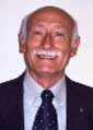 Alfio Ferlito合作论文数ENT Clinic, University of Udine School of Medicine/International Head and Neck Scientific Group27
 Primož Strojan合作论文数24
 William M. Mendenhall合作论文数Department of Radiation Oncology, College of Medicine, University of Florida21Alessandra Rinaldo合作论文数Department of Surgical Sciences, ENT Clinic, University of Udine21
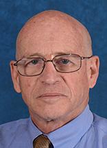 Avraham Eisbruch合作论文数Department of Radiation Oncology, University of Michigan20Belinda Vangelov合作论文数15
 June Corry合作论文数Division of Radiation Oncology Locked Bag No 1, Peter MacCallum Cancer Centre15
 Jonathan J Beitler合作论文数Medical College of Wisconsin14Janet K. Williams合作论文数14
 Lee, WMA (李詠梅)合作论文数Department of Clinical Oncology, School of Clinical Medicine, LKS Faculty of Medicine, University of Hong Kong14Gp Bridger合作论文数12
 Carlos Suárez合作论文数Serv Otorrinolaringol, Hosp Univ Cent Asturias12Damian P Kotevski合作论文数12Williams Janet R合作论文数12Rodrigo Juan P合作论文数10
 J.A. (Hans) Langendijk合作论文数Faculty of Medical Sciences, University of Groningen10
 Antti Mäkitie合作论文数Department of Ophthalmology and Otorhinolaryngology, Faculty of Medicine, University of Helsinki;Department of Clinical Science, Intervention and Technology, Karolinska Institutet;Helsinki University Hospital10Kathryn Broadley合作论文数9
 Nabil Saba合作论文数Department of Hematology and Medical Oncology, School of Medicine, Emory University;Winship Cancer Institute, Emory University9Ross Darius Farhadieh合作论文数9
 Sandra Nuyts合作论文数Department of Radiation Oncology, UZ Leuven;Department of Oncology, KU Leuven, University of Leuven9Judith Bauer合作论文数8
 Pamela J. Russell合作论文数8
 Maria Kangas合作论文数Centre for Emotional Health, Department of Psychology, Macquarie University8
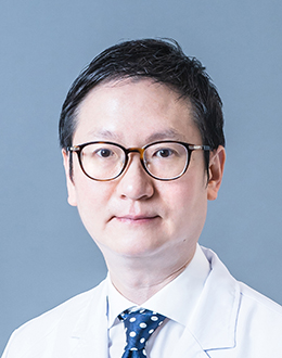 Wai Tong Ng (吴伟棠)合作论文数Li Ka Shing Faculty of Medicine, The University of Hong Kong;Department of Clinical Oncology, School of Clinical Medicine, The University of Hong Kong7Arash Salardini合作论文数Yale University7Rachelle A. Robinson合作论文数6
 Molly Barnhart合作论文数Sch Hlth & Rehabil Sci, Univ Queensland6Andrés Coca-Pelaz合作论文数6
 Elizabeth Ward合作论文数School of Health and Rehabilitation Sciences, Faculty of Health and Behavioural Sciences, The University of Queensland6Matthew Field合作论文数South Western Sydney Clinical School, Faculty of Medicine, The University of New South Wales6
 Daniel Aaron Moses合作论文数Graduate School of Biomedical Engineering, Faculty of Engineering, The University of New South Wales6
 R. de Bree合作论文数Department of Head and Neck Surgical Oncology, University Medical Center Utrecht6
 Michael Veness合作论文数Faculty of Medicine and Health, University of Sydney6
 Jia-Lin Yang合作论文数Oncology Research Centre, Prince of Wales Hospital6G Patrick Bridger合作论文数5Heleen Bollen合作论文数5
 Meagher Nicola S合作论文数Prince of Wales Clinical School, University of New South Wales5Michael Baldwin合作论文数5
 Claire Vajdic合作论文数Kirby Institute, University of New South Wales5
 Marcus Stoodley合作论文数Macquarie Medical School, Faculty of Medicine, Health and Human Sciences, Macquarie University5Sophie J. Chandler合作论文数4
 Fernando Lopez合作论文数4Virginia A Simms合作论文数4
 Richard Gallagher合作论文数Dept Med, Univ Notre Dame4
 David Goldstein合作论文数Department of Otolaryngology-Head and Neck Surgery, Temerty Faculty of Medicine, University of Toronto;Institute of Medical Science, Temerty Faculty of Medicine, University of Toronto;Department of Surgical Oncology, Princess Margaret Cancer Centre, University Health Network;Women's College Hospital4
 Kevin Thomas Robbins合作论文数4
 Robert Takes合作论文数Radboud University Medical Center4Balasubramanian Deepak合作论文数4




