Introduction:Whole blood viscosity (WBV), reflecting the intrinsic resistance of blood flow, is an established predictor of stroke events in individuals. This study aims to correlate the WBV at different shear rates with the outcome of mechanical thrombectomy, known to be an effective treatment for large vessel occlusion (LVO) stroke. Method:This is a single-center retrospective study conducted at our comprehensive stroke center. The charts of 317 patients who underwent mechanical thrombectomy within 6 h of LVO stroke presentation were reviewed. The modified Rankin score (mRS) at discharge was used as the outcome measure, with individuals categorized as low (0-2) or high (3-6). WBV at different shear rates was calculated using De Simone's Formula. The T-test and Chi-square test were used to compare baseline continuous and categorical data, respectively, amongst the mRS study groups. We utilized multivariable logistic regression analyses to identify the independent risk factors associated with the outcome of interest following mechanical thrombectomy. In addition, Spearman rank order correlation was used to assess for r value between mRS and WBV at different shear rates. Results:Baseline group characteristics, including demographics and medical history, were similar among the two study groups. Of note, our study found no significant differences in clinical outcomes between the two groups with WBV at high shear rate (OR 0.969, 95% CI 0.77-1.204, p = 0.780) and low shear rate (OR 0.998, 95% CI 0.988-1.008, p = 0.779) following mechanical thrombectomy. Spearman rank order correlation between mRS at discharge with WBV at high shear rate (r = 0.058, p = 0.123) and low shear rate (r = 0.048, p = 0.128) was non-significant. Discussion:There is limited information of the effect of WBV at high and low shear rates on the clinical outcome following mechanical thrombectomy in patients with LVO. Our results revealed that WBV at high and low shear rates did not impact the functional outcome of mechanical thrombectomy. This result might be affected by the potential limitation of the formula used to derive the given shear rates. Despite this lack of association observed in our study, other contributors of viscosity may still potentially play a significant role in outcome following mechanical thrombectomy.
Introduction To evaluate patients acute cerebral ischemia in order to assess for factors which may help to differentiate patients with small vessel involvement from those with large vessel involvement in an effort to determine diagnostic yield of vascular imaging. Material and Method We prospectively and consecutively evaluated all acute ischemic stroke patients at our medical center from May 16, 2021 to December 10, 2021. Distinction between small vessel and large vessel involvement was based upon clinical presentation, the results of brain imaging and either computed tomographic angiography, in the vast majority, or magnetic resonance angiography. Patient demographics and risk factors for stroke as well as therapeutic intervention was assessed. Conclusion and Result Of the 90 patients studied, 59 had large vessel ischemia (66%) with 26 (44%) having large vessel occlusion and one had symptomatic high-grade middle cerebral artery stenosis. Conversely, none of the 31 patients with small vessel presentation (34%) had large vessel occlusion or high-grade stenosis. In addition, 19 out of 59 (32%) large vessel patients compared to 2 of 31 (6%) of the small vessel patients had atrial fibrillation identified as a potential mechanism with a p-value of 0.01 by univariate analysis and 0.17 by multivariate analysis. The routine use of vascular imaging in acute ischemic stroke is of very low yield in small vessel presentation with the presence of potential cardiogenic emboli is also relatively low. Efforts at accelerated identification of a small vessel mechanism, to avoid unnecessary testing, should provide significant value from both a patient management and cost standpoint.
Objective: To determine if elevated inflammatory markers are associated with worse functional outcomes in SARS-CoV-2 positive patients presenting with acute cerebrovascular events. Background: The association of acute Coronavirus disease 2019 (COVID-19) with both arterial and venous thromboembolic events is well-established. One of the several proposed mechanisms is excessive inflammatory response. We aim to assess if higher inflammatory markers are associated with worse functional outcomes Design/Methods: Medical records of adult patients admitted to our institution between 3/2020 and 8/2021 with positive SARS-COV-2 and acute cerebrovascular events were reviewed (n=29). Demographics, neuroradiological and pertinent lab findings were collected. Functional outcomes were assessed using binarized mRS scores (worse 4–6 vs better 0–3) ascertained on discharge day. A series of logistic regression models were utilized to assess the association of inflammatory markers with stroke outcomes. Variables assessed were; [admission day heart rate, temperature and white blood cell count], binarized maximum procalcitonin ( > 0.1 ng/mL vs </= 0.1 ng/mL) and maximum ferritin level. All analyses were conducted using SAS software. Results: Among 29 patients (mean age 63.6 +/− 13.4 years, 65.5% males), tested parameters of inflammation were not shown to be significantly associated with stroke outcomes in an adjusted logistic regression model. When assessing stroke outcome via binarized modified Rankin stroke scale, binarized maximum procalcitonin level showed 1.95 times the odds of a poor outcome (OR: 1.95, 95% CI: 0.05, 84.3, p-value: 0.73). Additionally, temperature on admission showed 1.59 times the odds of a poor outcome (OR: 1.59, 95% CI: 0.42, 6.07, p-value: 0.17). Conclusions: Among patients with acute cerebrovascular events associated with positive SARS-COV-2 result, no significant associations of worse functional outcomes in those with elevated inflammatory markers were observed. A trend of higher mRS scores on discharge in those with elevated procalcitonin and those with fever on admission was noted. Further research is needed to confirm these findings Disclosure: Dr. Elsekaily has nothing to disclose. Dr. Ansari has nothing to disclose. Dr. Sheikh has nothing to disclose. Dr. Ayub has nothing to disclose. Dr. Triay has nothing to disclose. Miss Angelette has nothing to disclose. Ms. Gaudet has nothing to disclose. Dr. Kelley has nothing to disclose.
Objective: To analyze the effects of co-morbidities established as stroke risk factors in the setting of coronavirus disease 2019 (COVID-19). Background: Given the protean sequelae of COVID-19 and its emerging interplay on chronic disease states, the effects of well-established stroke risk factors must be analyzed in this context. Design/Methods: We conducted a retrospective analysis on 29 patients who presented with acute stroke and were found to be COVID-19 positive from 3/2020 through 8/2021. Information was collected on demographic variables, medical history, and pertinent clinical parameters. Stroke outcomes were assessed via the following: binarized modified Rankin scale score (mRSS) on discharge (4–6: poor outcome vs 0–3: good outcome [ref]), and whether mortality resulted. A series of logistic regression models adjusted for age, gender, body mass index, presence of diabetes mellitus (DM), presence of hyperlipidemia, prior history of coronary artery disease and/or cerebrovascular disease, and mean arterial pressure at admission were run to assess the association of these parameters with stroke outcomes in a cohort of COVID-19 positive stroke patients. All analyses were conducted using SAS Studio OnDemand for Academics (SAS Institute Inc., Cary, NC). Results: Among 29 COVID-19 patients (mean age 63.6 ± 13.4 years, 65.5% males), tested clinical parameters were not shown to be significantly associated with stroke outcomes across a series of logistic regression models. Regarding mortality, presence of DM showed 5.89 times the odds of mortality (OR: 5.89; 95% CI: 0.67, 52.47; p-value: 0.11). When assessing for binarized mRSS at discharge, presence of DM showed 2.97 times the odds of a poor outcome (OR: 2.97, 95% CI: 0.27, 32.58; p-value: 0.37). Conclusions: Analysis via adjusted logistic regression models revealed a statistically insignificant positive association between DM and both outcomes: worse mRSS and mortality. Further research is needed with larger patient cohorts and employing matched controls without COVID. Disclosure: Dr. Sheikh has nothing to disclose. Dr. Ansari has nothing to disclose. Dr. Elsekaily has nothing to disclose. Dr. Ayub has nothing to disclose. Dr. Triay has nothing to disclose. Miss Angelette has nothing to disclose. Ms. Gaudet has nothing to disclose. Dr. Kelley has nothing to disclose.
Objective: To prospectively compare recovery outcomes, presentation with NIH stroke scale and modified ranking scale (MRS) in the study population with follow up at discharge. Background: Earlier studies suggested lacunar stroke has a better prognosis compared to other strokes, but recent studies show prognosis of lacunar stroke is simiar or worse when compared to large strokes. Design/Methods: We prospectively collected demographic information, admission NIH stroke scale, and modified ranking scale with serial measurements during hospitalized course in single tertiary care hospital in six-month period(n=74). We also collected information on discharge disposition between lacunar and non-lacunar stroke groups. Two groups adjusted for sex and race. Independent t-test is used for comparing continuous variables and chi-square test for comparing categorical variables. Results: There were 30 lacunar stroke patients and 44 non-lacunar stroke patients with mean age of 60 years for lacunar stroke patients and 70 for non-lacunar stroke patients. Mean admission NIH stroke scale was 11 in the non-lacunar group with MRS of 0.4 compared to 6 (p= 0.009) and 1(p=0.006), respectively in the lacunar group. Discharge NIH and MRS values were 7 and 2.3 respectively for the non-lacunar group compared to 4(p=0.03) and 2.2(p=0.88) for the lacunar group. ΔNIH (admit-discharge) is 4 and 2 for non-lacunar and lacunar group, respectively. ΔMRS (admit-discharge) is −1.9 and −1.3 for non-lacunar and lacunar group, respectively. Discharge to home was 46.7% & 37% (p=0.03) in non-lacunar group and lacunar group, respectively. Discharge to Skilled nursing facility was 13.3% and 18.5%(p=0.03) in non-lacunar group and lacunar group, respectively. Conclusions: Despite the relatively small volume of infarct associated with lacunar stroke compared to typically large infarcts seen with non-lacunar stroke, we found that the outcome was similar if not worse in terms of recovery for lacunar type stroke based upon prospective information collected in our six-month database extending from May 2021 to November Disclosure: Dr. Buchhanolla has nothing to disclose. Dr. PANDEY has nothing to disclose. Dr. Bir has nothing to disclose. Dr. Javalkar has nothing to disclose. Dr. Kelley has nothing to disclose.
To identify and analyze the clinical characteristics of the patients that had stroke-like symptoms (stroke mimics) on presentation and received IV tPA but subsequently were found not to have an ischemic stroke.
Objective: To assess the effect of pertinent co-morbidities and imaging findings on functional outcomes in patients with Atrial fibrillation (AF) taking oral anticoagulation (OAC) therapy who developed acute ischemic stroke (AIS). Background: Per current guidelines, most patients with AF are treated with OACs. However, these patients can still develop AIS. Exploring the underlying factors associated with worse functional outcomes is crucial. Design/Methods: We retrospectively reviewed 33 adult patients with AF on long-term OACs presenting with AIS. Information collected included demographic variables, medical history, and pertinent clinical and imaging parameters. A series of logistic regression models adjusted for age, gender, hypertension, diabetes, hyperlipidemia, and LVO were run to analyze the association of these parameters with AIS outcomes, assessed as binarized modified Rankin scale score (mRSS) (4–6: worse outcome vs 0–3: better outcome [ref]) and binarized NIH stroke scale (NIHSS) (>15: worse outcome vs 0–15: better outcome [ref]) at discharge. All analyses were conducted using SAS Studio OnDemand for Academics (SAS Institute Inc., Cary, NC). Results: Among 33 patients (mean age 74 + 13.6 years, 51.5% males) assessed for binarized mRSS, presence of LVO showed 21.1 times the odds of a poor outcome (OR: 21.1, 95% CI: 1.68, 265.27, p=0.02). When assessed for NIHSS at discharge, presence of LVO showed 3.82 times the odds of a poor outcome (OR: 3.82, 95% CI: 0.37, 39.03, p=0.26). When assessed for binarized mRSS outcome, hyperlipidemia showed 9.87 times the odds of a poor outcome (OR: 9.87, 95% CI: 0.9, 108.13, p=0.06). When assessed for binarized NIHSS at discharge, hyperlipidemia showed 18.4 times the odds of a poor outcome (OR: 18.4, 95% CI: 1.3, 261.28, p=0.03). Conclusions: Our analysis revealed significant associations between the presence of LVO and worse mRSS at discharge and between HLD and worse NIHSS at discharge. However, further research is needed by employing matched controls. Disclosure: Dr. Ayub has nothing to disclose. Dr. Ansari has nothing to disclose. Dr. Sheikh has nothing to disclose. Dr. Elsekaily has nothing to disclose. Dr. Buchhanolla has nothing to disclose. Dr. Neshatfar has nothing to disclose. Dr. Chappidi has nothing to disclose. Dr. Parvathaneni has nothing to disclose. Dr. Triay has nothing to disclose. Ms. Gaudet has nothing to disclose. Nimer Abushehab has nothing to disclose. Hugo Cuellar-Saenz has received personal compensation in the range of $5,000-$9,999 for serving as a Consultant for Microvention. Dr. Kelley has nothing to disclose. Dr. Javalkar has nothing to disclose.
A 50-year-old male with human immunodeficiency virus (HIV) infection, on highly active antiretroviral therapy, presented with new-onset generalized tonic-clonic seizures and right hemiparesis. On physical examination, no skin rash was noted, and the patient denied any prior history of skin lesions or rashes. The neurological examination demonstrated right upper and lower extremity weakness (Medical Research Council grade 3/5) but was otherwise unremarkable. Laboratory work revealed pancytopenia, Creactive protein of 14 mg/L, CD4+ of 8, and a viral load of 102,491. The magnetic resonance imaging (MRI) of the brain with and without contrast demonstrated multiple ring-enhancing lesions in the bilateral cerebral hemispheres (right > left) on the T1 post-contrast sequence (Figure 1). The findings on the diffusion weighted imaging and the apparent diffusion coefficient sequences were not consistent with ischemic strokes. The gradient echo sequence did not demonstrate any hemorrhagic strokes. The computed tomography angiography was unremarkable with no segmental stenosis or occlusions. Serum Toxoplasma gondii IgM and IgG were negative. Cerebrospinal fluid (CSF) analysis revealed a white blood cell count of 1, red blood cell count of 8, glucose of 51, protein of 68, and oligoclonal bands of 0. CSF polymerase chain reaction was positive for varicella-zoster virus (VZV) and negative for herpes simplex virus 1/2, Epstein-Barr virus, John Cunningham virus, Cryptococcus neoformans, Toxoplasma gondii, Enterococcus faecalis, and Mycobacterium tuberculosis. Intravenous acyclovir therapy was initiated. Repeat CSF analysis and MRI of the brain after intravenous acyclovir therapy revealed clearance of the VZV infection and an interval decrease in the multiple ringenhancing lesions (Figure 1), respectively. A biopsy of the ring-enhancing lesion was offered to the patient for a definitive diagnosis; however, the patient refused. The patient was discharged on maintenance acyclovir therapy with no further reported recurrences. Discussion
Carotid stenosis is an important contributor to ischemic stroke risk with resultant significant impact on neurological disability and death in adults and with worldwide implications. Management of carotid stenosis is impacted by whether there are associated symptoms along with the degree of stenosis. Understanding of the pathogenesis of carotid atherosclerosis or stenosis is important in management of carotid stenosis. Atherosclerotic plaque formation is a chronic insidious process with a number of potential contributors to the formation of such a plaque. The definition of atherosclerosis is not simply limited to abnormal deposition of lipid but also includes a chronic, complex, inflammatory process. Molecularly, in atherosclerosis, there is decreasing nitric oxide (NO) bioavailability, activity and/or expression of endothelial NO synthase, or increasing degradation of NO secondary to enhanced superoxide production. These above changes cause endothelial dysfunction leading to formation of foam cell followed by formation on lipid plaque. After lipid plaque formation, stable or unstable atherosclerotic plaque is formed depending on the calcium deposition over the lipid plaque. It continues to be clearly established that carotid intervention for symptomatic high-grade carotid stenosis is best managed with intervention either by carotid endarterectomy or carotid stenting. However, asymptomatic carotid stenosis is the subject of considerable controversy in terms of optimal management. This review of carotid atherosclerosis is an attempt to incorporate the information provided by more recent studies on pathogenesis and management which may help in the decision-making process for optimal management for protection against stroke.
To determine the potential prevalence of thrombophilia and its association in cerebral infarction (CI) in a nationwide database, as well as to assess the potential effect of antithrombotic therapy on patients with identified hypercoagulability and CI.
Objective: The identification of hypercoagulability with acute ischemic stroke can impact on management and potential outcome. We sought to determine the potential effect of antithrombotic therapy on outcome in patients with identified hypercoagulability and cerebral infarct in a nationwide data bank of hospital admissions. Method: We assessed data from the Nationwide Inpatient Sample (NIS) from 2016 to 2018. We looked for the potential association of hypercoagulability, including protein C, protein S and antithrombin III deficiency, the diagnostic code for cerebral infarction, the use of various forms of antithrombotic therapy, and outcome in reference to inpatient mortality, average length of stay (LOS), and discharge to either a rehabilitation center (RC) or a skilled nursing facility (SNF). A P value of <0.05 was considered significant. Results: Among 124,902 patients coded for hypercoagulability, 35.1%, 2.1 %, and 61.4% patients were on anticoagulants, antiplatelets, and without antithrombotic group, respectively. The prevalence of cerebral infarct was significantly lower in the anticoagulant group, OR 0.61, CI-0.57-0.66, P<0.001 and this was significantly higher than in the antiplatelet group, OR 2.2, CI-1.92-2.59, P <0.001 and those without antithrombotics, OR 1.40, CI-1.32-1.52, P <0.001. The LOS (8 vs. 5 days, p=0.001) and discharge to either RC or SNF (40% vs. 35%, p=0.003) was higher in the patients without antithrombotics compared to the anticoagulant group. There was a trend toward reduced inpatient mortality rate in the anticoagulant group, but this was not significant (3.5% vs. 4.5%, p=0.22). Conclusion: Patients with hypercoagulability in association with cerebral infarct have a better outcome with anticoagulant therapy than either antiplatelet therapy or no antithrombotic therapy. The worse outcome in the antiplatelet group might reflect a higher risk population for which anticoagulant therapy is not felt to be appropriate or safe.
This Special Issue of Biomedicines highlights recent advances in stroke evaluation and management and provides some pertinent information about potential new directions of stroke intervention in the research realm [...].
(1) Background: COVID-19 infection is responsible for the ongoing pandemic and acute cerebrovascular disease (CVD) has been observed in COVID-19 patients. (2) Methods: We conducted a retrospective, observational study of hospitalized adult patients admitted to our hospital with SARS-CoV-2 and acute cerebrovascular disease. All clinical data were reviewed including epidemiology, clinical features, laboratory data, neuroradiological findings, hospital management and course from 32 patients hospitalized for COVID-19 management with acute cerebrovascular disease. (3) Results: Acute CVD with COVID-19 was associated with higher NIH stroke scale on discharge compared to non-COVID-19 CVDs. Seizures complicated the hospital course in 16% of COVID-19 patients with CVD. The majority of the acute CVDs were ischemic (81%) in nature followed by hemorrhagic (22%). Acute CVD with COVID-19 resulted in average hospital stays greater than twice that of the control group (13 days in COVID-19, 5 days in control). Acute CVD with COVID-19 patients had worse clinical outcomes with 31% patient deaths and 6% discharged to hospice. In the control group, 6% of patients died. (4) Conclusions: Acute CVD associated with COVID-19 tends to be more complicated with unique and adverse clinical phenotype, longer hospital admissions, and worse clinical outcomes.
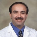 Alireza Minagar合作论文数35
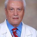 Eduardo Gonzalez Toledo合作论文数27
 Vijayakumar Javalkar合作论文数Department of Neurosurgery, Louisiana State University Health Sciences Center20
 Oleg Y. Chernyshev合作论文数15
 Bir Shyamal C合作论文数Dept Neurosurg, LSU Hlth Shreveport12
 J. Steven Alexander合作论文数Department of Molecular & Cellular Physiology, School of Graduate Studies, Louisiana State University Health Shreveport;Arganum Transplant Solutions12
 Prabandh Buchhanolla合作论文数9
 Junaid Ansari合作论文数Hlth Sci Ctr Shreveport, Louisiana State Univ9Alexandra Gaudet合作论文数8Rachel Triay合作论文数8Alexis Angelette合作论文数7
 Bharat Guthikonda合作论文数Department of Neurosurgery, LSU Health Shreveport7
 Hugo Cuellar合作论文数Department of Neurosurgery, Louisiana State University Health Sciences Center7
 Okkes Kuybu合作论文数Department of Neurology, University of Pittsburgh6
 Sandeep Kandregula合作论文数Dept Neurosurg, LSU Hlth Shreveport6
 Romy Hoque合作论文数Department of Neurology, Louisiana State University Health Sciences Center6Omar Elsekaily合作论文数5Abdallah Amireh合作论文数5Mohammad Sheikh合作论文数5Uma Menon合作论文数5Muhammad Ayub合作论文数5
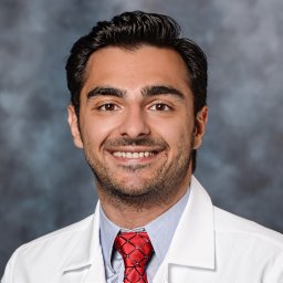 Maghzi Amir-Hadi合作论文数Isfahan Univ Med Sci5Vivek Misra合作论文数5
 Robert N Schwendimann合作论文数5Irena Adamashvili合作论文数5Meghan K. Harris合作论文数5Stacy J. Smith合作论文数4
 Etemadifar Masoud合作论文数Dept Neurol, Isfahan Univ Med Sci4
 Mansoureh Barzegar合作论文数Department of Biochemistry and Molecular Biology, Louisiana State University Health Sciences Center4Wenche Jy合作论文数4Meghan Kenner合作论文数4Karen Y. Stokes合作论文数4Halim Fadil合作论文数4Steven C. Karceski合作论文数3Brian P. Kelley合作论文数3
 Aimee Borazanci合作论文数3Jeanie McGee合作论文数3
 Tasneem F Hasan合作论文数Dept Neurol, Ochsner Louisiana State Univ3
 Steven Craig Schachter合作论文数Beth Israel Deaconess Medical Center, Massachusetts General Hospital3Riel-Romero Rosario Maria S合作论文数3Elena Korniychuk合作论文数3
 Felicity Gavins合作论文数Department of Life Sciences, College of Health, Medicine and Life Sciences, Brunel University of London;Centre for Inflammation Research and Translational Medicine, Brunel University of London;Imperial College London;Louisiana State University Health Sciences Center Shreveport3Nadejda Alekseeva合作论文数3Lawrence L. Horstman合作论文数3Yeon S. Ahn合作论文数3
 Carlos J Bidot合作论文数3
 Robert Zivadinov合作论文数Department of Neurology, Jacobs School of Medicine and Biomedical Sciences, University at Buffalo, The State University of New York;Translational Imaging Center, Clinical Translational Research Center, Jacobs School of Medicine & Biomedical Sciences, University at Buffalo, The State University of New York;College of Medicine, Florida International University;Buffalo Neuroimaging Analysis Center, Jacobs Neurological Institute3Nimer Abushehab合作论文数2




