The progress made over the years in electron microscopy has pushed the instrument abilities towards new frontiers. Higher beam intensities, smaller spot sizes, fast and accurate detectors – all these features enable sample imaging and elemental analysis at atomic scale resolution in real time. Often equipped with one or more Energy Dispersive X-Ray (EDX) detectors, the analytical capabilities of the electron microscopes have gained more and more significance over the last years. For high resolution electron microscopes like TEMs or high resolution SEMs, the distance between the pole piece and the sample plane (so-called working distance) is often very small; thus when using an EDX detector of a given size, the maximum attainable solid angle for collecting the x-ray signal is limited by the minimum distance at which the detector can approach the sample point. For these cases, using an oval-shaped detector of reduced size in the vertical dimension enables a closer positioning of the detector with respect to the sample point (see Figure 1a) and therefore a significant increase of the solid angle. The solid angle can be further increased by using a multi-detector configuration. Figure 2b shows a dual EDX detector system with two oval-shaped detectors arranged at 180° around the polepiece. At PNDetector we have been working since many years on the optimization of the detector geometry with respect to the maximum achievable solid angle. One of the first detectors especially designed for high solid angle application is the annual multi-channel detector configuration with central hole called Rococo2. This detector configuration has been presented several times in the past [1] and is not the subject of this contribution. More recently, several single-channel detector configurations of oval shape have been developed for high solid angle EDX analysis in TEM and SEM. Figure 2 shows three such configurations of the so-called SDD Oval-Line with active areas of 60 mm, 100 mm and 200 mm. The 100 mm oval shaped detector is already in use for more than five years. Depending on the poleshoe configuration, solid angle values of up to 1 sr can be achieved. Combining the detector with the SDD-FET technology [2], energy resolution values down to 125 eV at 5.9 keV x-ray energy are measured (see plot in Figure 3). Using the detector in a windowless configuration, the sensitivity for light elements greatly improves, enabling light element detection down to Si-L lines (90 eV). In this contribution we will presents new results obtained with the various oval-shaped detector configurations. The advantages (large solid angle, excellent optimum energy resolution) and the limitations (e.g. ballistic deficit due to the large aspect ratio) of these detectors will be discussed.
With its long experience in the development and manufacturing of the SDD chips and modules, PNDetector is continuously working on the optimization of the detector performance in terms of energy resolution, detection efficiency, detector geometry or solid angle coverage. Owing the unique feature of directly integrating the first FET onto the detector chip which leads to a very small value of the total input capacitance, the energy resolution of the SDD with integrated FET has been pushed continuously towards the theoretical limit set by the statistical fluctuations in the detector material (Fano noise, for silicon: fwhm 119 eV at Mn-Kα).
Journal Article Solid State Backscattered Electron Detectors with Improved Image Contrast and Detection Speed Get access A Liebel, A Liebel Search for other works by this author on: Oxford Academic Google Scholar R Eckhardt, R Eckhardt Search for other works by this author on: Oxford Academic Google Scholar A Niculae, A Niculae Search for other works by this author on: Oxford Academic Google Scholar H Soltau H Soltau Search for other works by this author on: Oxford Academic Google Scholar Microscopy and Microanalysis, Volume 20, Issue S3, 1 August 2014, Pages 1148–1149, https://doi.org/10.1017/S1431927614007478 Published: 27 August 2014
Since several years pnCCDs are well known as radiation detectors for spectroscopic imaging in many fields of science: X-Ray Fluorescence analysis (XRF), X-ray astronomy, X-ray Free Electron Laser science, synchrotrons and in the visible range as wave front sensors in adaptive optics systems. For direct electron detection in the focal plane of (S)TEMs they have delivered remarkable results in an energy range from 20 keV up to 300 keV.
An abstract is not available for this content so a preview has been provided. As you have access to this content, a full PDF is available via the ‘Save PDF’ action button.
An abstract is not available for this content so a preview has been provided. As you have access to this content, a full PDF is available via the ‘Save PDF’ action button.
Transmission Electron Microscopes (TEM) have become over the last years very powerful analytical tools for a various applications ranging from material analysis in the semiconductor industry to biological cell microscopy in life sciences. Very often the TEM instruments are equipped with Energy Dispersive X-Ray spectroscopy (EDX) tools, which in STEM mode enable fast and accurate elemental analysis on atomic level.
Extended abstract of a paper presented at Microscopy and Microanalysis 2013 in Indianapolis, Indiana, USA, August 4 – August 8, 2013.
Extended abstract of a paper presented at Microscopy and Microanalysis 2013 in Indianapolis, Indiana, USA, August 4 – August 8, 2013.
Extended abstract of a paper presented at Microscopy and Microanalysis 2013 in Indianapolis, Indiana, USA, August 4 – August 8, 2013.
Extended abstract of a paper presented at Microscopy and Microanalysis 2012 in Phoenix, Arizona, USA, July 29 – August 2, 2012.
Extended abstract of a paper presented at Microscopy and Microanalysis 2012 in Phoenix, Arizona, USA, July 29 – August 2, 2012.
Extended abstract of a paper presented at Microscopy and Microanalysis 2012 in Phoenix, Arizona, USA, July 29 – August 2, 2012.
Extended abstract of a paper presented at Microscopy and Microanalysis 2011 in Nashville, Tennessee, USA, August 7–August 11, 2011.
Extended abstract of a paper presented at Microscopy and Microanalysis 2011 in Nashville, Tennessee, USA, August 7–August 11, 2011.
Extended abstract of a paper presented at Microscopy and Microanalysis 2011 in Nashville, Tennessee, USA, August 7–August 11, 2011.
An abstract is not available for this content so a preview has been provided. Please use the Get access link above for information on how to access this content.
Extended abstract of a paper presented at Microscopy and Microanalysis 2008 in Albuquerque, New Mexico, USA, August 3 – August 7, 2008
Extended abstract of a paper presented at Microscopy and Microanalysis 2010 in Portland, Oregon, USA, August 1 – August 5, 2010.
The Miniaturised Mössbauer Spectrometers MIMOS II on board the two Mars Exploration Rovers (MER) have now been collecting valuable scientific data for more than five years. Mössbauer Spectrometers are part of two future missions: Phobos Grunt (Russian Space Agency) and a joint ESA—NASA Rover in 2018. The new advanced MIMOS IIA instrument described in this paper uses Silicon Drift Detectors (SDD) allowing also X-ray fluorescence chemical analysis (XRF) simultaneously to Mössbauer acquisitions. This paper highlights the features and technological improvements of the new spectrometer MIMOS IIA.
 Heike Soltau合作论文数28Lothar Strüder合作论文数Universität Siegen23Adrian Niculae合作论文数21A. Liebel合作论文数16Gerhard Lutz合作论文数15
 F. Schopper合作论文数13Peter Lechner合作论文数13Peter Holl合作论文数11G Schaller合作论文数10m bornschlegl合作论文数8O. Jaritschin合作论文数8Robert Hartmann合作论文数8s jeschke合作论文数7K. Hermenau合作论文数6joachim herrmann合作论文数5N. Meidinger合作论文数4Heinzinger, K.合作论文数4R. Richter合作论文数4Alimdar Simsek合作论文数4Johannes Treis合作论文数4S Bonerz合作论文数3J. Englhauser合作论文数3Ihle, Sebastian合作论文数3
 Günther Hasinger合作论文数Institute for Astronomy, University of Hawaii3
 Norbert Krause合作论文数Max-Planck-Institut für extraterrestrische Physik, Giessenbachstraße, 85741 Garching, Germany3
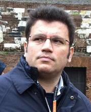 Carlo Fiorini合作论文数Polytechnic University of Milan,Department of Electronics, Information and Bioengineering3A. Bechteler合作论文数3Olaf Halker合作论文数3M. Schnecke合作论文数3
 Joachim Trümper合作论文数Max-Planck-Institut Für Extraterrestrische Physik;University of Munich3Ch Thamm合作论文数3H Brauninger合作论文数2christian koitsch合作论文数2
 Gerhard Schaller合作论文数2Rainer Richter合作论文数2Sven Herrmann合作论文数2Ladislav Andricek合作论文数2Robert Andritschke合作论文数2
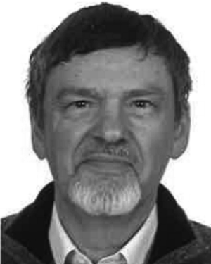 R.H. Richter合作论文数2A. Schöning合作论文数1b schweinfest合作论文数1s bjeoumikhova合作论文数1J. Brückner合作论文数1M. Blumers合作论文数1p bodin合作论文数1Damien Rambaud合作论文数1N. Kimmel合作论文数1j girones lopez合作论文数1




