Im Arterieneystem treten an den« jenigen Orten Reflexionen der Pulswelle auf f eil denen sich der Wellenwider stand (Z) ändert oder an denen der Abschlußwiderstand (Re) nicht gleich dem Wellenwiderstand ist. Werden arterieller Druck (p) und arterielle Stromstärke (i) am gleichen Meßort registriert, so ist es bei Kenntnis des Wellenwiderstandes möglich, die Summe aller peripherwärt s (pd, i*) und zentralwärt s (p*» i*) laufenden Vollen auf einfache Weise zu berechnen. Die Wellen des Druckes und der Stromstärke überlagern sich in der Welse, daß sich der registrierte Druck (p) aus der Summe (p-Pi+P2)* die registrierte Stromstärke (i; aus der Differenz (i«ii-i2) der Summe aller rechtund rückläufigen Wellen ergibt. Der Wellenwiderst and ist unter der Voraussetzung gleicher Laufrichtung als das Verhältnis von Wellendruck zu Wellenstromstärke definiert (Z· Pl/ilP2/i2) · ann §üt nach von Kries (1892) für die Summe aller rechtläufigen Wellen pWp+i-Z)/2 bzw. i<f(p/2+i)/2 und für die Summe der rückläufigen Wellen p2-(P-i'Z)/2 , N bzw. i2-(P/Z-i)/2 (2). Die Anwendung dieser Gleichungen zur Analyse von Pulswellen verschiedener Laufrichtung wird an im Tierversuch blutig registrierten Druckund Strompulsen demonstriert· Der Wellenwiderstand wird hierbei aus der Messung des Gefäßquerschnittes (Q), der Pulswellengeschwindigkeit (o) und der Dichte des Blutes (?) nach der Gleichung Z«e*c/Q näherungsweise ermittelt und vereinfachend als reell angenommen· Es ergibt sich, daß die Anstiege von po und i2 gegenüber p-j und i-j zeitlich verspätet beginnen· Die Kurven der recht lauf igen Druckund Strompulse haben übereinstimmende Form, ebenso die der rückläufigen Druckund Strompulse· Die Pulswellen beider Richtungen werden daher vollständig durch die Kurvenverläufe von pt und £2 oder von i^ und i2 dargestellt. Am Menschen können mittels Sphygmographie und Ultraschall-Doppler (USD)-Strömungsregistrierung unblutig Pulse gewonnen werden, deren Form dem Verlauf des arteriellen Druckes bzw. der arteriellen Stromstärke entspricht. Strompulse, die mit ausreichend hoher Grenzfrequenz des Tiefpaßfilters im Ausgang des USD-Gerätes registriert sind, können nicht als solche zur Berechnung herangezogen werden, da ihnen störende Zacken und Schwingungen überlagert sind. Unter der Voraussetzung eines stationären Kreislaufzustandes liefert die elektronische Mittelung(Averaging) einer Serie von Einzelpulsen einen weitgehend geglätteten Strömungspuls, der hinsichtlich Registriertreue dem blutig registrierten sehr nahe kommt (1).
Nitric oxide (NO) is known to counteract apoptosis by S-nitrosylation of protein thiol groups. NO is generated and stored in erythrocytes, which may undergo eryptosis, a suicidal cell death similar to apoptosis of nucleated cells. Eryptosis is triggered by increased cytosolic Ca2+ activity and/or ceramide and characterized by cell shrinkage and phosphatidylserine exposure at the cell surface. The present study explored whether nitric oxide could interfere with the machinery underlying eryptosis. To this end, erythrocyte phosphatidylserine exposure (annexin V-binding) and cell volume (forward scatter) were determined by flow cytometry. The Ca2+ ionophore ionomycin (0.1 μM) increased cytosolic Ca2+ activity, triggered annexin binding, and decreased forward scatter. The annexin binding and decrease of forward scatter but not the increase of cytosolic Ca2+ activity were reversed by the NO-donor nitroprusside (1 μM) and papanonoate (100 μM). Higher concentrations of nitroprusside (0.1 and 1 mM) stimulated eryptosis. Glucose depletion, exposure to C6-ceramide (3 μM), hypertonic (addition of 550 mM sucrose), and isotonic (replacement of Cl− with gluconate) cell shrinkage all triggered annexin V binding, effects all reversed by nitroprusside (1 μM). Dibutyryl–cGMP (1 mM) blunted the ionomycin- but not the ceramide-induced annexin V binding. Ionomycin decreased protein nitrosylation and thioredoxin activity, effects reversed by the NO-donor papanonoate. Clearance of erythrocytes from circulating blood was significantly faster in eNOS knockout mice than in their wild-type littermates. In conclusion, nitric oxide participates in the regulation of erythrocyte survival, an effect partially mimicked by cGMP and paralleled by alterations of protein nitrosylation and thioredoxin activity.
Review Articles| November 13 2008 Regulation of NO Synthesis in Endothelial Cells Subject Area: Cardiovascular System , Nephrology Rudi Busse; Rudi Busse Institut für Kardiovaskuläre Physiologie, Klinikum der J.W. Goethe–Universität, Frankfurt am Main, Deutschland Search for other works by this author on: This Site PubMed Google Scholar Ingrid Fleming Ingrid Fleming Institut für Kardiovaskuläre Physiologie, Klinikum der J.W. Goethe–Universität, Frankfurt am Main, Deutschland Search for other works by this author on: This Site PubMed Google Scholar Kidney Blood Press Res (1998) 21 (2-4): 264–266. https://doi.org/10.1159/000025872 Article history Published Online: November 13 2008 Content Tools Views Icon Views Article contents Figures & tables Video Audio Supplementary Data Peer Review Share Icon Share Facebook Twitter LinkedIn Email Tools Icon Tools Get Permissions Cite Icon Cite Search Site Citation Rudi Busse, Ingrid Fleming; Regulation of NO Synthesis in Endothelial Cells. Kidney Blood Press Res 1 July 1998; 21 (2-4): 264–266. https://doi.org/10.1159/000025872 Download citation file: Ris (Zotero) Reference Manager EasyBib Bookends Mendeley Papers EndNote RefWorks BibTex toolbar search Search Dropdown Menu toolbar search search input Search input auto suggest filter your search All ContentAll JournalsKidney and Blood Pressure Research Search Advanced Search Keywords: Nitric oxide synthase, Endothelial cells, Shear stress This content is only available via PDF. 1998Copyright / Drug Dosage / DisclaimerCopyright: All rights reserved. No part of this publication may be translated into other languages, reproduced or utilized in any form or by any means, electronic or mechanical, including photocopying, recording, microcopying, or by any information storage and retrieval system, without permission in writing from the publisher.Drug Dosage: The authors and the publisher have exerted every effort to ensure that drug selection and dosage set forth in this text are in accord with current recommendations and practice at the time of publication. However, in view of ongoing research, changes in government regulations, and the constant flow of information relating to drug therapy and drug reactions, the reader is urged to check the package insert for each drug for any changes in indications and dosage and for added warnings and precautions. This is particularly important when the recommended agent is a new and/or infrequently employed drug.Disclaimer: The statements, opinions and data contained in this publication are solely those of the individual authors and contributors and not of the publishers and the editor(s). The appearance of advertisements or/and product references in the publication is not a warranty, endorsement, or approval of the products or services advertised or of their effectiveness, quality or safety. The publisher and the editor(s) disclaim responsibility for any injury to persons or property resulting from any ideas, methods, instructions or products referred to in the content or advertisements. Article PDF first page preview Close Modal You do not currently have access to this content.
Objectives— Basic fibroblast growth factor (bFGF) stimulates vascular smooth muscle cell (SMC) migration. We determined whether bFGF increases SMC reactive oxygen-species (ROS) and studied the role of ROS for SMC migration. Methods and Results— bFGF rapidly increased rat SMC ROS formation and migration through pathways sensitive to inhibition of NADPH oxidases, PI3-kinase, protein kinase C, and Rac-1. SiRNA directed against the NADPH oxidase Nox4 impaired basal but not bFGF-induced ROS formation and did not affect migration. In contrast, siRNA against Nox1 blocked the agonist-induced ROS generation as well as the bFGF-induced migration. Agonist-induced migration was also attenuated in SMC derived from Nox1 y/− mice and transduction of Nox1 restored normal migration. Likewise, SMC outgrowth in response to bFGF was attenuated in aortic segments from Nox1 y/− mice as compared with Nox1 y/+ mice. bFGF activated JNK but not Src in a Nox1-dependent manner. Consequently, phosphorylation of the adaptor protein paxillin, which is central for migration and secretion of matrix-metalloproteinases, were dependent on Nox1 as well as JNK but not Src. Conclusions— These data demonstrate that bFGF activates the Nox1-containing NADPH oxidase and that bFGF through a pathway involving ROS and JNK stimulates SMC migration.
AIMS Phosphorylation of forkhead box O (FoxO) transcription factors induces their nuclear exclusion and proteosomal degradation. Here, we investigated the effect of fluid shear stress on FoxO1a in primary cultures of human endothelial cells and the kinases that regulate its phosphorylation. METHODS AND RESULTS Shear stress (12 dynes/cm2) elicited the phosphorylation, nuclear exclusion, and degradation of FoxO1a. Inhibition of Akt signalling using either a dominant negative (DN) mutant of Akt or downregulation of Gab1 largely failed to affect the shear stress-induced changes in FoxO1a, while a DN-AMP-activated protein kinase (AMPK) abrogated its shear stress-induced phosphorylation and degradation. Similar effects were observed using the AMPK inhibitor compound C. Moreover, in an in vitro assay, the AMPK directly phosphorylated FoxO1a. As FoxO1a regulates the expression of angiopoietin-2 (Ang-2), we determined the role of shear stress and the AMPK in this phenomenon. Not only did the DN-AMPK increase the expression of Ang-2 in cells maintained under static conditions, it also abrogated the shear stress-induced decrease in FoxO1a and Ang-2 protein levels. Functionally, Ang-2 sensitizes endothelial cells to the effects of tumour necrosis factor (TNF)-alpha, and DN-AMPK increased basal endothelial cell E-selectin expression and permeability as well as the increase induced by TNF-alpha. CONCLUSION These data indicate that the AMPK activated by fluid shear stress is a novel regulator of FoxO1a phosphorylation and protein levels. Moreover, as the AMPK-dependent phosphorylation and degradation of FoxO1a attenuates Ang-2 expression and protects against the pro-inflammatory actions of TNF-alpha, this kinase may be a useful target to prevent the progression of vascular diseases.
Background and Purpose— Cerebral ischemia/reperfusion is associated with reactive oxygen species (ROS) generation, and NADPH oxidases are important sources of ROS. We hypothesized that NADPH oxidases mediate blood-brain barrier (BBB) disruption and contribute to tissue damage in ischemia/reperfusion. Methods— Ischemia was induced by filament occlusion of the middle cerebral artery in mice for 2 hours followed by reperfusion. BBB permeability was measured by Evans blue extravasation. Monolayer permeability was determined from transendothelial electrical resistance of cultured porcine brain capillary endothelial cells. Results— BBB permeability was increased in the ischemic hemisphere 1 hour after reperfusion. In NADPH oxidase–knockout (gp91phox−/−) mice, middle cerebral artery occlusion–induced BBB disruption and lesion volume were largely attenuated compared with those in wild-type mice. Inhibition of NADPH oxidase by apocynin prevented BBB damage. In porcine brain capillary endothelial cells, hypoxia/reoxygenation induced translocation of the NADPH oxidase activator Rac-1 to the membrane. In vivo inhibition of Rac-1 by the 3-hydroxy-3-methylglutaryl coenzyme A reductase inhibitor atorvastatin or Clostridium difficile lethal toxin B also prevented the ischemia/reperfusion–induced BBB disruption. Stimulation of porcine brain capillary endothelial cells with H2O2 increased permeability, an effect attenuated by inhibition of phosphatidyl inositol 3-kinase or c-Jun N-terminal kinase but not blockade of extracellular signal–regulated kinase-1/2 or p38 mitogen-activated protein kinase. Inhibition of Rho kinase completely prevented the ROS-induced increase in permeability and the ROS-induced polymerization of the actin cytoskeleton. Conclusions— Activation of Rac and subsequently of the gp91phox containing NADPH oxidase promotes cerebral ROS formation, which then leads to Rho kinase–mediated endothelial cell contraction and disruption of the BBB. Inhibition of NAPDH oxidase is a promising approach to reduce brain injury after stroke.
Während zerebraler Ischämie und Reperfusion (I/R) kommt es zur Bildung von reaktiver Sauerstoffspezies (ROS), von welcher angenommen wird, dass sie zum neuronalen Schaden beitragen. NADPH Oxidasen produzieren ROS. Wir untersuchten daher, ob und in welchem Maße NADPH Oxidasen am Gewebeschaden und insbesondere an der Schädigung der Blut- Hirn- Schranke (BHS) nach experimenteller I/R beteiligt sind.
The native CD34+/CD31- cell population present in the stroma-vascular fraction of human adipose tissue (hAT) displays progenitor cell properties since they exhibit adipocyte- and endothelial cell-like phenotypes under appropriate stimuli. To analyze the signals within hAT regulating their phenotypes, the influence of hAT-derived capillary endothelial cells (CECs) was studied on the chemotaxis and differentiation of the hAT-CD34+/CD31- cells. Conditioned medium from hAT-CECs led to a strong chemotaxis of the hAT-CD34+/CD31- cells that was inhibited with pretreatments with pertussis toxin, CXCR-4 antagonist, or neutralizing antibodies. Furthermore, hAT-CECs produced and secreted the CXCR-4 ligand, that is, the stromal derived factor-1 (SDF-1). Finally, hAT-CECs induced the differentiation of hAT-CD34+/CD31- cells toward an endothelial cell (EC) phenotype. Indeed, hAT-CECs and -CD34+/CD31- cell coculture stimulated in a two-dimensional system the expression of the EC CD31 marker by the hAT-progenitor cells and, in a three-dimensional approach, the formation of capillary-like structures via a SDF-1/CXCR-4 dependent pathway. Thus, the migration and differentiation of hAT progenitor cells are modulated by hAT-CEC-derived factors. SDF-1, which is secreted by hAT-derived CECs, and its receptor CXCR-4, expressed by hAT-derived progenitor cells, may promote chemotaxis and differentiation of hAT-derived progenitor cells and thus contribute to the formation of the vascular network during the development of hAT.
Background: Flow-mediated dilation (FMD) of human conduit arteries is, in part, related to shear stress-induced release of endothelium-derived nitric oxide (NO). However, NO synthase inhibitors do not completely abolish this FMD-response. Recently, a cytochrome P450 (CYP) epoxygenase of the 2C family was linked to NO- and prostacyclin-independent relaxation of conduit arteries. We therefore evaluated the contribution of CYP 2C9 to FMD in humans.Methods and results: FMD of the radial artery was determined in 12 healthy volunteers by high-resolution ultrasound and analyzed before and after intra-arterial infusion of sulfaphenazole, a specific CYP 2C9 inhibitor, L-NMMA (NO synthase inhibitor) and co-infusion of both. Endothelium-independent vasodilation was characterized after intra-arterial infusion of SNP. FMD was reduced after sulfaphenazole (11.5 +/- 0.87% vs. 7.4 +/- 0.95%, p < 0.01), after L-NMMA (6.0 +/- 0.71%; p < 0.01), and after co-infusion 3.9 +/- 0.73% (p < 0.05 VS. L-NMNIA; p < 0.01 vs. sulfaphenazole). Sulfaphenazole had no effect on endothelium-independent vasodilation. In patients with chronic heart failure, the portion of FMD blocked by sulfaphenazole was not affected. CYP 2C was detected by immunohistochemistry in radial artery samples obtained from patients undergoing coronary bypass surgery.Conclusions: FMD in human conductance arteries is reduced after inhibition of CYP 2C9, supporting the concept that CYP 2C metabolites contribute to endothelium-mediated vasodilation of peripheral conduit arteries in vivo. In patients with heart failure, the CYP-dependent FMD appears to be preserved. (c) 2007 European Society of Cardiology. Published by Elsevier B.V. All fights reserved.
Objective— An initial step in endothelium-derived hyperpolarizing factor-mediated responses is endothelial cell hyperpolarization. Here we address the mechanisms by which cytochrome P450 (CYP)-derived epoxyeicosatrienoic acids (EETs) contribute to this effect in native and cultured endothelial cells. Methods and Results— In native CYP2C-expressing endothelial cells, bradykinin elicited a Ca 2+ influx that was potentiated by the soluble epoxide hydrolase inhibitor, 1-adamantyl-3-cyclohexylurea (ACU), and attenuated by CYP inhibition. Similar effects were observed in cultured endothelial cells overexpressing CYP2C9, but not in CYP2C9-deficient cells, and were prevented by the EET antagonist 14,15-epoxyeicosa-5(Z)-enoic acid as well as by the cAMP antagonist, Rp-cAMPS. The effects on Ca 2+ were mirrored by prolongation of the bradykinin-induced hyperpolarization. Ruthenium red and the combination of charybdotoxin and apamin prevented the latter effect, suggesting that Trp channel activation increases Ca 2+ influx and prolongs the activation of Ca 2+ -dependent K + (K Ca ) channels. Indeed, overexpression of CYP2C9 enhanced the agonist-induced translocation of a TrpC6-V5 fusion protein to caveolin-1–rich areas of the endothelial cell membrane, which was prevented by Rp-cAMPS and mimicked by 11,12-EET. Conclusions— Elevated EET levels regulate Ca 2+ influx into endothelial cells and the subsequent activation of K Ca channels, via a cAMP/PKA-dependent mechanism that involves the intracellular translocation of Trp channels.
In this study, we investigated the effects of prolonged administration of the selective COX-2 inhibitors celecoxib and rofecoxib and the non-selective NSAID naproxen on the initiation and progression of atherosclerosis. ApoE(-/-) mice, as well as corresponding wild-type mice, were fed either a normal chow or a high fat Western diet with or without addition of the respective drugs over a period of 16 weeks. Thereafter, aortic lesion size, plasma lipid levels, and COX-2 expression in the plaques were determined. The results showed that neither the COX-2 selective inhibitors nor naproxen had a significant impact on the initiation and progression of atherosclerosis in diet-fed ApoE(-/-) mice, although both celecoxib and rofecoxib showed a tendency to reduce plaque size. This slight effect may be due to selective inhibition of COX-2 activity because the COX-2 expression was not altered in the plaque. Plasma lipid levels were also not significantly influenced by these drugs. Interestingly, in ApoE(-/-) mice that have been fed with normal chow, we found an increased incidence of plaque formation after treatment with celecoxib and rofecoxib, indicating that coxibs may promote the initiation of atherosclerosis. This effect was probably masked in diet-fed mice by the more pronounced effects of the high cholesterol diet. In conclusion, the reduction in diet-induced plaque size in animals fed a high fat diet and the promotion of atherosclerosis in mice on a normal diet indicate a dual role of the coxibs. In advanced stages of atherosclerosis, they may exert antithrombotic properties due to their COX-2 inhibiting activity, whereas in very early stages they may favor the initiation of atherogenesis. However, because these results were only observed in ApoE(-/-) and not in wild-type animals, coxibs may increase the risk of thrombosis in patients with a predisposition for thrombotic complications.
The rate-limiting enzyme for cholesterol synthesis, the hydroxy-methylglutaryl coenzyme A reductase (HCR), is phosphorylated by the AMP-activated protein kinase (AMPK). As shear stress activates the AMPK in endothelial cells, we determined whether it affects HCR activity and subsequent HCR-dependent signaling. Shear stress (12 dynes cm −2 ) rapidly increased the phosphorylation and activity (6.5- and 4-fold, respectively) of the AMPK in cultured endothelial cells and the activated AMPK phosphorylated the HCR in vitro. Moreover, shear stress and the AMPK activator 5-aminoimidazole-4-carboxamide ribonucleoside (AICAR) attenuated endothelial HCR activity by 37% and 33%, respectively. Inhibition of NO production attenuated the acute shear stress–induced phosphorylation of the AMPK and the decrease in HCR activity. Prolonged shear stress (18 hours) led to a significant (50%) decrease in HCR mRNA expression that was dependent on NO, AMPK, and the subsequent phosphorylation and degradation of FoxO1a. Correspondingly, the downregulation of FoxO (small interfering RNA) decreased HCR expression. Prolonged shear stress also attenuated the bradykinin-induced activation of Ras and extracellular signal-regulated kinase 1/2, a phenomenon that was comparable to the effects of cerivastatin and that was reversed by mevalonate and thus attributed to HCR inhibition. A decrease (35%) in HCR expression was also detected in femoral arteries from mice following voluntary exercise, and the bradykinin-induced vasodilatation of the mouse hindlimb was attenuated by both exercise and the HCR inhibitor cerivastatin. These data indicate that fluid shear stress regulates the activity and expression of the HCR in endothelial cells and determines responsiveness to stimuli, such as bradykinin via a mechanism involving NO, AMPK, FoxO1a, and p21Ras.
Sphingosine-1-phosphate (S1P) is a bioactive lipid involved in multiple biological processes; it is stored in platelets and released upon activation. Although S1P is known to exert extracellular effects on platelets such as shape change and aggregation, the receptor that mediates these effects and the molecular mechanisms involved are poorly understood. The aim of the present study was to characterize the S1P receptor expressed in platelets as well as the molecular mechanism underlying S1P-mediated platelet activation and eventual changes associated with the development of type 2 diabetes. We found that washed human platelets from healthy volunteers expressed mainly the S1P2 receptor and to a lesser extent the S1P1 receptor. Stimulation of these platelets with exogenous S1P led to a concentration-dependent increase in intracellular calcium as well as to platelet aggregation. The latter response was accompanied by the translocation of RhoA to the membrane and was inhibited (by 50%, P<0.01) by the Rho kinase inhibitor Y27632 (1 μmol/L). Neither the PKC inhibitor, Ro-318220, the MAPK inhibitor, PD98059, nor the tyrosine kinase inhibitor, tyrphostin A-23, affected the S1P-mediated platelet aggregation. Serum obtained from patients with type 2 diabetes contained 1.5-fold higher levels (P<0.01) of S1P that from healthy individuals. However, platelets from patients with type 2 diabetes demonstrated an attenuated aggregability to S1P as well as decreased levels of the S1P2 protein. The latter was found to be a target of the calcium-dependent protease μ-calpain which we found to be highly activated in platelets from patients with type 2 diabetes. Finally, stimulation of platelets from healthy donors with high concentration of S1P led to the activation of μ-calpain. Taken together, our results show that S1P can elicit platelet aggregation by activating the S1P2 receptor and the RhoA-Rho kinase pathway. In platelets from patients with type 2 diabetes, in which the circulating concentration of S1P is elevated, responses to S1P are damped, via a phenomenon that correlates with the activation of μ-calpain and the subsequent degradation of the S1P2 receptor. The latter may represent an auto-protective mechanism against exaggerated platelet activation in diabetes.
AIMS/HYPOTHESIS:Increased visceral white adipose tissue (WAT) is linked to the risk of developing diabetes.METHODS/RESULTS:We showed by fluorescence activated cell sorting analysis that human visceral WAT contains macrophages, the proportion of which increased with obesity. Selective isolation of mature adipocytes and macrophages from human visceral WAT by CD14 immunoselection revealed that macrophages expressed higher levels of chemokines (monocyte chemotactic protein 1, macrophage inflammatory protein 1alpha, IL-8) and the adipokines resistin and visfatin than did mature adipocytes, as assessed by real-time PCR analysis. Moreover, resistin and visfatin proteins were found to be released predominantly by visceral WAT macrophages. Macrophage-derived secretory products stimulated phosphorylation of protein kinase B in human hepatocytes.CONCLUSIONS/INTERPRETATION:Resistin and visfatin might be considered to be proinflammatory markers. The increased macrophage population in obese human visceral WAT might be responsible for the enhanced production of chemokines as well as resistin and visfatin.
In addition to its function as dipeptidase, ACE can act as a signal transduction molecule following the binding of ACE inhibitors. The “ACE signaling pathway” affects endothelial gene expression via phosphorylation of ACE on Ser1270 and activation of the JNK/cJun-pathway. Since it is unclear how ACE inhibitors initiate these intracellular signaling events we determined whether ACE can dimerize and whether ACE dimer formation is required for ACE signaling. Native gel electrophoresis revealed that ACE exists in its monomeric form and as a dimer of 520 kDa in endothelial cells. ACE dimerization was confirmed using the split-ubiquitin assay as well as by chemical crosslinking. ACE inhibitors elicited a rapid, concentration-dependent increase in ACE dimerization that correlated with the ACE inhibitor induced ACE phosphorylation. ACE dimerization in vitro depends on an N-terminal carbohydrate recognition domain, but neither carbohydrates nor N-terminus shielding ACE antibodies were able to affect ACE dimerization in endothelial cells. However, inactivation of the C-domain active centre of ACE by mutation of the two Zn2+-complexing histidines prevented the basal and ramiprilat-induced ACE dimerization as well as ACE phosphorylation on Ser1270 and the subsequent activation of JNK. Mutation of the N-domain active centre was without effect on ACE dimerization or the initiation of signaling. Taken together, our data suggest that ACE inhibitors, most probably by binding to the C-domain active centre, initiate the “ACE signaling pathway” by promoting the formation of ACE dimers.
 Ingrid Fleming合作论文数Institute for Vascular Signalling, Goethe University Frankfurt am Main168
 Beate Fisslthaler合作论文数66
 Ralf Brandes合作论文数Institute for Cardiovascular Physiology, Goethe-Universität54Eberhard Bassenge合作论文数51A Mülsch合作论文数46M Hecker合作论文数43U Pohl合作论文数34V. Schini-Kerth合作论文数32Rd Bauer合作论文数30Johann Bauersachs合作论文数30
 Ruediger Popp合作论文数29Anne Bouloumié合作论文数Institut des Maladies Metaboliques et Cardiovasculaires, Universite de Toulouse26Andreas Schabert合作论文数26Andreas Lückhoff合作论文数23Y. Summa合作论文数22E Wetterer合作论文数20U R Michaelis合作论文数18
 Juergen Holtz合作论文数12
 Ulrich Pohl合作论文数Walter-Brendel-Centre of Experimental Medicine, Ludwig-Maximilians-University12Alexander Mulsch合作论文数11Karin Kohlstedt合作论文数11Coralie Sengenès合作论文数Université de Toulouse, Université P. Sabatier10Daniel Lamontagne合作论文数10Steffen Bassus合作论文数10Agnes Gorlach合作论文数10Agnieszka T. Bara合作论文数10
 Voahanginirina Randriamboavonjy合作论文数9
 Ulrich Förstermann合作论文数Institut für Pharmakologie, Universitätsmedizin der Johannes Gutenberg-Universität Mainz;Department of Pharmacology, Johannes Gutenberg University Medical Center9Nicola Kronemann合作论文数9Jan Galle合作论文数9Cm Kirchmaier合作论文数8M Boese合作论文数8
 Stefanie Dimmeler合作论文数Institute of Cardiovascular Regeneration, Goethe-University Frankfurt;Institute for Molecular Medicine, Goethe-University Frankfurt;German Centre for Cardiovascular Research;Cardiopulmonary Institute8Th. Pasch合作论文数8H Korner合作论文数7Ca Curat合作论文数7A Miranville合作论文数7
 Andreas M. Zeiher合作论文数Goethe Universität Frankfurt7P Mordvintcev合作论文数6Hal Dietz合作论文数6W Sperling合作论文数6
 Charles J. Lowenstein合作论文数School of Medicine, Johns Hopkins University;Division of Cardiology, Johns Hopkins University;Johns Hopkins Heart and Vascular Institute6
 Gerda Breitwieser合作论文数Charles E Schmidt College of Medicine, Florida Atlantic University6
 Roberto Bolli合作论文数Institute of Molecular Cardiology, Division of Cardiovascular Medicine, Department of Medicine, School of Medicine, University of Louisville6
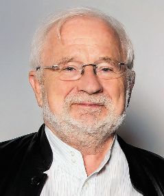 Paul M. Vanhoutte合作论文数Department of Pharmacology, Li Ka Shing Faculty of Medicine, University of Hong Kong6
 John R Falck合作论文数University of Texas Southwestern Medical Center6
 David Kass合作论文数Department of Biomedical Engineering, Whiting School of Engineering, Johns Hopkins University;Institute of CardioScience, School of Medicine, Johns Hopkins University6
 Toren Finkel合作论文数Division of Cardiology, University of Pittsburgh;Department of Medicine, University of Pittsburgh;Coloma Therapeutics6
 Werner Müller-Esterl合作论文数Institute for Biochemistry II, University of Frankfurt Medical School6




