Supplementary Table 4 shows a summary of differential expression analysis in NDI-associated genes and differentially expressed in CRC tumors of TCGA-COAD, and discordant for direction of effect in analysis of metformin treated healthy colon organoid lines.
AbstractBackground: Greater neighborhood deprivation, as defined by the neighborhood deprivation index (NDI), has been associated with increased colorectal cancer risk. However, the biological mechanisms underlying this association remain unclear. Methods: Normal colorectal biopsies (matched right and left colon and rectum) from African American [AA (n = 34)] and European American [EA (n = 23)] adults undergoing colonoscopy were collected for a cross-sectional study in Cleveland, Ohio. NDI-associated differentially expressed genes (DEG) were identified from RNA sequencing using limma. Normal colon organoid lines were derived from biopsies of AA (n = 4) and EA (n = 6) individuals in Charlottesville, Virginia, treated daily with metformin for 72 hours, and metformin-DEGs were identified in limma. Results: A total of 237 DEGs were found to be commonly associated with NDI in AA and EA populations across individual-matched biopsy triplets (right and left colon and rectum). Of the 237 NDI-associated DEGs, 82 overlapped with colorectal cancer tumor DEGs from a publicly available dataset (P = 2.21E−05), and 9 were prioritized as therapeutic targets, including MYC overexpression [Benjamini–Hochberg (BH) = 2.48E−12]. Metformin exposure in colon organoids also altered the expression of 28 of these genes, with ∼79% showing the opposite direction, including reduced MYC expression (BH = 0.095). Conclusions: NDI is associated with colorectal cancer–related transcriptional differences. The observed reversal of gene expression by metformin, including at MYC, suggests a potential role for metformin in reducing colorectal cancer risk by modifying NDI-high related gene expression. Impact: NDI-associated colorectal cancer risk genes provide a novel opportunity for future biomarkers or early therapeutic targets in at-risk populations.
Supplementary Table 3 shows a summary of differential expression analysis in NDI-associated genes and differentially expressed in MYC knockdown. Positive fold changes correspond to increased expression of genes in cells where MYC expression has been reduced. "NS" corresponds to genes that were present within the dataset after filtering, but were not-significantly associated with MYC exposure status. "-" reflects genes that were not present in the genome annotation or were too lowly expressed to be analyzed.
Supplementary Table 1 shows a summary of meta-analysis for differential expression of NDI.
Supplementary Figure 1 shows the enrichment of pathways identified in a meta-analysis of NDI-associated DEGs.
BACKGROUND & AIMS:Tuft cells play protective roles in infection, inflammation, and tumorigenesis through the secretion of cytokines and eicosanoids. Tuft cells are known for their tall, blunt microvilli, thought to be analogous to mechanosensory hair cell stereocilia; however, a functional role for the microvillar apparatus has not been identified. POU Class 2 Homeobox 3 (POU2F3) is the master regulator transcription factor for tuft cells, yet how POU2F3 drives formation of this unique structure is unknown. Here, we aimed to identify POU2F3 target genes and commonalities between tuft and hair cells to better understand this unique structure. METHODS:POU2F3 chromatin immunoprecipitation sequencing was performed on tuft cells and compared with the hair cell transcriptome. Tuft cell RNA sequencing datasets were interrogated for hair cell structural and mechanosensory genes; expression was validated. Intestinal and gallbladder tuft cells were examined using multiple light and electron microscopy modalities. Protocadherin 20 (PCDH20) was knocked down in mouse models, and ultrastructural analyses were performed. The tuft cell cytoskeleton was modeled using AlphaFold3 prediction. RESULTS:Genes encoding structural and mechanosensory proteins common to both tuft and hair cells, including Pcdh20, were identified. Imaging localized PCDH20 to tuft cell microvilli and hair cell stereocilia. Genetic ablation of Pcdh20 in mice resulted in structural defects in tuft cell microvilli, including loss of rigidity and organization. Molecular modeling suggests PCDH20 homodimers link adjacent microvilli. CONCLUSIONS:Pcdh20 is a POU2F3 target gene in tuft cells, critical to maintain the rigid microvillar apparatus. These findings, together with the shared expression of mechanosensory components like transmembrane channel-like protein 1, support the hypothesis that tuft cells could have mechanosensory capabilities analogous to cochlear hair cells.
Supplementary Table 2 shows a summary of differential expression analysis in NDI-associated genes that were found to be differentially expressed in CRC tumors of TCGA-COAD.
Tuft cells are solitary chemosensory cells known for their distinct tall, blunt microvilli, thought to be analogous to mechanosensory hair cell stereocilia. Identification of commonalities between tuft and hair cells could identify a role for tuft cells in mechanotransduction. Transcription factor POU2F3 is the master regulator of tuft cell formation, however how POU2F3 drives formation of this unique cell and the functional role of the microvillar apparatus is unknown. POU2F3 ChIP-seq was performed on isolated tuft cells and compared to the cochlear hair cell transcriptome. Structural genes common to both tuft and hair cells, including protocadherin 20 (PCDH20), were identified. Immunogold labeling and imaging localized PCDH20 to extensive intermicrovillar linkages in tuft cells. Knockdown of PCDH20 in mice resulted in impaired microvilli formation and a disruption in structure. Altogether, PCDH20 is a POU2F3 target gene in tuft cells critical to maintain the rigid microvillar apparatus, which may function in mechanotransduction.
Intestinal epithelial barrier integrity is essential for human health, and its disruption induces and exacerbates intestinal inflammatory disorders. While the epithelial cytoskeleton is critical for maintaining gut barrier-integrity, the role of septins - a family of GTP-binding, cytoskeletal proteins - is largely unknown. This highlights an important knowledge gap, as dysfunction of septins, and specifically septin 9 (SEPT9), is associated with intestinal pathologies. We determined that SEPT9 localizes to the apical junctions of intestinal epithelial cells (IECs), overlapping with both tight and adherens junctions. IEC-specific ablation of SEPT9 in mice resulted in leaky gut, due to mislocalization of junctional proteins, and increased susceptibility to experimental colitis. Consistently, SEPT9 expression was significantly reduced in intestinal mucosa of patients with inflammatory bowel disease (IBD). Using affinity-purification mass spectrometry, super-resolution imaging, and genetic KO, we determined that SEPT9 interacts with and is necessary to recruit nonmuscle myosin IIC (NMIIC) to the IEC perijunctional actomyosin belt. Loss of NMIIC also caused IEC barrier disruption. In summary, SEPT9 regulates intestinal barrier integrity by supporting the assembly of tight and adherens junctions through NMIIC recruitment to the actomyosin belt. The septin cytoskeleton safeguards the intestinal mucosa during acute inflammation, and its disruption in IBD suggests a loss of this protective function.
BACKGROUND:An increasing body of evidence has linked fructose intake to colorectal cancer (CRC). African-American (AA) adults consume greater quantities of fructose and are more likely to develop right-side colon cancer than European American (EA) adults. OBJECTIVES:We examined the hypothesis that fructose consumption leads to epigenomic and transcriptomic differences associated with CRC tumor biology. METHODS:Deoxyribonucleic acid methylation data from this cross-sectional study was obtained using the Illumina Infinium MethylationEPIC kit (GSE151732). Right and left colon differentially methylated regions (DMRs) were identified using DMRcate through analysis of Food Frequency Questionnaire data on fructose consumption in normal colon biopsies (n = 79) of AA adults undergoing screening colonoscopy. Secondary analysis of CRC tumors was carried out using data derived from The Cancer Genome Atlas Colon Adenocarcinoma, GSE101764, and GSE193535. Right colon organoids derived from AA (n = 5) and EA (n = 5) adults were exposed to 4.4 mM of fructose for 72 h. Differentially expressed genes (DEGs) were identified using DESeq2. RESULTS:We identified 4263 right colon fructose-associated DMRs [false-discovery rates (FDR) < 0.05]. In contrast, only 24 DMRs survived multiple testing corrections (FDR < 0.05) in matched, left colon. Almost 50% of right colon fructose-associated DMRs overlapped regions implicated in CRC in ≥1 of 3 data sets. Highly significant enrichment was also observed between genes corresponding to right colon fructose-associated DMRs and DEGs associated with fructose exposure in right colon organoids of AA individuals (P = 3.28E-30). Overlapping and significant enrichments for fatty acid metabolism, glycolysis, and cell proliferation pathways were also found. Cross-referencing genes within these pathways to DEGs in CRC tumors reveal potential roles for ankyrin repeat domain containing protein 23 and phosphofructokinase, platelet in fructose-mediated CRC risk for AA individuals. CONCLUSIONS:Our data support that dietary fructose exerts a greater CRC risk-related effect in the right than left colon among AA adults, alluding to its potential role in contributing to racial disparities in CRC.
The pleiomorphic structure and dynamic behavior of cellular endomembrane systems have been extensively studied using classical electron microscopy. However, fixation and staining constraints limit the in situ visualization of transient interactions, such as membrane fusion, scission, and intraluminal vesicle formation, potentially overlooking intermediate structures like membrane hemifusion. Using in situ cryo-electron tomography in four mammalian cell lines, we identify heterotypic hemifused vesicles featuring an extended hemifusion diaphragm consistently associated with a 42-nanometer proteolipid nanodroplet (PND). We designate these vesicular organelle complexes as "hemifusomes." Hemifusomes constitute up to 10% of vesicular organelles at the cell periphery but do not engage in canonical endocytic pathways. These structures exhibit diverse conformations and frequently contain intraluminal vesicles. Building on the continuum of related morphologies observed, we propose that hemifusomes serve as platforms for vesicular biogenesis, mediated by the PND. These findings provide direct in situ evidence of long-lived hemifused vesicle complexes and introduce an ESCRT-independent model for multivesicular body (MVB) formation.
Membrane remodeling drives a broad spectrum of cellular functions, and it is regulated through mechanical forces exerted on the membrane by cytoplasmic complexes. Here, we investigate how actin filaments dynamically tune their structure to control the active transfer of membranes between cellular compartments with distinct compositions and biophysical properties. Using intravital subcellular microscopy in live rodents we show that a lattice composed of linear filaments stabilizes the granule membrane after fusion with the plasma membrane and a network of branched filaments linked to the membranes by Ezrin, a regulator of membrane tension, initiates and drives to completion the integration step. Our results highlight how the actin cytoskeleton tunes its structure to adapt to dynamic changes in the biophysical properties of membranes.
The complex, pleiomorphic membrane structure of the vesicular components within the endolysosomal system has been appreciated through decades of classical electron microscopy. However, due to the heavy fixation and staining required in these approaches, in situ visualization of fragile intermediates between early endosomes, late endosomes and ultimately multivesicular bodies (MVBs), remains elusive, raising the likelihood that other structures may have also been overlooked. Here, using in situ cryo-electron tomography in four mammalian cell lines, we discover heterotypic hemifused vesicles that share an extended hemifusion diaphragm, associated with a 42nm proteolipid nanodroplet (PND). We term this previously undescribed vesicular organelle-complex, “hemifusome”. Hemifusomes make up approximately 10% of the organelle pool of the endolysosomal system, but do not participate directly in transferrin-mediated endocytosis. Hemifusomes exist in compound conformations and also contain intraluminal vesicles. Based on their range of morphologies, and the consistent presence of the PND at sites of compound hemifused vesicles, we propose that hemifusomes function as platforms for vesicular biogenesis mediated by the PND. These findings offer direct in situ evidence for a long-lived hemifusion diaphragm, and a new, ESCRT-independent model for the formation of late endosomes containing intraluminal vesicles and ultimately MVBs.
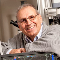 Bechara Kachar合作论文数Section on Structural Cell Biology, National Institute on Deafness and Other Communications Disorders, National Institutes of Health19
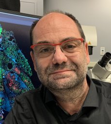 Roberto Weigert合作论文数National Institute of Dental and Craniofacial Research, National Institutes of Health Intracellular Membrane Trafficking Unit15
 Masedunskas Andrius合作论文数Natl Inst Dent & Craniofacial Res, NIH7Cui Runjia合作论文数National Institute for Deafness and Communications Disorders, National Institute of Health7
 Gaizun Hu合作论文数5Desu Chen合作论文数Lab Cellular & Mol Biol, NCI5Krystofiak Evan S合作论文数5Millis Bryan合作论文数5
 Edna C. Hardeman合作论文数Muscle Development Unit;Medical Research Institute;Muscle Development Unit, Children ' s Medical Research Institute5Marco Heydecker合作论文数4
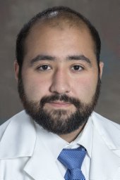 Tora Muhibullah合作论文数National Institute of Dental and Craniofacial Research, National Institutes of Health4
 Akiko Shitara合作论文数4
 Peter Gunning合作论文数School of Medical Sciences, Faculty of Medicine and Health, The University of New South Wales4A. Shitara合作论文数3Lenka Malec合作论文数3
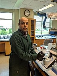 Amirrasoul Tavakoli合作论文数Dept Chem & Biochem, Baylor Univ3Sun Xiangqing合作论文数3Shiqiong Hu合作论文数3
 Milberg Oleg合作论文数3Tran Duy T合作论文数3Cynthia Yoshida合作论文数3
 Uri Manor合作论文数National Institute on Deafness and Other Communication Disorders, National Institutes of Health3
 Leonardo Rodrigues Andrade合作论文数Instituto de Ciências Biomédicas, Universidade Federal do Rio de Janeiro3
 Steve Powell合作论文数3Samyukta Venkatesh合作论文数2mark appaduray合作论文数2
 Jian Liu合作论文数2
 Jorge Luis Galeano Niño合作论文数Human Biology Division, Fred Hutchinson Cancer Center2W Sharon Zheng合作论文数2Stephen Eaton合作论文数2
 Angela Ballesteros Morcillo合作论文数National Institute on Deafness and Other Communication Disorders, National Institutes of Health2
 Zoellner Jamie M合作论文数Virginia Tech, Blacksburg, VA, USA.2Baird Michelle A合作论文数Natl High Magnet Field Lab, Florida State Univ2Ng Yeap合作论文数2
 Daniel J. Weisenberger合作论文数Keck School of Medicine, University of Southern California2
 Devall Matthew合作论文数University of Exeter Medical School2
 Robert Simon Adelstein合作论文数Laboratory of Molecular Cardiology, National Heart, Lung, and Blood Institute, National Institutes of Health2Casey Graham合作论文数2
 Sun ww合作论文数2Lijin Dong合作论文数2
 Robin Maytum合作论文数2Mary Anne Conti合作论文数2
 Christopher Karl Ernst Bleck合作论文数Division of Cell Biology, Deutsches Krebsforschungszentrum2
 Kedar Narayan合作论文数2
 Matthew W. Hoffman合作论文数Google DeepMind2
 Gregory Cooper合作论文数Department of Biomedical Informatics, University of Pittsburgh2
 Kathleen Delgiorno合作论文数Department of Pharmacology, Stony Brook University2Joseph Willis合作论文数2D. Feliciano合作论文数1




