Ribosome recycling factor is proposed to be flexible, and that flexibility is believed to be important to its function. Here we use molecular dynamics to test the flexibility of Escherichia coli RRF (ecRRF) with and without decanoic acid bound to a hydrophobic pocket between domains 1 and 2, and Thermus thermophilus RRF (ttRRF) with and without a mutation in the hinge between domains 1 and 2. Our simulations show that the structure of ecRRF rapidly goes from having an interdomain angle of 124 degrees to an angle of 98 degrees independently of the presence of decanoic acid. The simulations also show that the presence or absence of decanoic acid leads to changes in ecRRF flexibility. Simulations of wild-type and mutant ttRRF (R32G) show that mutating Arg-32 to glycine decreases RRF flexibility. This was unexpected because the range of dihedral angles for arginine is limited relative to glycine. Furthermore, the interdomain angle of wild-type T. thermophilus goes from 81 degrees to 118 degrees whereas the R32G mutant remains very close to the crystallographic angle of 78 degrees . We propose that this difference accounts for the fact that mutant ttRRF complements an RRF deficient strain of E. coli whereas wild-type ttRRF does not. When the ensemble of RRF structures is modeled into the ribosomal crystal structure, a series of overlaps is found that corresponds with regions where conformational changes have been found in the cryoelectron microscopic structure of the RRF/ribosome complex, and in the crystal structure of a cocomplex of RRF with the 50S subunit. There are also overlaps with the P-site, suggesting that RRF flexibility plays a role in removing the deacylated P-site tRNA during termination of translation.
The order in which proteins bind to 16S rRNA, the assembly map, was determined by Nomura and co-workers in the early 1970s. The assembly map shows the dependencies of binding of successive proteins but fails to address the relationship of these dependencies to the three-dimensional folding of the ribosome. Here, using molecular mechanics techniques, we rationalize the order of protein binding in terms of ribosomal folding. We determined the specific contacts between the ribosomal proteins and 16S rRNA from a crystal structure of the 30S subunit (1FJG). We then used these contacts as restraints in a rigid body Monte–Carlo simulation with reduced-representation models of the RNA and proteins. Proteins were added sequentially to the RNA in the order that they appear in the assembly map. Our results show that proteins nucleate the folding of the head, platform, and body domains, but they do not strongly restrict the orientations of the domains relative to one another. We also examined the contributions of individual proteins to the formation of binding sites for sequential proteins in the assembly process. Binding sites for the primary binding proteins are generally more ordered in the naked RNA than those for other proteins. Furthermore, we examined one pathway in the assembly map and found that the addition of early binding proteins helps to organize the RNA around the binding sites of proteins that bind later. It appears that the order of assembly depends on the degree of pre-organization of each protein's binding site at a given stage of assembly, and the impact that the binding of each protein has on the organization of the remaining unoccupied binding sites.
Cryo-EM density maps showing the 70S ribosome of E. coli in two different functional states related by a ratchet-like motion were analyzed using real-space refinement. Comparison of the two resulting atomic models shows that the ribosome changes from a compact structure to a looser one, coupled with the rearrangement of many of the proteins. Furthermore, in contrast to the unchanged inter-subunit bridges formed wholly by RNA, the bridges involving proteins undergo large conformational changes following the ratchet-like motion, suggesting an important role of ribosomal proteins in facilitating the dynamics of translation.
 Sengupta Jayati合作论文数10M. Valle合作论文数9
 Rajendra K. Agrawal合作论文数Department of Biomedical Sciences, State University of New York at Albany7
 Steve Harvey合作论文数Georgia Institute of Technology6S.C. Harvey合作论文数5R.C. Nielsen合作论文数5P. Van Roey合作论文数5A Sali合作论文数5
 Andrei A Korostelev合作论文数Univ Massachusetts, Sch Med, Dept Biochem & Mol Pharmacol, RNA Therapeut Inst, Worcester, MA 01605 USA5
 Michael S. Chapman合作论文数Department of Biochemistry, College of Agriculture, Food and Natural Resources, University of Missouri;School of Medicine, University of Missouri5
 Andrey Zavialov合作论文数International Center for Aging and Cancer, Hainan Medical University5Narayanan Eswar合作论文数5
 Poul Nissen合作论文数Department of Molecular Biology and Genetics, Aarhus University;Faculty of Natural Sciences, Aarhus University5Mans Ehrenberg合作论文数5S.T. Harvey合作论文数4J. Frank合作论文数4SC HARVEY合作论文数4J.H. Mendez合作论文数3
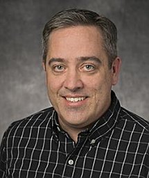 Jason A Mears合作论文数NIDDK/NIH3H. Gao合作论文数2
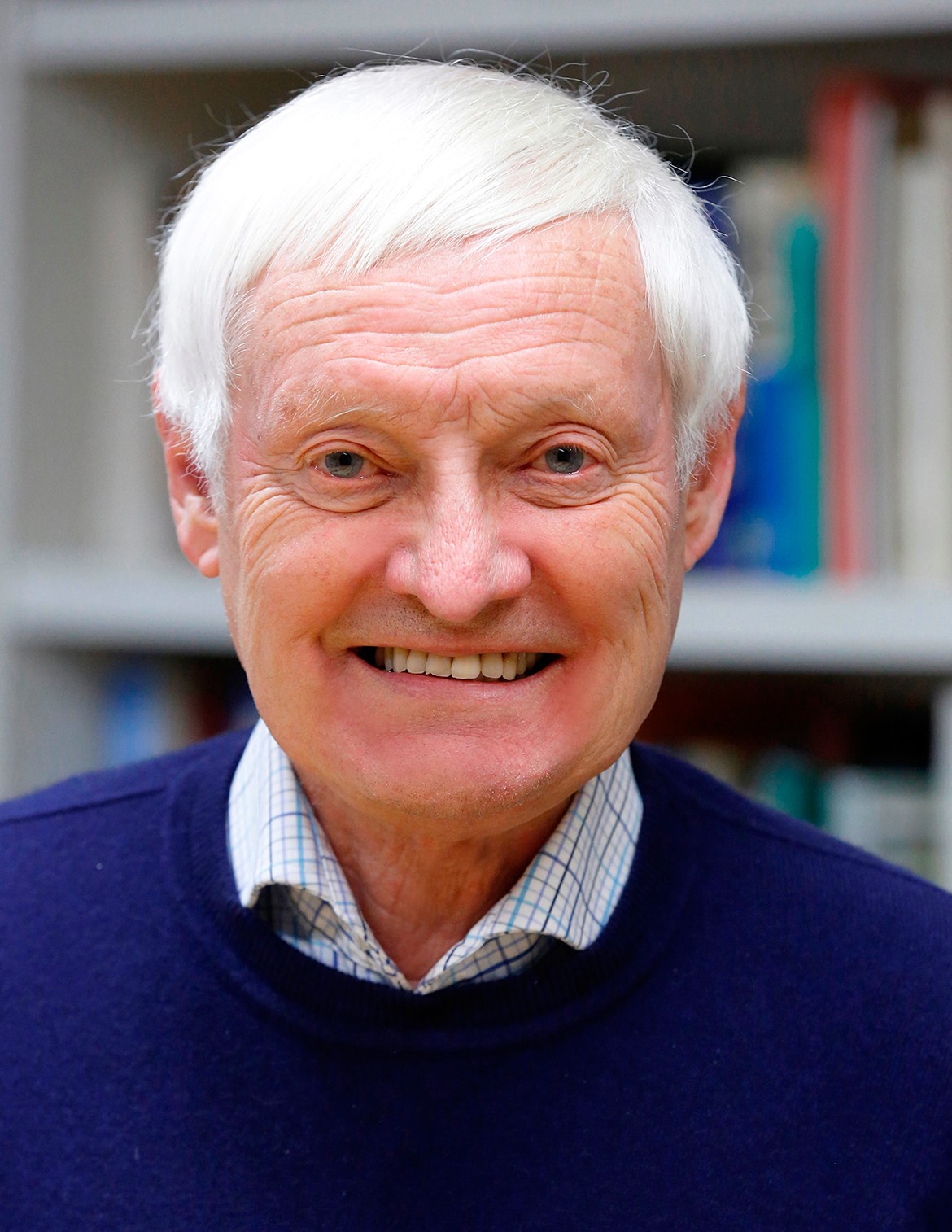 Joachim Frank (阿希姆·弗兰克)合作论文数Howard Hughes Medical Institute, Department of Biochemistry and Molecular Biophysics, College of Physicians and Surgeons, Columbia University;Department of Biological Sciences, Columbia University2Mikel Valle合作论文数2J J Cannone合作论文数1E.D. Hoffer合作论文数1A. J. Johnson合作论文数1J Audhya合作论文数1
 Dolly Al Koborssy合作论文数Team "Olfaction: From Coding to Memory,"Lyon Neuroscience Center, University Lyon 1 Lyon1
 Paul J. Hagerman合作论文数Department of Biochemistry and Molecular Medicine, School of Medicine, UC Davis Health;Division of Biological Chemistry, Medical Center, UC Davis Health1
 Robin R. Gutell合作论文数Institute for Cellular and Molecular Biology, University of Texas1
 Job Dekker合作论文数Department of Systems Biology, University of Massachusetts Chan Medical School1Margaret S. Vanloock合作论文数1
 Debra A Fadool合作论文数1
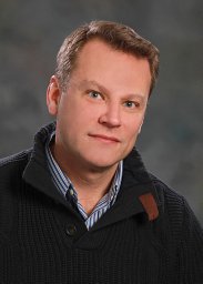 Ken Keiler合作论文数Department of Molecular Biology, College of Natural Sciences, The University of Texas at Austin1Haixiao Gao合作论文数1Niranjan Y. Sardesai合作论文数1Ashley A. Frazer-Abel合作论文数1
 Paul Schimmel合作论文数Department of Molecular Medicine, The Scripps Research Institute1




