The YPEL family genes are highly conserved across a diverse range of eukaryotic organisms and thus potentially involved in essential cellular processes. Ypel4, one of five YPEL family gene orthologs in mouse and human, is highly and specifically expressed in late terminal erythroid differentiation (TED). In this study, we investigated the role of Ypel4 in murine erythropoiesis, providing for the first time an in-depth description of a Ypel4-null phenotype in vivo. We demonstrated that the Ypel4-null mice displayed a secondary polycythemia with macro- and reticulocytosis. While lack of Ypel4 did not affect steady-state TED in the bone marrow or spleen, the anemia-recovering capacity of Ypel4-null cells was diminished. Furthermore, Ypel4-null red blood cells (RBC) were cleared from the circulation at an increased rate, demonstrating an intrinsic defect of RBCs. Scanning electron micrographs revealed an ovalocytic morphology of Ypel4-null RBCs and functional testing confirmed reduced deformability. Even though Band 3 protein levels were shown to be reduced in Ypel4-null RBC membranes, we could not find support for a physical interaction between YPEL4 and the Band 3 protein. In conclusion, our findings provide crucial insights into the role of Ypel4 in preserving normal red cell membrane integrity.
Production of red blood cells relies on proper mitochondrial function, both for their increased energy demands during differentiation and for proper heme and iron homeostasis. Mutations in genes regulating mitochondrial function have been reported in patients with anemia, yet their pathophysiological role often remains unclear. PGC1β is a critical coactivator of mitochondrial biogenesis, with increased expression during terminal erythroid differentiation. The role of PGC1β has however mainly been studied in skeletal muscle, adipose and hepatic tissues, and its function in erythropoiesis remains largely unknown. Here we show that perturbed PGC1β expression in human hematopoietic stem/progenitor cells from both bone marrow and cord blood results in impaired formation of early erythroid progenitors and delayed terminal erythroid differentiation in vitro, with accumulations of polychromatic erythroblasts, similar to MDS-related refractory anemia. Reduced levels of PGC1β resulted in deregulated expression of iron, heme and globin related genes in polychromatic erythroblasts, and reduced hemoglobin content in the more mature bone marrow derived reticulocytes. Furthermore, PGC1β knock-down resulted in disturbed cell cycle exit with accumulation of erythroblasts in S-phase and enhanced expression of G1-S regulating genes, with smaller reticulocytes as a result. Taken together, we demonstrate that PGC1β is directly involved in production of hemoglobin and regulation of G1-S transition and is ultimately required for proper terminal erythroid differentiation.
Massive expansion of erythroid progenitor cells is essential for surviving anemic stress. Research towards understanding this critical process, referred to as stress-erythropoiesis, has been hampered due to lack of specific marker-combinations enabling analysis of the distinct stress-progenitor cells capable of providing radioprotection and enhanced red blood cell production. Here we present a method for precise identification and in vivo validation of progenitor cells contributing to both steady-state and stress-erythropoiesis, enabling for the first time in-depth molecular characterization of these cells. Differential expression of surface markers CD150, CD9 and Sca1 defines a hierarchy of splenic stress-progenitors during irradiation-induced stress recovery in mice, and provides high-purity isolation of the functional stress-BFU-Es with a 100-fold improved enrichment compared to state-of-the-art. By transplanting purified stress-progenitors expressing the fluorescent protein Kusabira Orange, we determined their kinetics in vivo and demonstrated that CD150+CD9+Sca1- stress-BFU-Es provide a massive but transient radioprotective erythroid wave, followed by multi-lineage reconstitution from CD150+CD9+Sca1+ multi-potent stem/progenitor cells. Whole genome transcriptional analysis revealed that stress-BFU-Es express gene signatures more associated with erythropoiesis and proliferation compared to steady-state BFU-Es, and are BMP-responsive. Evaluation of chromatin accessibility through ATAC sequencing reveals enhanced and differential accessibility to binding sites of the chromatin-looping transcription factor CTCF in stress-BFU-Es compared to steady-state BFU-Es. Our findings offer molecular insight to the unique capacity of stress-BFU-Es to rapidly form erythroid cells in response to anemia and constitute an important step towards identifying novel erythropoiesis stimulating agents.
•Erythroid-specific deletion of pRb results in MDS-like anemia.•There are increased progenitors in BM and a block at the orthochromatic erythroblast stage.•Mitochondrial gene expression and function are disrupted (decreased OXPHOS, disturbed heme production and iron transport).•Enhancement of mitochondrial function through the PPAR-γ pathway rescues MDS-like anemia.
The hematopoietic system is responsible for transporting oxygen and nutrients, fighting infections, and repairing tissue damage. Hematopoietic system dysfunction therefore causes a range of serious health consequences. Lifelong hematopoiesis is maintained by repopulating multipotent hematopoietic stem cells (HSCs) that replenish shorter -lived, mature blood cell types. A prokaryotic mechanism of immunity, the Clustered Regularly Interspaced Short Palindromic Repeats (CRISPR)/Cas9 nuclease system, has been recently "repurposed" to mutate mammalian genomes efficiently and in a sequence -specific manner. The application of this genomeediting technology to hematology has afforded new approaches for functional genomics and even the prospect of "correcting" dysfunctional HSCs in the treatment of serious genetic hematological diseases. In this Perspective, we provide an overview of three recent CRISPR/ Cas9 methods in hematology: gene disruption, gene targeting, and saturating mutagenesis. We also summarize the technical considerations and advice provided during the May 2017 International Society of Experimental Hematology New Investigator Committee webinar on the same topic. (C) 2017 ISEH Society for Hematology and Stem Cells. Published by Elsevier Inc. All rights reserved.
Hematopoietic stem cells (HSCs) reside in specialized microenvironments known as niches. The niche is essential to support HSC function and to maintain a correct balance between self renewal and differentiation. Recent advances in defining different mesenchymal and endothelial bone marrow cell populations, as well as hematopoietic stem and progenitor cells, greatly enhanced our understanding of these niches and of the molecular mechanisms by which they regulate HSC function. In addition to the role in maintaining HSC homeostasis, the niche has also been implicated in the pathogenesis of blood disorders including hematological malignancies. Characterizing the extrinsic regulators and the cellular context in which the niches interact with HSCs will be crucial to define new strategies to enhance blood regeneration. Furthermore, a better understanding of the role of the niche in leukemia development will open new possibilities for the treatment of these disorders by using therapies aiming to target the leukemic niche specifically. To update on recent findings on this topic, the International Society for Experimental Hematology (ISEH) organized a webinar, presented by Prof. Sean J. Morrison and Dr. Simon Mendez-Ferrer and moderated by Dr. Cristina Lo Celso, entitled "The evolving view of the hematopoietic stem cell niche," which we summarize here. Published by Elsevier Inc. on behalf of ISEH - International Society for Experimental Hematology.
In the active debate regarding blood development architecture, the progenitors with erythroid and megakaryocytic potential are often neglected since their immunophenotypes are not completely defined, and mature erythrocytes and platelets are difficult to trace in vivo after transplantation. Precise identification and in vivo validation of progenitor cells contributing to steady-state and stress-megakaryo- /erythropoiesis is important in order to study the regulation of these processes. Here we demonstrate that the combination of surface markers CD150, CD9 and Sca1 defines a hierarchy of splenic stress-progenitors during irradiation-induced stress recovery, and provides high purity isolation of the earliest erythroid progenitor Burst Forming Unit-Erythroid (BFU-Es) both in steady-state bone marrow and in the spleen during stress-recovery. By transplanting sorted stress-progenitor populations from transgenic mice expressing Kusabira Orange in all cells, including erythrocytes and platelets, we have for the first time determined their kinetics and full differentiation potential in vivo. We demonstrate that splenic CD150+CD9+Sca1- stress-BFU-Es provide a massive but transient erythroid wave, followed by multi-lineage reconstitution from CD150+CD9+Sca1+ multi-potent stem/progenitor cells. Our findings offer a method for high purity isolation of stress-erythroid progenitors mediating recovery from severe anemia, with a 100-fold improved enrichment compared to state-of-the-art. This provides a novel possibility to perform in-depth molecular characterization of these cells, and we are currently analyzing genome wide differences in transcriptome and chromatin availability between highly pure populations of stress-progenitors and steady-state BFU-Es using RNA sequencing and ATAC sequencing. The potential to identify new mechanisms regulating steady-state and stress erythropoiesis is an important step towards understanding and treating anemia.
1. Wistar rats of both sexes daily received an ethanol solution of ammonium metavanadate (AMV) of 0.3 mg V/cm3 5% ethanol concentration as sole drinking liquid, for a period of 4 weeks.2. The reference groups received for drinking aqueous AMV solution, or 5% ethanol, or water.3. In animals drinking both water and ethanol AMV solution a decrease in the erythrocyte count and haemoglobin level was noted together with an increase of the percentage of reticulocytes and polychromatophilic erythrocytes in the peripheral blood.4. A small rise of the percentage of polychromatophilic and orthochromatic erythroblasts was at the same time noted in the bone marrow of animals receiving ethanol AMV solution.5. In the group of animals drinking 5% ethanol a fall of the erythrocyte count was observed and a rise of the leukocyte count, particularly of lymphocytes.6. Substitution of water by 5% ethanol solution as solvent for AMV did not have any distinct influence on the toxicity of the tested compound.
Erythropoiesis is intimately coupled to cell division, and deletion of the cell cycle regulator Retinoblastoma protein (pRb) causes anemia in mice. However, at what stage of erythroid development pRb is important, and the precise underlying mechanisms, remain elusive. To further elucidate its importance in red blood cell (RBC) development, pRb was conditionally deleted in an erythroid-specific manner (Epor-Cre pRbfl/fl). This resulted in macrocytic anemia, in spite of elevated levels of Epo and accumulation of erythroid progenitors in the bone marrow, a phenotype strongly resembling the refractory anemia seen in Myelodysplastic syndromes (MDS). We have now identified a block in terminal erythropoiesis at the orthochromatic stage, and transcriptional profiling on purified populations immediately around the block revealed that final maturation stages lacking pRb failed to up-regulate large clusters of genes critical for mitochondrial function, heme synthesis and iron metabolism, including Pgc1b, Alas2, Abcb7, and Tmem14c. In accordance, these cells displayed disturbed mitochondrial membrane potential and heme production. Notably, de-regulated ABCB7 is known to cause ring sideroblastic anemia in MDS. And PGC1b, a critical co-activator of mitochondrial biogenesis, is heterozygously lost in the most prevalent cytogenetic abnormality of MDS; del5q. Since heme synthesis and iron metabolism are crucial for production of functional RBCs, we asked if enhanced mitochondrial biogenesis could ameliorate the anemia. To this end, we transplanted stem/progenitor cells from wildtype and erythroid specific pRb-deficient mice overexpressing Pgc1b into lethally irradiated recipients, which resulted in a normalization of both RBC counts and red cell size. In conclusion, we demonstrate that lack of pRb results in MDS-like anemia with a block in orthochromatic erythroblasts due to failure to up-regulate mitochondrial function and heme synthesis. While mitochondrial mutations have been reported in MDS, their relevance for refractory anemia remains elusive. We are currently investigating the role of mitochondrial function in development of human refractory anemia associated with MDS.
Erythroid cell commitment and differentiation proceed through activation of a lineage-restricted transcriptional network orchestrated by a group of well characterized genes. However, the minimal set of factors necessary for instructing red blood cell (RBC) development remains undefined. We employed a screen for transcription factors allowing direct lineage reprograming from fibroblasts to induced erythroid progenitors/precursors (iEPs). We show that Gata1, Tal1, Lmo2, and c-Myc (GTLM) can rapidly convert murine and human fibroblasts directly to iEPs. The transcriptional signature of murine iEPs resembled mainly that of primitive erythroid progenitors in the yolk sac, whereas addition of Klf1 or Myb to the GTLM cocktail resulted in iEPs with a more adult-type globin expression pattern. Our results demonstrate that direct lineage conversion is a suitable platform for defining and studying the core factors inducing the different waves of erythroid development.
Transplantation of hematopoietic stem cells (HSCs) to treat hematologic disorders is routinely used in the clinic. However, HSC therapy is hindered by the requirements of finding human leukocyte antigen (HLA)-matched donors and attaining sufficient numbers of long-term HSCs in the graft. Therefore, ex vivo expansion of transplantable HSCs remains one of the "holy grails" of hematology. Without the ability to maintain and expand human HSCs in vitro, two complementary approaches involving cellular reprogramming to generate transplantable HSCs have emerged. Reprogrammed HSCs represent a potentially inexhaustible supply of autologous tissue. On March 18th, 2015, Dr. George Q. Daley and Dr. Derrick J. Rossi, two pioneers in the field, presented and discussed their most recent research on these topics in a webinar organized by the International Society for Experimental Hematology (ISEH). Here, we summarize these seminars and discuss the possibilities and challenges in the field of hematopoietic specification.
Transcription factor-based direct lineage reprogramming is a powerful tool to discover and study factors determining cell lineage fate. We propose that this methodology can be used to define the core transcriptional program directing red blood cell development. The aim of this study was therefore to identify the minimal set of transcription factors that allows direct lineage reprogramming of murine fibroblasts to erythroid progenitor cells.
Objective: To identify how the gp130-signaling cytokine oncostatin M (OSM), acting alone or in concert with IL-1 beta or TNF alpha, affects synovial fibroblast expression of genes relevant to inflammation and bone erosion in inflammatory arthritis.Methods: Synovial fibroblasts (SFs) were isolated from non-arthritic wild type (WT) or OSM receptor deficient (OSMR-/-) mice and stimulated with OSM, IL-1 beta or TNF alpha and their combinations. Cytokine gene expression was assessed by quantitative RT-PCR. ELISA, flow cytometry and immunohistochemistry identified protein expression. Gene expression patterns were confirmed in SFs isolated from patients with osteoarthritis (OASFs) and rheumatoid arthritis (RASFs).Results: Expression of OSM and its receptors, gp130, OSMR and LIFR, was increased in synovial tissue from the mouse antigen-induced arthritis model. In isolated WT mouse synovial fibroblasts OSM alone, or in synergy with IL-1 beta, or together with TNF alpha, potently induced expression of the pro-inflammatory cytokine IL-6. OSM also induced a sustained increase in mRNA levels of the pro-osteoclastic cytokine RANKL. Combining OSM with IL-1 beta, but not with TNF alpha, further increased RANKL expression. Importantly these effects of OSM were all dependent on the expression of OSMR. Furthermore, OSM also increased expression of its own receptors, gp130 and OSMR and the IL-1 receptor, IL1-R1; the latter effects were also observed in both human OASFs and RASFs.Conclusion: Together our data suggests that OSM signaling via OSMR in SFs has the potential to contribute significantly to joint destruction in inflammatory arthritis. It not only induces expression of pro-inflammatory and pro-osteoclastic cytokines but can also augment its own actions and that of IL-1 by inducing expression of OSMR and IL1-R1. (C) 2014 Elsevier Ltd. All rights reserved.
The proto-oncogene SKI is highly expressed in human myeloid leukemia and also in murine hematopoietic stem cells. However, its operative relevance in these cells remains elusive. We have over-expressed SKI to define its intrinsic role in hematopoiesis and myeloid neoplasms, which resulted in a robust competitive advantage upon transplantation, a complete dominance of the stem and progenitor compartments, and a marked enhancement of myeloid differentiation at the expense of other lineages. Accordingly, enforced expression of SKI induced gene signatures associated with hematopoietic stem cells and myeloid differentiation. Here we provide detailed experimental methods and analysis for the gene expression profiling described in our recently published study of Singbrant et al. (2014) in Haematologica. Our data sets (available at http://www.ncbi.nlm.nih.gov/geo/query/acc.cgi?acc=GSE39457) provide a resource for exploring the underlying molecular mechanisms of the involvement of the proto-oncogene SKI in hematopoietic stem cell function and development of myeloid neoplasms.
The proto-oncogene SKI is highly expressed in human myeloid leukemia and also in murine hematopoietic stem cells. However, its operative relevance in these cells remains elusive. We have over-expressed SKI to define its intrinsic role in hematopoiesis and myeloid neoplasms, which resulted in a robust competitive advantage upon transplantation, a complete dominance of the stem and progenitor compartments, and a marked enhancement of myeloid differentiation at the expense of other lineages. Accordingly, enforced expression of SKI induced a gene signature associated with hematopoietic stem cells and myeloid differentiation, as well as hepatocyte growth factor signaling. Here we demonstrate that, in contrast to what has generally been assumed, the significant impact of SKI on hematopoiesis is independent of its ability to inhibit TGF-beta signaling. Instead, myeloid progenitors expressing SKI are partially dependent on functional hepatocyte growth factor signaling. Collectively our results demonstrate that SKI is an important regulator of hematopoietic stem cell activity and its overexpression leads to myeloproliferative disease.
Erythropoietin (Epo) has been used in the treatment of anemia resulting from numerous etiologies, including renal disease and cancer. However, its effects are controversial and the expression pattern of the Epo receptor (Epo-R) is debated. Using in vivo lineage tracing, we document that within the hematopoietic and mesenchymal lineage, expression of Epo-R is essentially restricted to erythroid lineage cells. As expected, adult mice treated with a clinically relevant dose of Epo had expanded erythropoiesis because of amplification of committed erythroid precursors. Surprisingly, we also found that Epo induced a rapid 26% loss of the trabecular bone volume and impaired B-lymphopoiesis within the bone marrow microenvironment. Despite the loss of trabecular bone, hematopoietic stem cell populations were unaffected. Inhibition of the osteoclast activity with bisphosphonate therapy blocked the Epo-induced bone loss. Intriguingly, bisphosphonate treatment also reduced the magnitude of the erythroid response to Epo. These data demonstrate a previously unrecognized in vivo regulatory network coordinating erythropoiesis, B-lymphopoiesis, and skeletal homeostasis. Importantly, these findings may be relevant to the clinical application of Epo.
Oral Presentations / Osteoarthritis and Cartilage 19S1 (2011) S7-S52 nicely correlated with thickening of the synovial lining layer comprising activated macrophages.When collagenase-induced-osteoarthritis was elicited in S100A9 -/-mice, significantly lower synovial activation was observed when compared to WT mice.Synovial activation was 62% lower at day 42.Cartilage destruction was significantly lower in all surfaces and ranged from a 45% reduction in the lateral tibia to 73% reduction in the medial femur.When primary mouse chondrocytes were stimulated with S100A8 or S100A9, a strong upregulation of particularly MMP-3 mRNA level was found indicating a direct role of S100A8/A9 in cartilage destruction.Conclusions: Alarmins S100A8/S100A9 are expressed by phagocytes in biopsies of early OA patients.S100A8/A9 play a crucial role in synovial activation and cartilage destruction in an osteoarthritis model that shows clear synovial involvement.S100A8/A9 expression in the synovium causes pathology probably by stimulating MMP-mediated damage in the cartilage matrix.
 Stefan Karlsson合作论文数Hogskolan i Skovde12
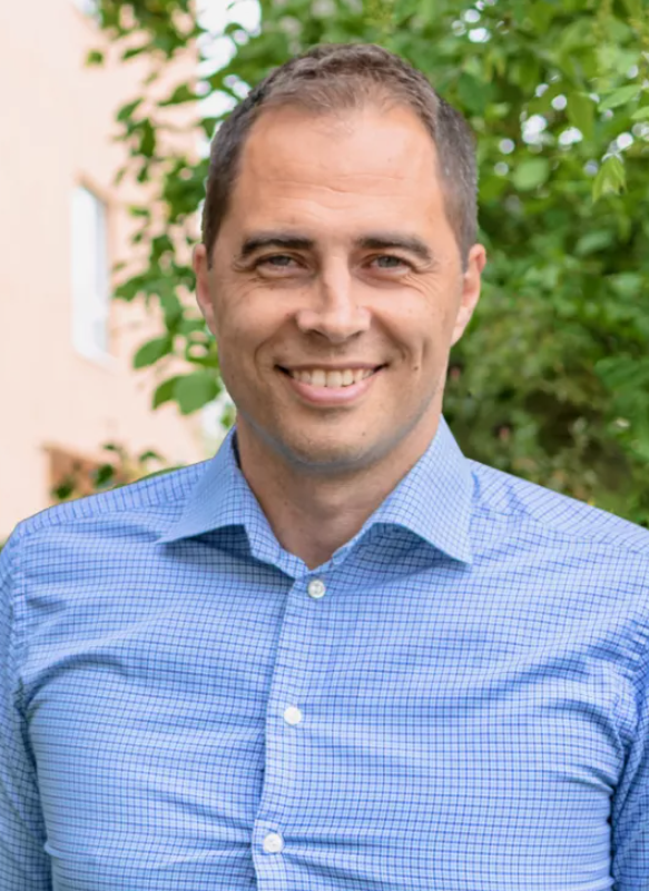 Göran Karlsson合作论文数Division of Molecular Hematology, Department of Laboratory Medicine, Faculty of Medicine, Lund University;Lund Stem Cell Center, Faculty of Medicine, Lund University;Nygen Analytics AB12
 Carl R. Walkley合作论文数Harvard Stem Cell Institute, Harvard Medical School9
 Johan Flygare合作论文数8Shamit Soneji合作论文数6Jennifer L. Moody合作论文数5
 Alexander Mattebo合作论文数5Ulrich Blank合作论文数5
 Jennie L Moody合作论文数Department of Environmental Sciences, University of Virginia5Taha Sen合作论文数4
 Thomas Martin合作论文数Faculty of Medicine, Dentistry and Health Sciences, University of Melbourne;Department of Medicine, St Vincent's Hospital;St Vincent’s Institute4Natalie a Sims合作论文数4
 Brian Liddicoat合作论文数3
 Vijay Sankaran合作论文数Division of Hematology/Oncology, Center for Life Sciences, Boston Children's Hospital;Dana-Farber Cancer Institute;Harvard Medical School3
 Magnus Gram合作论文数3Tonkin Brett A合作论文数3
 Alistair M Chalk合作论文数The Eskitis Institute for Cell and Molecular Therapies, Griffith University3Benoit Le Goff合作论文数3Kishori Dhulipala合作论文数2Sandra Capellera-Garcia合作论文数2Megan Russell合作论文数2Marie Aspling合作论文数2violeta rayonestrada合作论文数Laboratory of Lymphocyte Biology, The Rockefeller University, 1230 York Avenue, New York, NY 10065, USA2
 Nicole Corinne Walsh合作论文数2
 Jonas Larsson合作论文数2Purton Louise E合作论文数St Vincents Inst Med Res2
 Kavitha Siva合作论文数2Karin Olsson合作论文数2
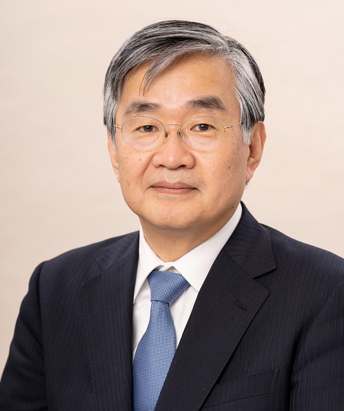 Kohei Miyazono (宮園浩平)合作论文数Graduate School of Medicine, The University of Tokyo;RIKEN2
 Mikael Sigvardsson合作论文数Department of Laboratory Medicine, Lund University;Lund University Cancer Centre;Department of Biomedical and Clinical Sciences, Linköping University2
 David Bryder合作论文数Institute of Laboratory Medicine, University Hospital of Lund2Ulrika Blank合作论文数2
 Tobias Strid合作论文数2Mats Ehinger合作论文数2Meaghan Wall合作论文数2Evan Romas合作论文数2Thomas Graf合作论文数1Sita R Dewamitta合作论文数1Taiju Utsugisawa合作论文数1
 Ramprasad Ramakrishnan合作论文数1Cr Walkley合作论文数1NC Walsh合作论文数1
 Maria Jassinskaja合作论文数University of York1Abdul Ghani Alattar合作论文数Lund Univ, Lund Strateg Ctr Stem Cell Biol, Dept Mol Med & Gene Therapy, Lund, Sweden1
 Daniel Lucas合作论文数Div Expt Hematol & Canc Biol, Cincinnati Childrens Hosp Med Ctr1Jun Chen合作论文数Hunan University(Hunan University,Hunan Univ.),Changsha,China1
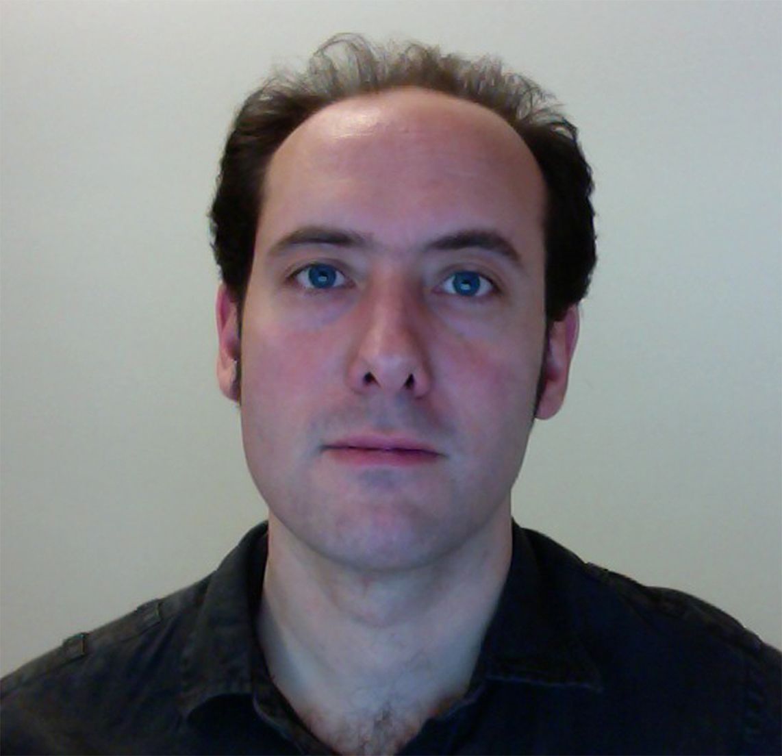 Matthias Stadtfeld合作论文数Weill Cornell Graduate School of Medical Sciences, Cornell University1




