Pre-mRNA binding to the yeast U2 small nuclear ribonucleoprotein (snRNP) during prespliceosome formation requires ATP hydrolysis, the highly conserved UACUAAC box of the branch point region of the pre-mRNA, and several factors. Here we analyzed the binding of a radiolabeled 2'-O-methyl oligonucleotide complementary to U2 small nuclear RNA to study interactions between the UACUAAC box, U2 snRNP, and Prp5p, a DEAD box protein necessary for prespliceosome formation. Binding of the 2'-O-methyl oligonucleotide to the U2 snRNP in yeast cell extract was assayed by gel electrophoresis. Binding was rapid, enhanced by ATP, and dependent on the integrity and conformation of the U2 snRNP. It was also stimulated by Prp5p that was found to associate physically with U2 snRNP. In vitro heat inactivation of the temperature-sensitive prp5-1 mutant extract decreased oligonucleotide binding to U2 and the ATP enhancement of binding by 3-fold. Furthermore, the temperature-sensitive prp5-1 mutation maps to the ATP-binding motif I within the helicase-like domain. Thus the catalytic activity of Prp5p likely promotes a conformational change in the U2 snRNP.
The mammalian 70K protein, a component of the U1 small nuclear ribonucleoprotein involved in pre-mRNA splicing, interacts with a number of proteins important for regulating constitutive and alternative splicing. Similar proteins that interact with the yeast homolog of the 70K protein, Snp1p, have yet to be identified. We used the two-hybrid system to find four U1-Snp1 associating (Usa) proteins. Two of these proteins physically associate with Snp1p as assayed by coimmunoprecipitation. One is Prp8p, a known, essential spliceosomal component. This interaction suggests some novel functions for Snp1p and the U1 small nuclear ribonucleoprotein late in spliceosome development. The other, Exo84p, is a conserved subunit of the exocyst, an eight-protein complex functioning in secretion. We show here that Exo84p is also involved in pre-mRNA splicing. A temperature-sensitive exo84 mutation caused increased ratios of pre-mRNA to mRNA for the Rpl30 and actin transcripts in cells incubated at the non-permissive temperature. The mutation also led to a defect in splicing and prespliceosome formation in vitro; an indication that Exo84p has a direct role in splicing. The results elucidate a surprising link between splicing and secretion.
The spliceosome is a large ribonucleoprotein that splices precursor messenger RNA (pre-mRNA) in the nuclei of eukaryotic cells (reviewed in refs. 1–3). It is composed of five small nuclear ribonucleoproteins (snRNPs) and numerous non-snRNP proteins. Each snRNP consists of a small nuclear RNA (snRNA) and several proteins.
Splicing of eukaryotic precursor messenger RNAs (pre-mRNAs) excises the intron from the precursor and ligates the two exons together to produce the mature mRNA. It occurs via a two-step mechanism (Fig. 1) (reviewed in ref. 1). In the first step the 2′ hydroxyl group of an intronic adenylyl residue initiates a transesterfication reaction at the 5′ splice site. The result is the cleavage of the 5′ splice site phosphodiester bond and the formation of a new, 2′–5′ phosphodiester bond between the adenylyl residue and the 5′ end of the intron. Because the intronic adenylyl residue has both 2′–5′ and 3′–5′ phosphodiester bonds, it is commonly referred to as the branch point nucleotide. The first splicing step yields two intermediates, “free” exon 1 and the lariat intermediate, which are not covalently linked together. In the second step, the 3′ hydroxyl of the “free” exon 1 initiates a second transesterification reaction at the 3′ splice site, resulting in the ligation of the two exons together to form the mRNA and the release of the lariat intron.
U1 small nuclear ribonucleoprotein (snRNP) may function during several steps of spliceosome assembly. Most spliceosome assembly assays, however, fail to detect the U1 snRNP. Here, I used a new native gel electrophoretic assay to find the yeast U1 snRNP in three pre-splicing complexes (delta, beta1, alpha2) formed in vitro. The order of complex formation is deduced to be delta --> beta1 --> alpha2 --> alpha1 --> beta2, the active spliceosome. The delta complex is formed when U1 snRNP binds to pre-mRNA in the absence of ATP. There are two forms of delta: a major one, deltaun, unstable to competitor RNA; and a minor one, deltacommit, committed to the splicing pathway. The other complexes are formed in the presence of ATP and contain the following snRNPs: beta1, the pre-spliceosome, has both U1 and U2; alpha2 has all five, however, U1 is reduced compared with the others; and alpha1 and beta2 have U2, U5, and U6. Prior work by others suggests that U1 is "handing off" the 5' splice site region to the U5 and U6 snRNPs before splicing begins. The reduced levels of U1 snRNP in the alpha2 complex suggests that the handoff occurs during formation of this complex.
We have analyzed the functions of several pre-mRNA processing (PRP) proteins in yeast spliceosome formation. Here, we show that PRP5 (a DEAD box helicase-like protein), PRP9, and PRP11 are each required for the U2 snRNP to bind to the pre-spliceosome during spliceosome assembly in vitro. Genetic analyses of their functions suggest that they and another protein, PRP21, act concertedly and/or interact physically with each other and with the stem-loop IIa of U2 snRNA to bind U2 snRNP to the pre-mRNA. Biochemical complementation experiments also indicate that the PRP9 and PRP11 proteins interact. The PRP9 and PRP11 proteins may be functioning similarly in yeast and mammalian cells. The requirement for ATP and the helicase-like PRP5 protein suggests that these factors might promote a conformational change (involving either the U1 or U2 snRNP) that is required for the association of U2 snRNP with the pre-mRNA.
Small, reversibly biotinylated RNAs as described here are versatile ligands for affinity chromatography of RNA-binding components. These RNAs can be attached to a solid support by binding to avidin and used as ligands, or they may be hybridized to another RNA which acts as the ligand. The incorporation of a disulfide bond in the linker arm connecting biotin to the RNA makes it possible to dissociate the RNA from avidin under mild conditions. Our results regarding the binding and elution of the biotinylated RNA may be applied to other, reversibly biotinylated molecules.
Splicing of nuclear precursor messenger RNA (pre-mRNA) occurs on a large ribonucleoprotein complex, the spliceosome. Several small nuclear ribonucleoproteins (snRNP's) are subunits of this complex that assembles on the pre-mRNA. Although the U1 snRNP is known to recognize the 5' splice site, its roles in spliceosome formation and splice site alignment have been unclear. A new affinity purification method for the spliceosome is described which has provided insight into the very early stages of spliceosome formation in a yeast in vitro splicing system. Surprisingly, the U1 snRNP initially recognizes sequences at or near both splice junctions in the intron. This interaction must occur before the other snRNP's (U2, U4, U5, and U6) can join the complex. The results suggest that interaction of the two splice site regions occurs at an early stage of spliceosome formation and is probably mediated by U1 snRNP and perhaps other factors.
When exposed to DNA-damaging agents, the yeast Saccharomyces cerevisiae induces the expression of at least six specific genes. We have previously identified one damage inducible (DIN) gene as a gene fusion (din-lacZ fusion) whose expression increases in response to DNA-damaging treatments. We describe here the identification of five additional DIN genes as din-lacZ fusions and the responses of all six DIN genes to DNA-damaging agents. Northern blot analyses of the transcripts of two of the DIN genes show that their levels increase after exposure to DNA-damaging agents. Five of the din-lacZ fusions are induced in S. cerevisiae cells exposed to UV light, gamma rays, methotrexate, or alkylating agents. One of the din-lacZ fusions is induced by either UV or methotrexate but not by the other agents. This finding suggests that there are sets of DIN genes that are regulated differently.
Publisher Summary This chapter discusses cloning regulated yeast genes from a pool of lacZ fusions. Gene fusions have been used during the past decade to study such diverse problems as transcriptional regulation, translational regulation, and protein localization. In prokaryotes, various genetic methods have been employed to create operon and protein fusions. The active, regulated gene fusions constructed by the procedures described below contain a yeast DNA fragment with sequences for the control and initiation of transcription and translation joined to a lacZ fragment from which the 5’ portion including the first few N-terminal amino acid codons of the gene have been deleted. A library of random yeast gene:lacZ fusions in yeast transformants was constructed and screened for regulated gene fusions.
This chapter describes the R looping and structural gene identification of recombinant deoxyribonucleic acid (DNA). The advent of recombinant DNA technology has made possible studies on the organization and expression of eukaryotic genes. In high concentrations of formamide, ribonucleic acid (RNA)–DNA duplexes have a higher thermal denaturation temperature than the corresponding DNA–DNA duplexes. When DNA and complementary RNA are incubated under these conditions, R loops are readily formed. For the formation of R loops with recombinant DNA, the procedure consists of incubating sufficient quantities of DNA with limited amounts of messenger RNA (mRNA) such that the rate of the reaction is DNA driven. The rate of R-loop formation is a sensitive function of the incubation temperature. The chapter discusses the R looping of histone plasmids.
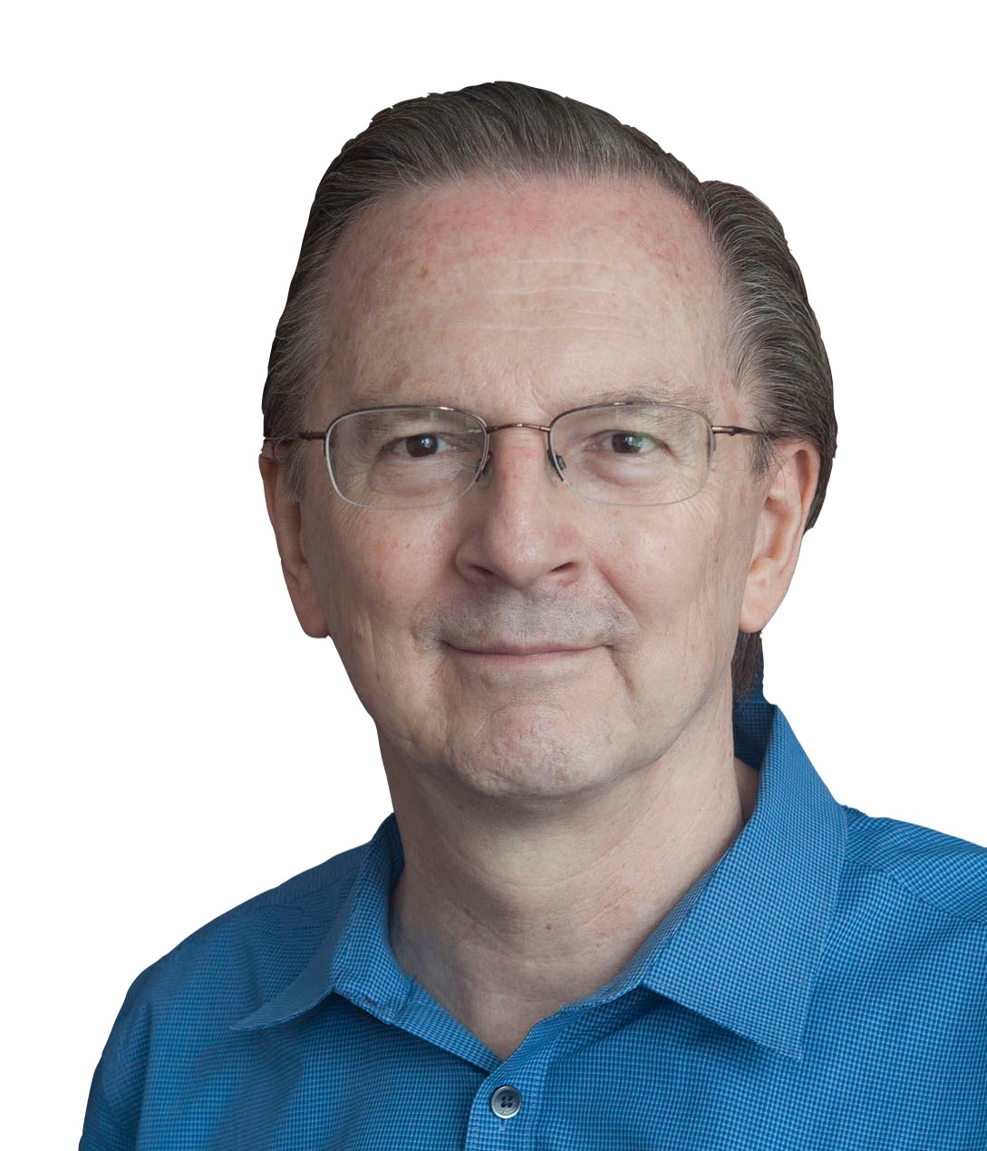 Jack W. Szostak合作论文数Department of Chemistry, The University of Chicago2
 John N. Abelson合作论文数Division of Biology and Biological Engineering, California Institute of Technology2
 Robert P. Ricciardi合作论文数Department of Basic & Translational Sciences, School of Dental Medicine, University of Pennsylvania1
 Andrew Murray合作论文数Department of Molecular and Cellular Biology, Harvard University;Rowland Institute, Harvard University;Howard Hughes Medical Institute1
 T.-H. Chang合作论文数1
 John L. Woolford, Jr.合作论文数Department of Biological Sciences, Mellon College of Science, Carnegie Mellon University1Lynna Hereford合作论文数1Zdenek Hostomsky合作论文数1
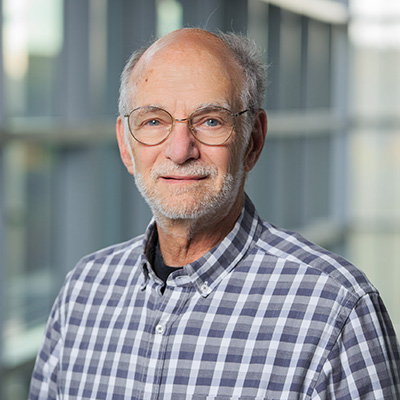 Michael Rosbash合作论文数Department of Biology, Brandeis University;Howard Hughes Medical Institute1




