This 20th anniversary of the RNA journal is a time to look back on what we have accomplished and perhaps more important to decide what is important for the future. I won't reiterate Olke Uhlenbeck's “Imperfect Account of the Founding of the RNA Society.” You can find that account on our website. I am glad Olke wrote it down when he did because you wouldn't want the aging founders to write it now. But I do remember quite well the elements I thought we needed. I thought it important that we have a society journal and that the journal should be rigorous in its review of the science but should not be a magazine. We even knew then that the criteria for a Science, Nature or Cell paper were not solely that the science be rigorous but that the material be “trendy.” Of course that is much worse now than it was then (or maybe we are no longer trendy). Serious peer reviewed publication of scientific results is important for getting a job, for academic advancement and for grant renewals. It is clear that we have accomplished what we set out to do in this regard. I thought that we should own the journal. The model for this is the JBC owned by ASBMB and the Protein Journal owned by the Protein Society. When the society owns the journal it can determine the policies of the journal and can maintain those policies. This would not necessarily be true if the journal is owned by a large publishing house. Furthermore I was aware that the ASBMB is wealthy because of its ownership of the JBC. We had been having various RNA processing meetings, first at Brookhaven and then for a number of years at Cold Spring Harbor. We wanted to formalize the nature and organization of an annual meeting. Our meetings have been a huge success because from the first the policy was that short talks be given by students (perhaps the early phage meetings were a model for this). But even more important, and this was never a written policy, was that the PIs be there and that they attend the talks. When a student gives a talk and he or she knows that the likes of Joan Steitz, Tom Cech, and Olke Uhlenbeck are sitting in the front row the talk takes on much more significance. All of the labs in this field work very hard with their students in preparing these talks and the sense of accomplishment in pulling it off is a priceless reward for the effort. For the future, I think we face a different and more difficult set of problems. We all know that the path the scientists of my era followed: Write a good thesis, do a good postdoc and get an academic job is much more difficult now. It is clear that our students understand this. I will not suggest solutions for this problem here but I have faith that our former students, now the leaders of the society, will take them on.
The ca. 2.45-2.22 Ga Turee Creek Group, Western Australia, contains carbonate-rich horizons that postdate earliest Proterozoic iron formations, bracket both Paleoproterozoic glaciogenic beds and the onset of the Great Oxidation Event (GOE), and predate ca. 2.2-2.05 Ga Lomagundi-Jatuli C-isotopic excursion(s). As such, Turee Creek carbonate strata provide an opportunity to characterize early Paleoproterozoic carbonate sedimentation and carbon cycle dynamics in the context of significant global change. Here, we report on the stratigraphy, sedimentology, petrology, carbon isotope chemostratigraphy, and stromatolite development for carbonate-rich successions within the pre-glacial part of the Kungarra Formation and the postglacial Kazput Formation.Kungarra carbonate units largely occur as laterally discontinuous beds within a thick, predominantly siliciclastic shelf deposit. While this succession contains thin microbialite horizons, most carbonates consist of patchy calcite overgrowths within a siliciclastic matrix. C-isotopic values show marked variation along a single horizon and even within hand samples, reflecting spatially and temporally variable mixing between dissolved inorganic carbon in seawater and isotopically light inorganic carbon generated via syn- and post-depositional remineralization of organic matter.In contrast, the Kazput carbonates consist of subtidal stromatolites, grainstones, and micrites deposited on a mixed carbonate-siliciclastic shelf. These carbonates exhibit moderate delta C-13 values of -2 parts per thousand to +1.5 parts per thousand and likely preserve a C-isotopic signature of seawater. Kazput carbonates, thus, provide some of the best available evidence that an interval of unexceptional C-isotopic values separates the Lomagundi-Jatuli C-isotopic excursion(s) from the initiation of the GOE as inferred from multiple sulfur isotopes (loss of mass independent fractionation). The Kazput Formation also contains unusual, m-scale stromatolitic buildups, which are composed of sub-mm laminae and discontinuous, convex upward lenticular precipitates up to a few mm in maximum thickness. Laminae, interpreted as microbial mat layers, contain quartz and clay minerals as well as calcite, whereas precipitate lenses consist of interlocking calcite anhedra, sometimes showing faint mm-scale banding. These cements formed either as infillings of primary voids formed by gas emission within penecontemporaneously lithified mats, or as local seafloor precipitates that formed on, or within, surface mats. It is possible that both mechanisms interacted to form the unique Kazput stromatolites. These microbialites speak to a distinctive interaction between life and environment early in the Paleoproterozoic Era. (C) 2015 Elsevier B.V. All rights reserved.
This chapter reviews what is known about the mechanism of precursor tRNA splicing: (i) the tRNA substrates for the splicing reaction, (ii) the enzymes involved in removing the introns to form the mature tRNA, (iii) interactions between these enzymes and their tRNA substrates and cofactors, (iv) the organization of tRNA splicing in the nucleus, (v) the identity of splicing mutants that affect the enzymatic machinery, (vi) current knowledge about the differences and similarities of tRNA splicing in systems of various organisms, and (vii) the possible function of tRNA introns.
The Cambrian Pedroche Formation comprises a mixed siliciclastic–carbonate succession recording subtidal deposition on a marine platform. Carbonate carbon isotope chemostratigraphy confirms previous biostratigraphic assignment of the Pedroche Formation to the Atdabanian regional stage of Siberia, correlative to Cambrian Series 2. At the outcrop scale, thrombolitic facies comprise ~60% of carbonate-normalized stratigraphy and coated-grains another ~10%. Petrographic point counts reveal that skeletons contribute at most 20% to thrombolitic inter-reef and reef-flank lithologies; on average, archaeocyath clasts make up 68% of skeletal materials. In contrast, petrographic point counts show that skeletons comprise a negligible volume of biohermal and biostromal thrombolite, associated nodular carbonate facies, and ooid, oncoid and peloid grainstone facies. As such, archaeocyathan reefal bioconstructions represent a specific and limited locus of skeletal carbonate production and deposition. Consistent with data from coeval, globally dispersed lower Cambrian successions, our analysis of the Pedroche Formation supports the view that lower Cambrian carbonates have more in common with earlier, Neoproterozoic deposits than with younger carbonates dominated by skeletal production and accumulation.
Valley-filling deposits of the Nama Group, southern Namibia, record two episodes of erosional downcutting and backfill, developed close together in time near the Ediacaran-Cambrian boundary. Geochronological constraints indicate that the older valley fill began 539.4 +/- 1 Ma or later; the younger of these deposits contains unusually well-preserved populations of the basal Cambrian trace fossil Treptichnus pedum. Facies analysis shows that T. pedum is closely linked to a nearshore sandstone deposit, indicating a close environmental or taphonomic connection to very shallow, mud-draped sandy seafloor swept by tidal currents. Facies restriction may limit the biostratigraphic potential of T. pedant in Namibia and elsewhere, but it also illuminates functional and ecological interpretation. The T. pedum tracemaker was a motile bilaterian animal that lived below the sediment-water interface-propelling itself forward in upward-curving projections that breached the sediment surface. The T. pedum animal, therefore, lived infaunally, perhaps to avoid predation, surfacing regularly to feed and take in oxygen. Alternatively, the T. pedum animal may have been a deposit feeder that surfaced largely for purposes of gas exchange, an interpretation that has some support in the observed association of T. pedum with mud drapes. Treptichnus pedum provides our oldest record of animals that combined anatomical and behavioral complexity. Insights from comparative biology suggest that basal Cambrian T. pedum animals already possessed the anatomical, neurological, and genetic complexity needed to enable the body plan and behavioral diversification recorded by younger Cambrian fossils.
Metabolic bone diseases like osteoporosis result from the disruption of normal bone mineral balance (BMB) resulting in bone loss. During spaceflight astronauts lose substantial bone. Bed rest provides an analog to simulate some of the effects of spaceflight; including bone and calcium loss and provides the opportunity to evaluate new methods to monitor BMB in healthy individuals undergoing environmentally induced-bone loss. Previous research showed that natural variations in the Ca isotope ratio occur because bone formation depletes soft tissue of light Ca isotopes while bone resorption releases that isotopically light Ca back into soft tissue (Skulan et al, 2007). Using a bed rest model, we demonstrate that the Ca isotope ratio of urine shifts in a direction consistent with bone loss after just 7 days of bed rest, long before detectable changes in bone mineral density (BMD) occur. The Ca isotope variations tracks changes observed in urinary N-teleopeptide, a bone resorption biomarker. Bone specific alkaline phosphatase, a bone formation biomarker, is unchanged. The established relationship between Ca isotopes and BMB can be used to quantitatively translate the changes in the Ca isotope ratio to changes in BMD using a simple mathematical model. This model predicts that subjects lost 0.25 +/- 0.07% (+/- SD) of their bone mass from day 7 to day 30 of bed rest. Given the rapid signal observed using Ca isotope measurements and the potential to quantitatively assess bone loss; this technique is well suited to study the short-term dynamics of bone metabolism.
The ca. 1.8Ga Duck Creek Formation, Western Australia, preserves 1000m of carbonates and minor iron formation that accumulated along a late Paleoproterozoic ocean margin. Two upward-deepening stratigraphic packages are preserved, each characterized by peritidal precipitates at the base and iron formation and carbonate turbidites in its upper part. Consistent with recent studies of Neoarchean basins, carbon isotope ratios of Duck Creek carbonates show no evidence for a strong isotopic depth gradient, but carbonate minerals in iron formations can be markedly depleted in 13C. In contrast, oxygen isotopes covary strongly with depth; δ18O values as positive as 2‰ VPDB in peritidal facies systematically decline to values of −6 to −16‰ in basinal rocks, reflecting, we posit, the timing of diagenetic closure. The Duck Creek Formation contains microfossils similar to those of the Gunflint Formation, Canada; they are restricted to early diagenetic cherts developed in basinal facies, strengthening the hypothesis that such fossils capture communities driven by iron metabolism. Indeed, X-ray diffraction data indicate that the Duck Creek basin was ferruginous throughout its history. The persistence of ferruginous waters and iron formation deposition in Western Australia for at least several tens of millions of years after the transition to sulfidic conditions in Laurentia suggests that the late Paleoproterozoic expansion of sulfidic subsurface waters was globally asynchronous.
Abundant tubular macrofossils occur in finely laminated siltstones and shales of the 548–542 Ma Schwarzrand Subgroup, Nama Group, Namibia. The Nama tubes occur in both the Vingerbreek and Feldschuhhorn members commonly in dense populations and always in fine-grained, lower shore-face lithologies deposited below fair-weather wave base. The tubes are preserved mostly as compressed casts and molds that range in width from 0.6 to 2.1 mm; apparently incomplete specimens reach lengths up to 10 cm. All specimens show sinuous bending and occasional brittle fracture, indicating an original construction of strong but flexible organic matter. Feldschuhhorn specimens preserve fine longitudinal pleats or folds that record pliant organic walls, but the older Vingerbreek populations do not. Similarly, some specimens in the Feldschuhhorn Member display branching, while Vingerbreek tubes do not. The abundant Feldschuhhorn tubes are assigned to the widespread Ediacaran problematicum Vendotaenia antiqua ; however, the distinctive Vingerbreek population remains in open nomenclature. The most abundant fossils in Nama rocks, these tubes resemble populations in Ediacaran successions from Russia, China, Spain, and elsewhere. Beyond their local importance, then, such tubes may turn out to be the most abundant record of Ediacaran life.
In Saceharomyces cerevisiae, the prp2l mutation causes accumulation of unspliced pre-mRNA at the nonpermissive temperature. We have cloned the PRP21 gene by complementation of its temperature-sensitive phenotype and found it to be the same as SPP91, an extragenic suppressor of the prp9 mutation previously studied in vivo by Chapon and Legrain [Chapon, C. & Legrain, P. (1992) EMBO J. 11, 3279-3288]. We have analyzed the effects ofthepprp2l mutation on splicing in vitro and have found that PRP21 is a splicing factor required for prespliceosome assembly. We also have analyzed the interaction of PRP21 with splicing complexes using anti-PRP21 antibodies and found that the RNA components of the prespliceosome-U1 and U2 small nuclear RNA (snRNA) particles and pre-mRNA-are specifically coimmunoprecipitated under splicing conditions in the presence of 0.2 M KCI. At higher KCI concentrations, Ul snRNP dissociates from splicing complexes; nevertheless, U2 snRNA and premRNA are still efficiently immunoprecipitated. Immunoprecipitation of both Ul and U2 snRNA as well as pre-mRNA is ATP-dependent and requires a pre-mRNA capable of supporting prespliceosome assembly. Analysis of the unbound complexes in native gels confirmed that prespliceosomes are specifically immunoprecipitated by anti-PRP21 antibodies. These results demonstrate that PRP21 is an integral component of the prespliceosome and establishes a stable interaction with U2 snRNP and/or pre-mRNA in that complex. Nuclear pre-mRNA splicing occurs in the spliceosome, a large multicomponent complex containing Ul, U2, U4, US, and U6 small nuclear ribonucleoprotein (snRNP) particles, a numberofnon-snRNPproteinfactors, andpre-mRNA. Spliceosome assembly is an ordered process that begins with binding of the U1 snRNP to the 5' end of the intron, forming a complex known as the commitment complex. Then, U2 snRNP binds to the conserved branch site sequence in an ATP-dependent manner to form the complex known as the prespliceosome. Subsequently, U4, U6, and U5 snRNPs are added to the complex to form the spliceosome. Splicing proceeds in two cleavage/ligation steps. In the first step, the 5' splice site is cleaved concomitant with the formation of a 2'-5' phosphodiester bond between the 5' end of the intron and a specific adenosine residue in the branch site sequence, producing a lariat molecule containing the intron and second exon. The second step involves the cleavage of the 3' splice site and ligation of the two exons to produce mature mRNA (for review see refs. 1-4 and references therein). Our current understanding of the splicing mechanism and its regulation comes largely from the identification and characterization of the factors involved. The generation of temperature-sensitive (ts) strains and other genetic approaches in Saccharomyces cerevisiae has greatly simplified the task of The publication costs of this article were defrayed in part by page charge payment. This article must therefore be hereby marked "advertisement" in accordance with 18 U.S.C. §1734 solely to indicate this fact. identification and cloning of a large number of genes that affect pre-mRNA processing (PRP genes; reviewed in refs. 1 and 4). Yeast strains carrying the prp2l mutation piesent a ts splicing defect causing accumulation ofunspliced pre-mRNA at the nonpermissive temperature (5). We have cloned the PRP21 gene and performed in vitro studies to investigate the role ofPRP21 in the splicing process. Our results indicate that PRP21 is required for prespliceosome assembly and that it interacts with U2 snRNP and/or pre-mRNA in the prespliceosome. MATERIALS AND METHODS Yeast Strains and Extracts. The wild-type (wt) yeast strains used in these studies were SS330 (MATa, ade2-101, his3A200, tyri, ura3-52) and EJ101 (MATa, trpl, prol-126, prbl-112, pep4-3, prcl-126). The prp2l strain was previously selected from a bank of ts mutants generated by in vivo ethyl methanesulfonate mutagenesis of SS330 (5). Yeast splicing extracts were prepared as described (6). Heat inactivation was performed by heating extract aliquots at 40°C for 6-7.5 min just before being used in splicing reactions. Cloning and Complementation. The PRP21 gene was isolated by transformation of the prp2l strain with a pYCP50 yeast genomic library (7). Plasmid DNA recovered from two transformants growing at 37°C was cut with various restriction enzymes, and the fragments were subcloned into the pPHY18 yeast CEN plasmid (8), transformed into the prp2l strain, and tested for growth at 37°C. All yeast transformations were carried out by the lithium acetate protocol (9). A 2-kilobase (kb) HindIII fragment* was sequenced by using a dideoxy sequencing kit as suggested by the manufacturer (United States Biochemical). The PRP21 gene was contained in a smaller 1.1-kb HindIII-Xho I fragment that complemented the prp2l growth defect. Generation of Anti-PRP21 Antiserum. By using standard polymerase chain reaction (PCR) and cloning techniques, the PRP21 open reading frame was subcloned into the Nde I site ofpETila (Novagen, Madison, WI) for expression under the control ofT7 polymerase in Escherichia coli (10). Expression of PRP21 in this system yielded a 33-kDa polypeptide in the form ofinsoluble inclusion bodies in quantities that amounted to about 30-50% of the total protein. PRP21 inclusion bodies were purified as described (11). PRP21 was further purified from solubilized inclusion bodies by preparative polyacrylamide/SDS gel electrophoresis. Rabbit antiserum against the electrophoretically pure PRP21 protein was prepared by Cocalico Biologicals (Reamstown, PA). Abbreviations: ts, temperature sensitive; PAS, protein A-Sepharose; PAS-Ab, PAS with bound antibody; snRNP, small nuclear ribonucleoprotein; wt, wild type. *The complete sequence of the HindIII fragment determined in this work has been deposited in the GenBank data base (accession no. L07744).
Elucidation of the three-dimensional (3D) structures of the two sequential active sites in spliceosomes is essential for understanding the mechanism of premessenger RNA splicing. The mechanism is predicted to be catalyzed by the small nuclear RNA (snRNA) components of spliceosomes. To obtain new tertiary constraints between the RNA components, we produced and mapped crosslinks between U6 snRNA and the proximal RNAs of active yeast spliceosomes ("yeast" in this report is Saccharomyces cerevisiae). Thus, specific sites in U6, when substituted with a photoreactive 4-thiouridine or 5-iodouridine, produced spliceosome-dependent crosslinks to U2 snRNA, or in one case, to the pre-mRNA substrate. One set of U2-U6 crosslinks formed before the Prp2p-dependent step of spliceosome assembly, whereas another set formed during or after this step but before the first chemical step of splicing. This latter set of crosslinks formed across U2-U6 helix I. Importantly, this set provides new tertiary constraints for developing 3D models of fully assembled yeast spliceosomes, which are poised for the first chemical step of splicing.
Stable addition of U2 small nuclear ribonucleoprotein (snRNP) to form the prespliceosome is the first ATP-dependent step in splicing, and it requires the DEXD/H box ATPase Prp5p. However, prespliceosome formation occurs without ATP in extracts lacking the U2 snRNP protein Cus2p. Here we show that Prp5p is required for the ATP-independent prespliceosome assembly that occurs in the absence of Cus2p. Addition of recombinant Cus2p can restore the ATP dependence of prespliceosome assembly, but only if it is added before Prp5p. Prp5p with an altered ATP-binding domain (Prp5-GNTp) can support growth in vivo , but only in a cus2 deletion strain, mirroring the in vitro results. Other Prp5 ATP-binding domain substitutions are lethal, even in the cus2 deletion strain, but can be suppressed by U2 small nuclear RNA mutations that hyperstabilize U2 stem IIa. We infer that the presence of Cus2p and stem IIa-destabilized forms of U2 small nuclear RNA places high demands on the ATP-driven function of Prp5p. Because Prp5p is not dispensable in vitro even in the absence of ATP, we propose that the core Prp5p function in bringing U2 to the branchpoint is not directly ATP-dependent. The positive role of Cus2p in rescuing mutant U2 can be reconciled with its antagonistic effect on Prp5 function in a model whereby Cus2p first helps Prp5p to activate the U2 snRNP for prespliceosome formation but then is displaced by Prp5p before or during the stabilization of U2 at the branchpoint.
The 59 and 39 domains of yeast U6 snRNA contain sequences that are thought to be important for binding to Prp24 and Lsm proteins. By extensive mutational analysis of yeast U6 snRNA, we confirmed that the 3 9 terminal uridine tract of U6 snRNA is important for U6 binding to Lsm proteins in yeast. Binding of Prp24 protein to U6 RNA is dependent on or is strongly enhanced by U6 binding of Lsm proteins. This supports a model for U6 snRNP assembly in which U6 RNA binds to the Lsm2–8 core prior to binding Prp24 protein. Using compensatory base-pairing analysis, we show that at least half of the recently identified U6 telestem as well as a nucleotide sequence in the other half of the telestem are important for binding of U6 RNA to Prp24 protein. Surprisingly, disruption of base pairing in the unconfirmed half of the telestem enhanced U6–Prp24 binding. Truncation of the entire 3 9 terminal domain or nearly the entire 5 9 terminal domain of yeast U6 allowed for detectable levels of splicing to proceed in vitro. In addition to gaining knowledge of the function of the 5 9 and 39 domains of yeast U6, our results help define the minimal set of requirements for yeast U6 RNA function in splicing. We present a revised secondary structural model of yeast U6 snRNA in free U6 snRNPs.
Pre-mRNA introns are spliced in a macromolecular machine, the spliceosome. For each round of splicing, the spliceosome assembles de novo in a series of ATP-dependent steps involving numerous changes in RNA-RNA and RNA-protein interactions. As currently understood, spliceosome assembly proceeds by addition of discrete U1, U2, and U4/U6.U5 snRNPs to a pre-mRNA substrate to form functional splicing complexes. We characterized a 45S yeast penta-snRNP which contains all five spliceosomal snRNAs and over 60 pre-mRNA splicing factors. The particle is functional in extracts and, when supplied with soluble factors, is capable of splicing pre-mRNA. We propose that the spliceosomal snRNPs associate prior to binding of a pre-mRNA substrate rather than with pre-mRNA via stepwise addition of discrete snRNPs.
In the pre-mRNA processing machinery of eukaryotic cells, U6 snRNA is located at or near the active site for pre-mRNA splicing catalysis, and U6 is involved in catalyzing the first chemical step of splicing. We have further defined the roles of key features of yeast U6 snRNA in the splicing process. By assaying spliceosome assembly and splicing in yeast extracts, we found that mutations of yeast U6 nt 56 and 57 are similar to previously reported deletions of U2 nt 27 or 28, all within yeast U2-U6 helix Ia. These mutations lead to the accumulation of yeast A1 spliceosomes, which form just prior to the Prp2 ATPase step and the first chemical step of splicing. These results strongly suggest that, at a late stage of spliceosome assembly, the presence of U2-U6 helix Ia is important for promoting the first chemical step of splicing, presumably by bringing together the 5' splice site region of pre-mRNA, which is base paired to U6 snRNA, and the branchsite region of the intron, which is base paired to U2 snRNA, for activation of the first chemical step of splicing, as previously proposed by Madhani and Guthrie [Cell, 1992, 71: 803-817]. In the 3' intramolecular stem-loop of U6, mutation G81C causes an allele-specific accumulation of U6 snRNP. Base pairing of the U6 3' stem-loop in yeast spliceosomes does not extend as far as to include the U6 sequence of U2-U6 helix Ib, in contrast to the human U6 3' stem-loop structure.
The 5' and 3' domains of yeast U6 snRNA contain sequences that are thought to be important for binding to Prp24 and Lsm proteins. By extensive mutational analysis of yeast U6 snRNA, we confirmed that the 3' terminal uridine tract of U6 snRNA is important for U6 binding to Lsm proteins in yeast. Binding of Prp24 protein to U6 RNA is dependent on or is strongly enhanced by U6 binding of Lsm proteins. This supports a model for U6 snRNP assembly in which U6 RNA binds to the Lsm2-8 core prior to binding Prp24 protein. Using compensatory base-pairing analysis, we show that at least half of the recently identified U6 telestem as well as a nucleotide sequence in the other half of the telestem are important for binding of U6 RNA to Prp24 protein. Surprisingly, disruption of base pairing in the unconfirmed half of the telestem enhanced U6-Prp24 binding. Truncation of the entire 3' terminal domain or nearly the entire 5' terminal domain of yeast U6 allowed for detectable levels of splicing to proceed in vitro. In addition to gaining knowledge of the function of the 5' and 3' domains of yeast U6, our results help define the minimal set of requirements for yeast U6 RNA function in splicing. We present a revised secondary structural model of yeast U6 snRNA in free U6 snRNPs.
This chapter discusses the process of pre-mRNA splicing in yeasts. The chapter outlines the history of pre-mRNA splicing. The genes for dozens of protein factors involved in the removal of pre-mRNA introns have been noted in the deeply rooted eukaryote Giardia lamblia, indicating that introns are present from the simpler eukaryotes to humans. Yeasts have only a few hundred introns, generally limited to one intron per intron-containing gene. For successful splicing, intronic sequences must be defined precisely to preserve the reading frame. The chapter presents a schematic of the products of each of the chemical steps of the splicing reaction. Although the signals are more divergent in humans, the chemistry of the pre-mRNA splicing reaction is conserved through evolution. The products of the first step of splicing are the 5' exon and the lariat intermediate, which contains the intron in the form of the branched lariat. The second step of splicing produces the mature mRNA and the lariat intron. The chapter discusses in vitro splicing extract preparation from Saccharomyces cerevisiae (S.cerevisiae) along with in vivo splicing reporter assays and native gel analysis of splicing complexes. The chapter also discusses various affinity purification techniques.
A sequential DNA-RNA hybridization procedure is described whereby RNA homologous to a target DNA region 35 to 140 base pairs in length can be purified up to 6700-fold from a complex in uitro transcript to a homogeneity sufficient for sequence analysis. Requirements for the procedure include: (a) uniform transcription over the target DNA region in uitro; (b) specialized transducing phages which carry genetic deletions defining the target region on either side; and (c) specialized transducing phages which carry the target DNA in opposite orientations. These requirements have been met for the genetic control region (promoter, operator) of the lactose operon of Escherichia coli, to which the method was applied. The procedure is independent of the activit.y of the genetic control signals under study and can therefore be applied without modification to the study of point mutations introduced into the template.
 Jennifer Normanly合作论文数Department of Biochemistry and Molecular Biology, College of Natural Sciences, University of Massachusetts Amherst11
 Usha Vijayraghavan合作论文数11Ren-Jang Lin合作论文数Division of Biology, California Institute of Technology Pasadena9
 Arthur J. Lustig合作论文数Department of Biochemistry and Molecular Biology, School of Medicine, Tulane University8Scott W Stevens合作论文数7
 Andrew Knoll合作论文数Department of Earth and Planetary Sciences, Faculty of Arts and Sciences, Harvard University7R RAUHUT合作论文数6Soo-Chen Cheng合作论文数6JR SAMPSON合作论文数6Me Saks合作论文数6
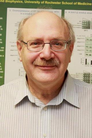 Eric M. Phizicky合作论文数Department of Biochemistry and Biophysics, University of Rochester Medical Center6
 John Grotzinger合作论文数Division of Geological and Planetary Sciences, California Institute of Technology6
 Arthur Landy合作论文数Brown University6
 Michael W. Clark合作论文数Chemistry and Biology;California Institute of Technology;Chemistry and Biology, California Institute of Technology6Daniel E Ryan合作论文数5J D Smith合作论文数5Westaway S K合作论文数5T.-H. Chang合作论文数5Chris L. Greer合作论文数California College of Medicine, University of California5
 Patrizia Fabrizio合作论文数5Chang Hee Kim合作论文数5
 Andrew J. Newman合作论文数Research Applications Laboratory, National Center for Atmospheric Research, Boulder, CO 80307-3000, USA5
 Vicente Mendoza Reyes合作论文数San Diego Supercomputer Center, University of California5LG KLEINA合作论文数4Mw Nowak合作论文数4Pc Kearney合作论文数4Rc Ogden合作论文数4
 Mohsen Company合作论文数4Andrew J Newman合作论文数4m simon合作论文数4
 Scott Emr合作论文数College of Agriculture and Life Sciences, Cornell University;Weill Institute for Cell and Molecular Biology, Department of Molecular Biology and Genetics, Cornell University4Jonathan Paul Wilson合作论文数4
 Markus Aebi合作论文数Institute of Microbiology, Eidgenössische Technische Hochschule Zürich;Department of Biology, Eidgenössische Technische Hochschule Zürich4
 Leroy Hood (莱诺·伊·胡德)合作论文数Department of Immunology, University of Washington;The Hood-Price Lab for Systems Biomedicine, Institute for Systems Biology;Providence St. Joseph Health;Department of Microbiology & Immunology, University of British Columbia;Keck Graduate Institute of Applied Life Sciences4George A. Komatsoulis合作论文数Departments of Mathematics, The University of Southern California4
 Dennis A. Dougherty合作论文数Division of Chemistry and Chemical Engineering, California Institute of Technology4
 Christine Guthrie合作论文数Department of Biochemistry and Biophysics, School of Medicine, University of California, San Francisco4
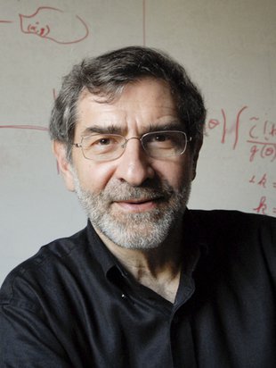 Henry A. Lester合作论文数Division of Biology and Biological Engineering, California Institute of Technology4
 Kevin Peter Hand合作论文数Jet Propulsion Laboratory, California Institute of Technology4
 Jeremy Thorner合作论文数Division of Biochemistry, Biophysics and Structural Biology, Department of Molecular and Cell Biology, College of Letters and Science, University of California, Berkeley4R Ogden合作论文数3S W Ruby合作论文数3D S Horowitz合作论文数3John H. Wilson合作论文数3soren lund jensen合作论文数3M Gefter合作论文数3JM MASSON合作论文数3john h richards合作论文数3Cg Labarca合作论文数3




