Leptomeningeal Metastases (LM) disease is the advanced stages of some complicated cancers. It Contaminates in the Cerebrospinal Fluid (CSF). Tumors might be in macroscopic or microscopic sizes. The medical operation is more risky than other cancers. Consequently, diagnosis of leptomeningeal metastases is important. Different methods are used to diagnose LM disease such as CSF examination and imaging systems Magnetic Resonance Imaging (MRI) or Computer Tomography (CT) examination. CSF examination result is more accurate compared to CT or MRI imaging systems. However imaging systems results are taken more early than CSF examination. Some details in MRI images are hidden and if the proper image enhancement method is used, the details will be revealed. Diagnosis of LM disease can be earlier with accurate results at that time. In this study, some image enhancement methods were used. The probability of result of Logarithmic Transformation (LT) method and Power-Law Transformation (PLT) method were almost the same and result was p=0.000 (p<0.001), and statistically high result was obtained. The probability of Contrast Stretching (CS) method was p=0.031 (p<0.05), and this result was statistically significant. The other four methods results were insignificant. These methods are Image Negatives Transformation (INT) method, thresholding transformations method; Gray-Level Slicing (GLS) method and Bit-Plane Slicing (BPS) method.
Estimation of the postmortem interval (PMI) has attracted the attention of many researchers. It is generally accepted as a challenging task in forensic medicine. Due to its difficulty, researchers have tried to estimate the PMI using different physical and chemical techniques. Since the PMI estimation accuracies of previous studies are not at the desired level, new methods should be developed to more accurately estimate the PMI. The development of opacity in the eye in the PMI might be an important breakthrough in this field. After death, corneal hydration occurs due to degenerated endothelial cells. The degenerated endothelial barrier of the cornea cannot prevent the flow of aqueous humor to the cornea, which results in opacity. The amount of aqueous humor in the cornea determines the level of opacity. Since the flow of aqueous humor to the cornea will continue for a while, opacity is expected to increase with the PMI. In this study, images of human eyes were investigated using computer-based image analysis. The corneal and non-corneal opacities of the recorded eye images increase during the experiment. The experimental results prove that there is a correlation between the elapsed time after death and the development of opacity in the corneal and non-corneal regions in human cases. Exponential curve fitting is employed to observe the decay of the opacity over time. A repeated ANOVA test is also used to show that the opacity development is statistically significant.
As a popular method to meathe complexity of images and generally signals, FD analyses have been used in neuroimaging studies to evaluate the morphological complexity of brain structures. The aim of this study is to perform an FD-based complexity analyses of cerebellar tissues, such as cerebellar white matter (WM), cerebellar gray matter (GM) and cerebrospinal fluid (CSF) spaces around the cerebellum, on magnetic resonance (MR) images of Chiari Malformation type-I (CM-I) patients and healthy controls. Besides, to determine the noise effects on complexity of sub cerebellar structures, two common nonlinear noise filters, median filter and bilateral filter, were applied to MR images and their performances were compared. Data of fourteen CM-I patients and sixteen normal subjects were used in this study. First, noise variance was estimated using a method based on skewness of the magnitude data. Second, as a preprocessing step, median and bilateral filters were applied on MR data separately to create different series of images for each filter. After the preprocessing, filtered brain images were segmented into three different tissues including WM, GM and CSF. Last, a 3D box-counting method was applied on segmented images to estimate the corresponding FD values. Our results showed that, while GM FD values was not significantly different between patients and controls (p = 0.051) in median filtering case, GM FD values in patients were found to be significantly lower than those in controls (p = 0.007) in bilateral filtering case. Additionally, in both cases, WM FD values in patients were found to be significantly lower than those in controls; however, this difference was more evident in bilateral filtering case (p = 0.0003) than that in median filtering case (p = 0.013). These outcomes indicated that bilateral filter was found to be more successful in discriminating CM-I patients from controls in cerebellar complexity analyses. In conclusion, results of this study revealed that noise removal is an important preprocessing step for a more successful analysis of digital images and bilateral filter is an effective filtering method for segmentation accuracy and FD analysis performance. (C) 2016 Elsevier Ltd. All rights reserved.
In this study, we investigated the effects of vibration of the whole lower leg on the content and the oxygenation of hemoglobin in the unloaded relaxed lateral gastrocnemius muscle. Vibration was applied orthogonal to and in parallel with leg axis to examine whether the extrusion of blood depends on an alignment of main vessel direction, axis of vibration and gravity.
Chiari malformation type I (CM-I), described by a descent of the cerebellar tonsils, is assumed to be a neurological developmental disorder. The aim of the present study was to investigate morphological variance in cerebellar sub-structures, including gray matter (GM), white matter (WM), and cerebrospinal fluid (CSF), using magnetic resonance (MR) images with three-dimensional (3D) fractal dimension (FD) analysis in patients with CM-I. MRI data of 16 patients and 15 control subjects were obtained, and structural complexity analyses were performed using a box-counting FD algorithm. Results showed that patients with CM-I had significantly reduced FD values for WM and CSF in comparison with controls, and statistically significant differences in cerebellar GM and CSF volumes between patients and controls were found. Moreover, a significant difference was not found between the WM volumes. This may suggest that there are changes in structural complexity in WM even when its volume is unaffected. We conclude that the findings of this preliminary study indicate the possibility of using FD analysis to understand the pathophysiology of CM-I in patients.
Objective: Cavitation and coagulation necrosis due to high temperature caused by application of the high intensity focused ultrasound (HIFU) to a focal area occurs.This feature makes HIFU possible to use in the destruction of cancerous tissue.In this study, it was aimed to determine the effect of HIFU on Balb/c mice with Ehrlich ascites tumor model to reach effective usage of HIFU with its clinical and experimental applications in various types of cancer.Methods: Ehrlich ascites tumor cells were obtained from the intraperitoneal region of a donor mouse.Fresh ehrlich ascites tumor cells were injected into 24 mice that were randomly distributed after tumor formation as a control (Group 1), sham treatment (Group 2) and HIFU treatment (Group 3).HIFU application was performed under ultrasound image until conversion of the hypoechoic tumor tissues to hyperechoic appearances.Tumor size and volumes were measured Results: Tumor sizes and volumes in all groups showed an increase after the treatment.Pathologically all tumors were solid carcinoma with an expansive growth pattern.Necrosis percentages in Group 1, Group 2 and Group 3 were 70±9.6,70±9.4 and 75.6±4.17,respectively.With regard to Volume 2, there were significant differences between Group 2 and both Group 1 and 3 (p=0.004and p=0.0001, respectively).Concluison: High intensity focused ultrasound is an effective method in Ehrlich ascites tumor model via prevention of tumor growth and production of necrosis.To achieve a reduction in tumor volume, increasing the duration of HIFU treatment and/or repeatedly application of the treatment is thought to be effective.
Summary Background: Heart rate variability (HRV) is a signal obtained from RR intervals of electro -cardiography (ECG) signals to evaluate the balance between the sympathetic nervous system and the parasympathetic nervous system; not only HRV but also pulse rate va -riability (PRV) extracted from finger pulse plethysmography (PPG) can reflect irregularities that may occur in heart rate and control procedures. Objectives: The purpose of this study is to compare the HRV and PRV during hypogly -cemia in order to evaluate the features that computed from PRV that can be used in detection of hypoglycemia. Methods: To this end, PRV and HRV of 10 patients who required testing with insulininduced hypoglycemia (IIHT) in Clinics of Endocrinology and Metabolism Diseases of Bezm-i Alem University (Istanbul, Turkey), were obtained. The recordings were done at three stages: prior to IIHT, during the IIHT, and after the IIHT. We used Bland-Altman analysis for comparing the parameters and to evaluate the correlation between HRV and PRV if exists. Results: Significant correlation (r > 0.90, p < 0.05) and close agreement were found between HRV and PRV for mean intervals, the root-mean square of the difference of successive intervals, standard deviation of successive intervals and the ratio of the low-to-high frequency power. Conclusions: In conclusion, all the features computed from PRV and HRV have close agreement and correlation according to Bland-Altman analyses’ results and features computed from PRV can be used in detection of hypoglycemia.
Complexity measures have been enormously used in schizophrenia patients to estimate brain dynamics. However, the conflicting results in terms of both increased and reduced complexity values have been reported in these studies depending on the patients' clinical status or symptom severity or medication and age status. The objective of this study is to investigate the nonlinear brain dynamics of chronic and medicated schizophrenia patients using distinct complexity estimators. EEG data were collected from 22 relaxed eyes-closed patients and age-matched healthy controls. A single-trial EEG series of 2 min was partitioned into identical epochs of 20 s intervals. The EEG complexity of participants were investigated and compared using approximate entropy (ApEn), Shannon entropy (ShEn), Kolmogorov complexity (KC) and Lempel-Ziv complexity (LZC). Lower complexity values were obtained in schizophrenia patients. The most significant complexity differences between patients and controls were obtained in especially left frontal (F3) and parietal (P3) regions of the brain when all complexity measures were applied individually. Significantly, we found that KC was more sensitive for detecting EEG complexity of patients than other estimators in all investigated brain regions. Moreover, significant inter-hemispheric complexity differences were found in the frontal and parietal areas of schizophrenics' brain. Our findings demonstrate that the utilizing of sensitive complexity estimators to analyze brain dynamics of patients might be a useful discriminative tool for diagnostic purposes. Therefore, we expect that nonlinear analysis will give us deeper understanding of schizophrenics' brain.
Introduction: Image segmentation has importance role for medical image analysis. Because increasing image-processing methods can provide more reliable information for the best interpretation of diseases. Therefore correct segmentation of the pathological and anatomical structures has critical importance in medical images. Additionally, CT scan interpretation requires lots of time and energy because the large number scans are generated for each patient. Method: For this reasons, in this study a robust and reliable fast algorithm was proposed for the lung segmentation from low-dose HRCT images. The images have been recruited from asthma subjects before and after two months treatment with low-dose limited HRCT scan. HRCT scans were collected in full inspiration for all patients, who were performed with stable and mild moderate asthma. Fuzzy Cmeans method, which is based on image histogram, was used in this study. Algorithm was evaluated on 20 asthma patients. Conclusion: The accuracy is approximately 93.7% and it takes almost a second for each slice. In future studies we are planning to study airway segmentation and measure airway values before and after treatment.
Electrogastrography (EGG) is a noninvasive technique to record the gastric electrical activity. In recent years, researchers have proved that EGG has great clinical potentials to evaluate the gastric disorders. However, it has still not been used in clinic to diagnoses of diabetic gastroparesis or some other disorders. One of the reasons of this can be related to methods using for EGG signal analyzing. Normal gastric myoelectrical activity consists of a slow wave or Electrical Control Activity (ECA) and spikes or Electrical Response Activity (ERA) potentials. Although ERA could be responsible for some changes in EGG signals, it is ignored and neither First furrier transform (FFT) based running spectral analysis (RSA) nor can discrete wavelet transform (DWT) reflect the ERA effect. Line length is an amplitude- and frequency-dependent linear feature therefore it reflects the waveform dimensionality changes. The aim of this study was to investigate the line length feature of the EGG whether it has potential for discriminate the healthy subjects (CT) from diabetic gastroparesis (GP) patients and discuss the line length capability to reflect the ERA effect on EGG. For this aim EGG signals were recorded from 20 CT and 20 GP patients who had delayed gastric emptying. The recordings were analyzed and compared in both groups' pre- and post-prandial states. The results we obtained from line length analysis of EGG signals proved that there are significant differences between the GP and CT groups is statistically significant for pre and post-prandial condition (p = 0,0298* for pre-prandial and p= 0,0032* for post-prandial). The proposed method confirmed that line length feature is useful for detecting delayed gastric emptying and using with some other features more successful computer-aided detection system can design.
Asthma is a chronic lung disease, which inflames and affects the airways, airway wall thickening, and lung density. In this study; we built up an automatic computerized method for the evaluation of the airway dimensions for asthma patients from low-dose 2D axial HRCT scans. Our proposed method concludes two frameworks. At first, right and left lung was eliminated from other structures. In the second step airway segmentation has been done using region growing and ellipse fitting method. Algorithm was evaluated on 10 mild asthma severity patients before and after drug using treatment and results were compared with respiratory function. We conclude that HRCT is a useful tool for airways evaluation in asthma patients. Because HRCT might be more sensitive for evaluate airway involvement, which is associated with airflow obstruction and diseases severity.
SummaryBackground: Spontaneous Spinal Cerebro -spinal Fluid Leaks (SSCFL) is a disease based on tears on the dura mater. Due to widespread symptoms and low frequency of the disease, diagnosis is problematic. Diagnostic lumbar puncture is commonly used for diagnosing SSCFL, though it is invasive and may cause pain, inflammation or new leakages. T2-weighted MR imaging is also used for diagnosis; however, the literature on T2-weighted MRI states that findings for diagnosis of SSCFL could be erroneous when differentiating the diseased and control. One another technique for diagnosis is CT-myelography, but this has been suggested to be less successful than T2-weighted MRI and it needs an initial lumbar puncture.Objectives: This study aimed to develop an objective, computerized numerical analysis method using noninvasive routine Magnetic Resonance Images that can be used in the evaluation and diagnosis of SSCFL disease.Methods: Brain boundaries were automatically detected using methods of mathematical morphology, and a distance transform was employed. According to normalized distances, average densities of certain sites were proportioned and a numerical criterion related to cerebrospinal fluid distribution was calculated.Results: The developed method was able to differentiate between 14 patients and 14 control subjects significantly with p = 0.0088 and d = 0.958. Also, the pre and post-treatment MRI of four patients was obtained and analyzed. The results were differentiated statistically (p = 0.0320, d = 0.853).Conclusions: An original, noninvasive and objective diagnostic test based on computerized image processing has been developed for evaluation of SSCFL. To our knowledge, this is the first computerized image processing method for evaluation of the disease. Discrimination between patients and controls shows the validity of the method. Also, post-treatment changes observed in four patients support this verdict.
Studies conducted in major depression (MD) patients have reported a high risk of cardiac morbidity as a result of the relationship between changed cardiovascular activity (CA) and autonomic dysfunctions. The investigation of heart rate variability (HRV) gives valuable idea about variances in autonomic CA of MD patients. To get this knowledge, frequency-domain HRV analysis is frequently performed using Fourier transformation (FT) or discrete-wavelet transformation (DWT) to decompose the data into high-frequency (HF) and low-frequency (LF) bands. Nevertheless, it has been reported that the FT is not useful for nonstationary HRV signals and the DWT does not ensure required frequency boundaries of each band. This study aims to compare the frequency-domain HRV features using wavelet-packet-transform (WPT) with absolutely excellent approximation to required band ranges between the controls and patients. In addition to LF and HF band energies, sympathovagal balance that indicates the variation of sympathetic and parasympathetic activities were compared between two groups. Patients had a significantly lower HF energy, higher values of LF energy and higher LF/HF ratio. Our results recommend that impairments in coordination between parasympathetic and sympathetic behavior in MD patients can be assessed by HRV analysis using WPT with high resolution decomposition for needed bands.
[Purpose] Repetitive upper extremity exercises result in overuse injuries. However, it is challenging to identify the specific causative movements. This study evaluated the effects of different shoulder positions on grip and electrophysiological activity of upper extremity muscles. [Subjects and Methods] Forty subjects ranging from 18 and 30 years of age were analyzed. Surface electromyography and hand grip strength were measured during a range of shoulder exercises and numerous signal processing methods were applied. [Results] The maximum electromyographic activity intensity was observed in the wrist extensors at various angles. Deltoid activity rose significantly during shoulder flexion. [Conclusion] Overhead work causes shoulder muscle fatigue and prevents effective hand function, which affects occupational health and efficiency. Overhead work involves a hazardous position and decreases efficiency. Ergonomic solutions should be developed to prevent muscle fatigue and decreased grip force.
In forensic medicine, estimation of the time of death (ToD) is one of the most important and challenging medico-legal problems. Despite the partial accomplishments in ToD estimations to date, the error margin of ToD estimation is still too large. In this study, electrical conductivity changes were experimentally investigated in the postmortem interval in human cases. Electrical conductivity measurements give some promising clues about the postmortem interval. A living human has a natural electrical conductivity; in the postmortem interval, intracellular fluids gradually leak out of cells. These leaked fluids combine with extra-cellular fluids in tissues and since both fluids are electrolytic, intracellular fluids help increase conductivity. Thus, the level of electrical conductivity is expected to increase with increased time after death. In this study, electrical conductivity tests were applied for six hours. The electrical conductivity of the cases exponentially increased during the tested time period, indicating a positive relationship between electrical conductivity and the postmortem interval.
In this study, A graphical user interface based on MATLAB® was developed for an easy filtering of digital images and comparison of several image denoising methods. As well as standard digital images, medical image formats can also be used with the help this program. Additionally, to evaluate the performances of different noise filtering methods, some measures such as signal to noise ratio are calculated by this application.
Objectives: Complexity measures have been enormously used in schizophrenia patients to estimate brain dynamics. However, the conflicting results in terms of both increased and reduced complexity values have been reported in these studies depending on the patients' clinical status or symptom severity or medication and age status. The objective of this study is to investigate the nonlinear brain dynamics of chronic, medicated schizophrenia patients and healthy control subjects using Katz's fractal dimension (FD). Moreover, in order to determine noise effect on complexity of EEG data, a noise elimination method based on wavelet and singular spectrum analysis (SSA) were assessed.Methods: Twenty-two schizophrenia patients and twenty-two age- and gender-matched control subjects underwent a resting state EEG examination with 120s. The discrete wavelet transform (DWT) was applied for EEG decomposition. Using a SSA approach, noise was removed and EEG reconstructed by inverse wavelet transform. The brain complexity of participants was investigated and compared using Katz's FD obtained from original and preprocessed EEG data.Results: Lower complexity values were found in schizophrenia patients. However, this difference was only statistically significant for each channel in preprocessed, noiseless EEG data. The most significant complexity differences between patients and controls were obtained in left frontal and parietal regions of the brain.Conclusion: Our findings demonstrate that the utilizing of complexity measures with preprocessing approaches on EEG data to analyze schizophrenics' brain dynamics might be a useful and discriminative tool for diagnostic purposes. Therefore, we expect that nonlinear analysis will give us more valuable results for understanding of schizophrenics' brain. (C) 2014 Elsevier Ltd. All rights reserved.
Chiari Malformations are serious neurological defects involving herniation of hindbrain tissues such as cerebellar tonsils, brainstem and IV ventricle into the spinal canal through the foramen magnum. By the severity of cerebellar descent, these malformations are classified into four different types. Clinically the least obvious and the mildest one is named as type I and defined as the descent of cerebellar tonsils into the cervical canal more than 5 mm. Magnetic Resonance Images (MRI) of brain in the sagittal plane provides the best clues in the diagnosis of the Chiari Malformation type I (CM-I). Previous studies investigated the morphological characteristics of cerebellum and nearby regions such as brain stem and fourth ventricle. Aim of this study is to analyze the cerebellar regions in chiari patients and healthy controls to search for the discriminative properties between the two groups. Sagittal brain MRI of eleven chiari patients and gender matched controls were used in order to examine the area of sub-cerebellar tissues such as gray matter (GM) and white matter (WM) and the area ratio between GM and WM. A graphical user interface (GUI) for implementing image processing techniques was developed using MATLAB environment. By means of GUI, the region embracing the whole cerebellum tissue on the mid-sagittal MR images were manually extracted. In addition, using Statistical Parametric Mapping (SPM) package the MRI slices were segmented into GM and WM tissues. Using the extracted cerebellum region as a mask, the cerebellar GM and WM tissues were achieved and the corresponding areas were computed by counting the number of pixels on each GM and WM slice. According to the statistical results, it has been found that cerebellar GM areas of the patients are significantly higher than the values of controls. As a consequence, this approach may provide a discriminative feature between patients with CM-I and health control subjects. Keywords : Chiari malformation, magnetic resonance imaging, segmentation, gray matter, white matter
[Purpose] Whole-body vibration (WBV) can induce reflex responses in muscles. A number of studies have reported that the physiological mechanisms underlying this type of reflex activity can be explained by reference to a stretch-induced reflex. Thus, the primary objective of this study was to test whether the WBV-induced muscular reflex (WBV-IMR) can be explained as a stretch-induced reflex. [Subjects and Methods] The present study assessed 20 healthy males using surface electrodes placed on their right soleus muscle. The latency of the tendon reflex (T-reflex) as a stretch-induced reflex was compared with the reflex latency of the WBV-IMR. In addition, simulations were performed at 25, 30, 35, 40, 45, and 50 Hz to determine the stretch frequency of the muscle during WBV. [Results] WBV-IMR latency (40.5 +/- 0.8 ms; 95% confidence interval [CI]: 39.0-41.9 ms) was significantly longer than T-reflex latency (34.6 +/- 0.5 ms; 95% CI: 33.6-35.5 ms) and the mean difference was 6.2 ms (95% CI of the difference: 4.7-7.7 ms). The simulations performed in the present study demonstrated that the frequency of the stretch signal would be twice the frequency of the vibration. [Conclusion] These findings do not support the notion that WBV-IMR can be explained by reference to a stretch-induced reflex.
In this study, the EEG signals of major depression (MD) patients and healthy control subjects were investigated using different chaotic features. The acquired signals during 3 minutes were compared using complexity measures such as Katz fractal, Higuchi fractal dimension, Lempel-Ziv complexity (LZC) and Kolmogorov complexity (KC) in MATLAB between two groups. In order to determine which complexity measure is more effective in discriminating MD patients from control subjects, statistical variance analyses were performed. As a result, it was found that patients had increased EEG complexity and better discrimination were obtained using the LZC and KC.
 Fatma Latifoglu合作论文数medical Research Group, Erciyes University26
 Salih Gunes合作论文数Department of Electrical and Electronics Engineering, Selcuk University19
 Sukru Okkesim合作论文数18
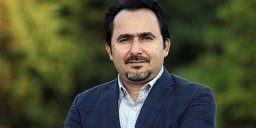 Vedat Bilgiç合作论文数16
 Saime Akdemir Akar合作论文数14
 Kemal Polat合作论文数Department of Electrical and Electronics Engineering, Abant Izzet Baysal University14
 Aysegul Guven合作论文数Department of Biomedical Engineering, Erciyes University13
 Adem Kiris合作论文数Faculty of Medicine, Firat University10engin akar合作论文数8
 Halil Ibrahim Cakar合作论文数Electr. & Electron. Eng., Fatih Univ.;stanbul;c;Electr. & Electron. Eng., Fatih Univ.8
 Hidayet Akdemir合作论文数Medical School, Erciyes University8Mustafa Okandan合作论文数Independent Biomedical Technology Consultant8
 Mustafa Yıldırım合作论文数Graduate School of Natural and Applied Sciences, Erciyes University7Fatma Dirgenali合作论文数7Sabri Altunkaya合作论文数Selçuk Üniversitesi;Alaeddin Keykubat Kampüsü6
 Yuksel Ozbay合作论文数Engineering and Architecture Faculty, Selcuk University5Murat Ceylan合作论文数Department of Electrical-Electronics Engineering, Selcuk University5
 Bekir Hakan Aksebzeci合作论文数Department of Electrical & Electronics Engineering, Erciyes University5
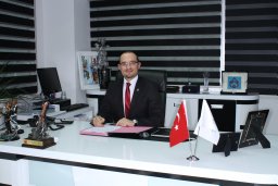 Tancan Uysal合作论文数Department of Orthodontics, Erciyes University5
 Saadetdin Herdem合作论文数Engineering and Architecture Faculty, Selcuk University5
 Musa H. Asyali合作论文数Biomedical Engineering Department, University of Southern California5
 Serap Aydin合作论文数Engineering Faculty, Ondokuz Mayıs University5Semra İçer合作论文数Department of Biomedical Devices Technology, Erciyes University5Rahime Ceylan合作论文数Engineering and Architecture Faculty, Selcuk University5Esma G Kaya合作论文数4
 Mehmet Güney合作论文数Dept Cardiovasc Surg, Dr Siyami Ersek Thorac & Cardiovasc Surg Training4
 Mehmet Akif Özçoban合作论文数Elektron. Teknolojisi Programi, Gedik Univ.;c;Elektron. Teknolojisi Programi, Gedik Univ.4
 Musa H Asyali合作论文数4Hatice Ozbilge合作论文数Faculty of Pharmacy, Erciyes University4
 Bayram Akdemir合作论文数Department of Electrical and Electronics Engineering, Selcuk University4
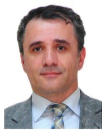 Ilhan Karacan合作论文数Cerrahpaşa Medical Faculty, Istanbul University4Niyazi Gormus合作论文数Cardiovascular Surgery, TR4Onur Toker合作论文数Department of Electrical and Computer Engineering, Florida Polytechnic University4
 Gulsah Kinali合作论文数3Sumeyra Agambayev合作论文数School of Biological and Health Sciences Engineering, Arizona State University3
 Omer Isik合作论文数3
 Erdal Korkmaz合作论文数Dept Elect & Elect Engn, Fatih Univ3
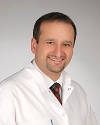 Yagci Ahmet合作论文数3Emine Doganay合作论文数3Kahraman, Y.合作论文数3
 Erkan Imal合作论文数Sch. of Eng., Sussex Univ.;c;Sch. of Eng., Sussex Univ.3Özgür Er合作论文数3Ali Salli合作论文数3Muharrem Cidem合作论文数3M. G. Kaya合作论文数2S Kemaloglu合作论文数2Gamze Çelik合作论文数2Sinem Aydin合作论文数Dept Biol, Giresun Univ2Kemal Sitki Türker合作论文数2




